. 2025 Sep 9;122(36):e2502640122.
doi: 10.1073/pnas.2502640122. Epub 2025 Sep 4.

12/15-lipoxygenase orchestrates murine wound healing via PPARγ-activating oxylipins acting holistically to dampen inflammation

Affiliations

12/15-lipoxygenase orchestrates murine wound healing via PPARγ-activating oxylipins acting holistically to dampen inflammation

Christopher P Thomas et al. Proc Natl Acad Sci U S A. .

Abstract

12/15-lipoxygenase (12/15-LOX, Alox15) generates bioactive oxygenated lipids during inflammation, however its homeostatic role(s) in normal healing are unclear. Here, the role of 12/15-LOX in resolving skin wounds was elucidated, focusing on how its lipids act together in physiologically relevant amounts. In mice, wounding caused acute appearance of 12/15-LOX-expressing macrophages and stem cells, coupled to early generation of ~12 monohydroxy-oxylipins and enzymatically oxidized phospholipids (eoxPL). Alox15 deletion increased collagen deposition, stem cell/fibroblast proliferation, IL6/pSTAT3, pSMAD3, and interferon (IFN)-γ levels. Conversely, CD206 expression, F480+ cells, and MMP9 and MMP2 activities were reduced. Alox15-/- skin was deficient in PPARγ/adiponectin activity. Furthermore, while pro-inflammatory genes were upregulated as normal during wounding, many including Il6, Il1b, ccl4, Cd14, Cd274, Clec4d, Clec4e, Csf3, Cxcl2, and miR-21 failed to revert to baseline during healing, indicating disruption of PPARγ's anti-inflammatory brake on NLRP3/inflammasome and TGF-β signaling. Reconstituting Alox15-/- wounds with a physiological mixture of Alox15-derived primary oxylipins generated by healing wounds restored MMP and dampened collagen deposition. The oxylipin mixture activated the PPARγ response element in vitro, while in vivo, its coactivator, Helz2, was significantly upregulated as well as several fatty acid and prostaglandin PPARγ ligands. Additional inflammatory and proliferative gene networks impacted by Alox15-/- included Elf4, Cebpb, and Tcf3. In summary, 12/15-LOX generates abundant monohydroxy oxylipins that act together via PPARγ. The identification of multiple gene alterations reveals several targets for treating nonhealing wounds. Our studies demonstrate that 12/15-LOX oxylipins act in concert, dampening inflammation in vivo, revealing the need to consider lipid signaling holistically.

Keywords: lipid; lipoxygenase; oxylipin; wound.


Conflict of interest statement

Competing interests statement:The authors declare no competing interest.

Figures
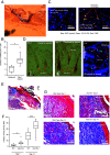
Fig. 1.
Fig. 1.
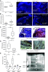
Wounding increases macrophage and hair follicle 12/15-LOX expression, and collagen levels. Panel (A) Representative image of wound architecture. Panel (B) Induction of macrophage 12/15-LOX by wounding 12/15-LOX+ve(green)/F4-80+ve(red)/DAPI+ve(blue) cells were measured at day 1 post wounding (n = 5/group), data were analyzed using one-way ANOVA with the Tukey post hoc test *P < 0.05, **P < 0.01. Panel (C) Representative images from Panel (B). Panels (D) Expression of 12/15-LOX in hair follicles near the wound edge postwounding. Hair shafts are shown on day 1, wild-type mice. The Left and Center panels show 12/15-LOX DAB+ve staining, and the Right panel shows 12/15-LOX+ve green fluorescent staining (with DAPI counterstain). Panel (E) Representative image of wound at low magnification showing region stained for collagen. Panel (F) Collagen is elevated in Alox15−/− on days 7 and 14. Wounds were harvested and analyzed for collagen using Masson’s Trichrome staining (collagen: blue, epithelial cells: deep red, nuclei: black, noncollagen structures: pink) and pixels counted (n = 10-14/group). Unpaired Student’s t test, comparing WT and Alox15−/− separately *P < 0.05, ***P < 0.005. Panel (G) Representative images from Panel (F).
Fig. 2.
Fig. 2.
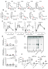
Alox15−/− wounds show elevated pSTAT3, pSMAD3, and INFγ but decreased CD206 and MMP activities. Panel (A) Alox15−/− wounds show elevated pSTAT3. pSTAT3 was measured using fluorescence immunohistochemistry. n = 5-6/group. Panel (B) Alox15−/ wounds show elevated pSMAD3. pSMAD3 was measured using fluorescence immunohistochemistry. n = 10-11/group. Panel (C) Alox15−/− wounds show elevated IFNγ. IFNγ was measured using DAB immunohistochemistry. n = 9/group (4 to 6 fields per wound). Panel (D) Alox15−/− wounds show reduced CD206 expression. CD206 was measured using DAB immunohistochemistry. n = 5/group (3 to 6 fields per wound). For all panels, data were analyzed an unpaired t test, *P < 0.05, **P < 0.01. Right panels show representative images for all the proteins analyzed. Panel (E) MMP activities are reduced in Alox15−/− wounds. MMP activities were measured using zymography. n = 6/group. The ladder shows proteins corresponding to 250, 148, 98, 64, and 50 kDa. Image J was used to calculate the density of each band. The gel is shown (Right). The impact of Alox12−/− was analyzed using an unpaired t test, mean ± SEM, *P < 0.05, **P < 0.01, ***P < 0.005.
Fig. 3.
Fig. 3.
Alox15−/− wounds show lower levels of many oxylipins and 12-HETE-PEs, while physiological levels of high oxylipins restore MMP activity. Panels (A and B) Oxylipins are rapidly elevated postwounding, but many are reduced in Alox15−/− wounds. Oxylipins were measured using LC/MS/MS as outlined in Methods. n = 6 samples/time point, with 4 wounds pooled/sample. For all panels, differences between groups were analyzed using two-way ANOVA (red stars), with the Bonferroni post hoc test between individual time points (black stars), mean ± SEM, *P < 0.05, **P < 0.01, ***P < 0.005. Panel (C) HETE-PEs elevate during healing, peaking on day 7, with significant loss of 12-HETE-PE isomers in Alox15−/− wounds. Oxidized phospholipids were measured using LC/MS/MS as outlined in Methods (n = 5 samples/time point, with 4 wounds pooled/sample). Unpaired t test, *P < 0.05, **P < 0.01, ***P < 0.005. Panel (D) High oxylipins restored MMP activities in wounds from Alox15−/− mice. Post wounding, lipids were added to wounds as indicated in Methods every second day. Wounds were harvested and analyzed for MMP activities using zymography as in Methods (n = 4, 4 to 5 wounds pooled/sample). Panel (E). A gel showing representative data from Panel (C). Panel (F) High oxylipins restore MMP activities to wild-type level. Wounds were harvested and analyzed for MMP activities using zymography as in Methods (n = 4, 4 to 5 wounds pooled/sample). For Panels (C and D) ANOVA with the Tukey post hoc test was used, *P < 0.05, **P < 0.01, ***P < 0.005.
Fig. 4.
Fig. 4.
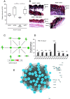
High oxylipins dampen collagen deposition, while unwounded Alox15−/− skin shows reduced PPARγ activity but a normal pro-inflammatory response to wounding, while a series of highly correlated genes fail to revert to baseline in Alox15−/− wounds at Day 7. Panel (A) Collagen generation is dampened by high oxylipins. Post wounding, lipids were added to wounds as indicated in Methods every second day. Wounds were harvested at Day 7 and analyzed for collagen using Masson’s Trichrome staining (collagen: blue, epithelial cells: deep red, nuclei: black, noncollagen structures: pink) and pixels counted (n = 5-6 wounds/group. Panel (B) Representative images from Panel (A). Panel (C) Unwounded Alox15−/− skin shows significantly reduced PPARγ/adiponectin expression/activity. RNA-seq was carried out as indicated in Methods on nonwounded skin (n = 3 to 4 samples per group, each sample was a pool of 4 wounds per animal with 2 animals per pool = 8 wounds per sample). All genes shown are significantly reduced in Alox15−/− skin and are either controlled by or regulate PPARγ/adiponectin. Panel (D) Upregulation of a series of canonical “inflammatory” genes is preserved in Alox15−/− wounds. Gene expression was normalized for each gene to its Day 0 mean value, then expressed as fold-change (n = 3 to 4 per group) each sample was a pool of 4 wounds per animal with 2 animals per pool = 8 wounds per sample). For Panel (A), ANOVA with the Tukey post hoc test. Panel (E) A large number of genes that are significantly different between wild-type and Alox15−/− wounds at Day 7 highly correlate across the whole timecourse, indicating coordinated regulation. Genes that were found to be significantly differentially expressed at Day 7 were analyzed in Cytoscape, using their expression levels for the entire time-course, with correlation [r] > 0.8 shown.
Fig. 5.
Fig. 5.
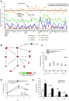
Genes that fail to revert at day 7 in Alox15−/− wounds remain 50% elevated above baseline, and many are controlled through NLRP3, IFNβ and IL-1, PPARγ expression is not upregulated during wounding, while Helz2 and unsaturated fatty acids are increased. Panel (A) All genes from the highly correlated network in Fig. 4E typically remain 50% elevated. Data from gene expression of highly correlated genes were averaged and normalized to day 4, wild-type mean (the inflammatory response level) (n = 3 to 4 per group). Panel (B) IPA network analysis of genes that are significantly different at Day 7 reveals significantly higher levels of Inflammasome, IFNb, and IL-1 pathways. Gene expression data from Day 7 were analyzed using IPA. Panel (C) Pparg expression are not increased during wounding. Transcriptional data on Pparg were compared across the timecourse (n = 3 to 4 per group). For all gene expression data, Student’s t test, followed by Benjamin Hochberg correction: *P < 0.05, **P < 0.01, ***P < 0.005. Panel (D) Data from gene expression are shown for WT and Alox15−/− wounds during the time course (n = 3 to 4 per group). For all gene expression data, Student’s t test, followed by Benjamin Hochberg correction: *P < 0.05, **P < 0.01, ***P < 0.005. Panel (E) UFA are significantly elevated in early wounds. Lipids were extracted from unwounded and wounded (day 1) skin, extracted and derivatized as outlined in Methods before UFA were quantified using LC/MS/MS (n = 3, unwounded, 4, day 1 wounded).

References

    1. Heydeck D., et al. , Interleukin-4 and -13 induce upregulation of the murine macrophage 12/15-lipoxygenase activity: Evidence for the involvement of transcription factor STAT6. Blood 92, 2503–2510 (1998). - PubMed
    1. Kuhn H., O’Donnell V. B., Inflammation and immune regulation by 12/15-lipoxygenases. Prog. Lipid Res. 45, 334–356 (2006). - PubMed
    1. Cyrus T., et al. , Disruption of the 12/15-lipoxygenase gene diminishes atherosclerosis in apo E-deficient mice. J. Clin. Invest. 103, 1597–1604 (1999). - PMC - PubMed
    1. Bleich D., et al. , Resistance to type 1 diabetes induction in 12-lipoxygenase knockout mice. J. Clin. Invest. 103, 1431–1436 (1999). - PMC - PubMed
    1. Anning P. B., et al. , Elevated endothelial nitric oxide bioactivity and resistance to angiotensin-dependent hypertension in 12/15-lipoxygenase knockout mice. Am. J. Pathol. 166, 653–662 (2005). - PMC - PubMed

MeSH terms

LinkOut - more resources