Multicenter Study
. 2025 Sep 23;105(6):e214075.
doi: 10.1212/WNL.0000000000214075. Epub 2025 Sep 4.

Deep Learning Modeling to Differentiate Multiple Sclerosis From MOG Antibody-Associated Disease

Rosa Cortese  1 Francesco Sforazzini  2 Giordano Gentile  1   2 Anna de Mauro  1 Ludovico Luchetti  1   2 Maria Pia Amato  3   4 Samira Luisa Apóstolos-Pereira  5 Georgina Arrambide  6 Barbara Bellenberg  7 Alessia Bianchi  1   8 Alvino Bisecco  9 Benedetta Bodini  10   11 Massimiliano Calabrese  12 Valentina Camera  12 Elisabeth G Celius  13 Carolina de Medeiros Rimkus  14 Yunyun Duan  15 Françoise Durand-Dubief  16 Massimo Filippi  17   18   19   20   21 Antonio Gallo  9 Claudio Gasperini  22 Cristina Granziera  23   24   25 Sergiu Groppa  26 Matthias Grothe  27 Mor Gueye  17   18   21 Matilde Inglese  28   29 Anu Jacob  30   31 Caterina Lapucci  29 Andrea Lazzarotto  10   11 Yaou Liu  15 Sara Llufriu  32 Carsten Lukas  7   33 Romain Marignier  16 Silvia Messina  34 Jannis Müller  23   24 Jacqueline Palace  34 Luisa Pastó  35 Friedemann Paul  36 Ferran Prados  8   37   38 Anne-Katrin Pröbstel  24 Àlex Rovira  39 Maria Assunta Rocca  17   18   21 Serena Ruggieri  22 Jaume Sastre-Garriga  6 Douglas Kazutoshi Sato  40 Ruth Schneider  7   33 Maria Sepulveda  32 Piotr Sowa  41 Bruno Stankoff  10   11 Carla Tortorella  22 Frederik Barkhof  42   43 Olga Ciccarelli  8   44 Marco Battaglini  1   2 Nicola De Stefano  1 MAGNIMS Study Group
Affiliations
Multicenter Study

Deep Learning Modeling to Differentiate Multiple Sclerosis From MOG Antibody-Associated Disease

Rosa Cortese et al. Neurology. .

Abstract

Background and objectives: Multiple sclerosis (MS) is common in adults while myelin oligodendrocyte glycoprotein antibody-associated disease (MOGAD) is rare. Our previous machine-learning algorithm, using clinical variables, ≤6 brain lesions, and no Dawson fingers, achieved 79% accuracy, 78% sensitivity, and 80% specificity in distinguishing MOGAD from MS but lacked validation. The aim of this study was to (1) evaluate the clinical/MRI algorithm for distinguishing MS from MOGAD, (2) develop a deep learning (DL) model, (3) assess the benefit of combining both, and (4) identify key differentiators using probability attention maps (PAMs).

Methods: This multicenter, retrospective, cross-sectional MAGNIMS study included scans from 19 centers. Inclusion criteria were as follows: adults with non-acute MS and MOGAD, with high-quality T2-fluid-attenuated inversion recovery and T1-weighted scans. Brain scans were scored by 2 readers to assess the performance of the clinical/MRI algorithm on the validation data set. A DL-based classifier using a ResNet-10 convolutional neural network was developed and tested on an independent validation data set. PAMs were generated by averaging correctly classified attention maps from both groups, identifying key differentiating regions.

Results: We included 406 MRI scans (218 with relapsing remitting MS [RRMS], mean age: 39 years ±11, 69% F; 188 with MOGAD, age: 41 years ±14, 61% F), split into 2 data sets: a training/testing set (n = 265: 150 with RRMS, age: 39 years ±10, 72% F; 115 with MOGAD, age: 42 years ±13, 61% F) and an independent validation set (n = 141: 68 with RRMS, age: 40 years ±14, 65% F; 73 with MOGAD, age: 40 years ±15, 63% F). The clinical/MRI algorithm predicted RRMS over MOGAD with 75% accuracy (95% CI 67-82), 96% sensitivity (95% CI 88-99), and specificity 56% (95% CI 44-68) in the validation cohort. The DL model achieved 77% accuracy (95% CI 64-89), 73% sensitivity (95% CI 57-89), and 83% specificity (95% CI 65-96) in the training/testing cohort, and 70% accuracy (95% CI 63-77), 67% sensitivity (95% CI 55-79), and 73% specificity (95% CI 61-83) in the validation cohort without retraining. When combined, the classifiers reached 86% accuracy (95% CI 81-92), 84% sensitivity (95% CI 75-92), and 89% specificity (95% CI 81-96). PAMs identified key region volumes: corpus callosum (1872 mm3), left precentral gyrus (341 mm3), right thalamus (193 mm3), and right cingulate cortex (186 mm3) for identifying RRMS and brainstem (629 mm3), hippocampus (234 mm3), and parahippocampal gyrus (147 mm3) for identifying MOGAD.

Discussion: Both classifiers effectively distinguished RRMS from MOGAD. The clinical/MRI model showed higher sensitivity while the DL model offered higher specificity, suggesting complementary roles. Their combination improved diagnostic accuracy, and PAMs revealed distinct damage patterns. Future prospective studies should validate these models in diverse, real-world settings.

Classification of evidence: This study provides Class III evidence that both a clinical/MRI algorithm and an MRI-based DL model accurately distinguish RRMS from MOGAD.


Conflict of interest statement

R. Cortese was awarded a MAGNIMS-ECTRIMS fellowship in 2019; received speaker honoraria/travel support from Roche, Merck Serono, UCB, Sanofi Genzyme, Alexion, Novartis, and Janssen; and received a research grant from the Italian Ministry of University and Research, Research Project of Relevant National Interest (PRIN), project code: 2022PR3PEY. M.P. Amato has served on Scientific Advisory Boards for Biogen, Novartis, Roche, Merck, Sanofi Genzyme, and Teva; received speaker honoraria from Biogen, Merck, Sanofi Genzyme, Roche, Novartis, and Teva; and received research grants for her Institution from Biogen, Merck, Sanofi Genzyme, Novartis, and Roche. G. Arrambide has received speaking honoraria; has received compensation for consulting services or participation in advisory boards from Merck, Roche, and Horizon Therapeutics; has received travel support for scientific meetings from Novartis, Roche, and ECTRIMS; is editor of Europe of the Multiple Sclerosis Journal – Experimental, Translational and Clinical; is a member of the International Women in Multiple Sclerosis (iWiMS) network executive committee; and is a member of the European Biomarkers in MS (BioMS-eu) consortium steering committee. A. Bisecco received speaker's honoraria and/or compensation for consulting service and/or speaking activities and/or travel grants from Biogen, Roche, Novartis, Merck, Alexion, Amgen, UCB, and Genzyme. B. Bodini has received traveling and speaker's honoraria from Novartis, Genzyme, Roche, and Merck Serono; and received research support from Biogen. M. Calabrese received speaker honoraria from Biogen, Bristol Myers Squibb, Celgene, Genzyme, Merck Serono, Novartis, and Roche; and receives research support from the Progressive MS Alliance and Italian Minister of Health. E.G. Celius has received honoraria for lecturing and advice from Biogen, Merck, and Roche, and grants and honoraria from Novartis and Sanofi Genzyme. C. de Medeiros Rimkus was awarded an MSIF-ECTRIMS fellowship in 2021; is an associate editor of Arquivos de Neuropsiquiatria, and has received speaker honoraria from Roche Pharma, Novartis, and Guerbet. M. Filippi is Editor-in-Chief of the Journal of Neurology, an associate editor of Human Brain Mapping, Radiology, and Neurological Sciences; has received compensation for consulting services from Alexion, Almirall, Biogen, Merck, Novartis, Roche, and Sanofi Genzyme; for speaking activities from Bayer, Biogen, Celgene, Chiesi Italia SpA, Eli Lilly, Genzyme, Janssen, Merck-Serono, Neopharmed Gentili, Novartis, Novo Nordisk, Roche, Sanofi Genzyme, Takeda, and TEVA; for participation in advisory boards for Alexion, Biogen, Bristol Myers Squibb, Merck, Novartis, Roche, Sanofi Aventis, Sanofi Genzyme, and Takeda; and for scientific direction of educational events for Biogen, Merck, Roche, Celgene, Bristol Myers Squibb, Lilly, Novartis, and Sanofi Genzyme; and receives research support from Biogen Idec, Merck-Serono, Novartis, Roche, Italian Ministry of Health, Fondazione Italiana Sclerosi Multipla, and ARiSLA (Fondazione Italiana di Ricerca per la SLA). C. Gasperini received fees as an invited speaker or travel expenses for attending meeting from Biogen, Merck‐Serono, Teva, Mylan, Sanofi Genzyme, and Novartis. The University Hospital Basel (USB), as the employer of C. Granziera, has received the following fees that were used exclusively for research support: (1) advisory board and consultancy fees from Actelion, Sanofi Genzyme, Novartis, GeNeuro, and Roche; (2) speaker fees from Sanofi Genzyme, Novartis, GeNeuro, and Roche; (3) research support from Siemens, GeNeuro, and Roche. C. Granziera is supported by the Swiss National Science Foundation (SNSF) grant PP00P3_176984, the Stiftung zur Förderung der gastroenterologischen und allgemeinen klinischen Forschung, and the EUROSTAR E!113682 HORIZON2020. M. Grothe has received compensation for serving on scientific advisory boards for Novartis, Roche, and Sanofi Genzyme; received speaker honoraria and travel support from Merck, Novartis, Roche, Sanofi Genzyme, and Teva; and received research support from Novartis. M. Inglese received grants from NIH, NMSS, FISM, and Ministero dell'Università e ricerca (MUR); and received fees for consultation from BMS, Janssen, Roche, Genzyme, Merck, Biogen, and Novartis. C. Lapucci has received honoraria for speaking, travel grants, and participating in advisory boards from Merck, Sanofi, Novartis, Roche, and Alexion. S. Llufriu received compensation for consulting services and speaker honoraria from Biogen Idec, Novartis, Bristol Myers Squibb, Sanofi, Johnson & Johnson, and Merck. C. Lukas received a research grant from the German Federal Ministry for Education and Research, BMBF, and German Competence Network Multiple Sclerosis (KKNMS, grant 01GI1601I); and has received consulting and speaker's honoraria from Biogen Idec, Bayer Schering, Daiichi Sanykyo, Merck Serono, Novartis, Sanofi, Genzyme, and TEVA. J. Müller has received funding from the Swiss National Science Foundation (grants P500PM_214230 and P5R5PM_225288). J. Palace has received support for scientific meetings and honoraria for advisory work from Merck Serono, Novartis, Chugai, Alexion, Roche, Medimmune, Argenx, UCB, Mitsubishi, Amplo, Janssen, and Sanofi; has received grants from Alexion, Roche, Medimmune, UCB, and Amplo Biotechnology; has patent ref. P37347WO and licence agreement Numares multimarker MS diagnostics Shares in AstraZeneca; and acknowledges partial funding by highly specialized services NHS England. F. Paul serves on scientific advisory boards for Novartis, Viela Bio, and Alexion; and has received speaker honoraria from Bayer, Teva, Merck, Viela, Alexion, Roche, and Novartis. F. Prados received a Guarantors of Brain fellowship 2017–2020 and is supported by National Institute for Health Research (NIHR), Biomedical Research Centre initiative at University College London Hospitals (UCLH). A.K. Pröbstel has participated as speaker in meetings sponsored by and received consulting fees and/or grant support from Biogen and Roche. A. Rovira serves/ed on scientific advisory boards for Novartis, Sanofi Genzyme, Synthetic MR, TensorMedical, Roche, and Biogen; has received speaker honoraria from Bayer, Sanofi Genzyme, Merck-Serono, Teva Pharmaceutical Industries Ltd., Novartis, Roche, Bristol Myers, and Biogen; is CMO and cofounder of TensorMedical; and receives research support from Fondo de Investigación en Salud (PI19/00950) from Instituto de Salud Carlos III, Spain. M.A. Rocca received consulting fees from Biogen, Bristol Myers Squibb, Eli Lilly, Janssen, and Roche; received speaker honoraria from AstraZeneca, Biogen, Bristol Myers Squibb, Bromatech, Celgene, Genzyme, Horizon Therapeutics Italy, Merck Serono SpA, Novartis, Roche, Sanofi, and Teva; receives research support from the MS Society of Canada, the Italian Ministry of Health, the Italian Ministry of University and Research, and Fondazione Italiana Sclerosi Multipla; and is an associate editor of Multiple Sclerosis and Related Disorders. J. Sastre-Garriga serves as a co-editor of Europe for the Multiple Sclerosis Journal and as Editor-in-Chief of Revista de Neurología; receives research support from Fondo de Investigaciones Sanitarias (19/950 and 22/750); and in the last 12 months, has served as a consultant/speaker for BMS, Roche, Sanofi, Janssen, and Merck. D.K. Sato received research support from CNPq/Brazil (425331/2016-4, 308636/2019-8, and 306016/2022-2), FAPERGS/Brazil (17/2551-0001391-3 and 21/2551-0000077-5), Teva, Merck, and Biogen for investigator-initiated studies and speaker honoraria from Horizon, Amgen, Alexion, AstraZeneca, Novartis, and Roche; and participated in advisory boards for Horizon, Roche, Alexion, and AstraZeneca. R. Schneider has received consulting and speaker honoraria from Merck, Biogen-Idec, GmbH, Bayer HealthCare, Alexion Pharma, Novartis Pharma, and Roche Pharma AG; and has received research scientific grant support from Novartis Pharma. M. Sepulveda received speaking honoraria from Roche and Biogen; and received travel reimbursement from Roche, Biogen, and Sanofi for national and international meetings. B. Stankoff has received grants and personal fees for lectures from Roche, Sanofi Genzyme, and Merck-Serono; and personal fees for lectures from Novartis and Janssen. C. Tortorella received honoraria for speaking and travel grants from Biogen, Sanofi-Aventis, Merck Serono, Bayer-Schering, Teva, Genzyme, Roche, Alexion, Horizon, Celgene, Almirall, and Novartis. F. Barkhof is a steering committee or data safety monitoring board member for Biogen, Merck, Eisai, and Prothena; is an advisory board member for Combinostics, Scottish Brain Sciences, and Alzheimer Europe; is a consultant for Roche, Celltrion, Rewind Therapeutics, Merck, and Bracco; has research agreements with ADDI, Merck, Biogen, GE Healthcare, and Roche; and is cofounder and shareholder of Queen Square Analytics LTD. O. Ciccarelli is an NIHR Research Professor (RP-2017-08-ST2-004); acts as a consultant for Biogen, Merck, Novartis, Roche, and Lundbeck; and has received research grant support from the MS Society of Great Britain and Northern Ireland, the NIHR UCLH Biomedical Research Centre, the Rosetree Trust, the National MS Society, and the NIHR-HTA. N. De Stefano has received honoraria from Biogen-Idec, Bristol Myers Squibb, Celgene, Genzyme, Immunic, Merck Serono, Novartis, Roche, and Teva for consulting services, speaking, and travel support; serves on advisory boards for Merck, Novartis, Biogen-Idec, Roche, Genzyme, and Immunic; and has received research grant support from the Italian MS Society. All other authors report no relevant disclosures. Go to Neurology.org/N for full disclosures.

Figures

Figure 1
Figure 1. Schematic Representation of the Study Design
The work was structured in 3 steps: (A) FLAIR/T2 sequences of the scans from the validation test set were assessed and scored considering the number of lesions and the presence of Dawson fingers (blue). (B) ResNet-10 architecture was trained using cross-entropy loss function and then accuracy was assessed on the validation test set (green). Then, individual attention maps were created using a guided backpropagation approach and then normalized using z-scores. Finally, 2 PAMs (1 for each disease group) were created by nonlinearly registering to MNI and then averaging all the correctly classified attention maps of RRMS and MOGAD, respectively (yellow). (C) The DL-based and clinical/MRI classifiers were combined to assess their performances in differentiating the 2 disorders (red). In brief, both the number of lesions and the presence of Dawson fingers were concatenated to the features extracted by the DL network and used to discriminate between the 2 classes. CNN = convolutional neural network; DL = deep learning; FLAIR = fluid-attenuated inversion recovery; MNI = Montreal Neurological Institute; MOGAD = myelin oligodendrocyte glycoprotein antibody–associated disease; MRI = magnetic resonance imaging; PAMs = probability attention maps; RRMS = relapsing remitting multiple sclerosis.
Figure 2
Figure 2. Topographical Distributions of the Areas Significantly Contributing to the Differentiation Between Patients With RRMS and With MOGAD
PAMs revealed the involvement of corpus callosum, right cingulate cortex, right thalamus, and left precentral gyrus for the identification of RRMS (A) while the relevant role of the brainstem, bilateral hippocampi, and parahippocampal gyri in the identification of MOGAD (B). Significant voxels are shown in a color scale from light blue to dark blue for RRMS and from yellow to red for MOGAD, from the most to the less significant, respectively (all p < 0.001). MOGAD = myelin oligodendrocyte glycoprotein antibody–associated disease; PAMs = probability attention maps; RRMS = relapsing remitting multiple sclerosis.
Figure 3
Figure 3. Voxel-Wise Comparison of RRMS and MOGAD Lesion Maps
The voxel-wise comparison between RRMS and MOGAD showed a highest probability of RRMS lesions in the periventricular area. Significant voxels are shown in a color scale from yellow to red for RRMS, from the most to the least significant, respectively (all p < 0.001). MOGAD = myelin oligodendrocyte glycoprotein antibody–associated disease; RRMS = relapsing remitting multiple sclerosis.
Figure 4
Figure 4. Performance of DL-Based Classifiers in Differentiating RRMS and MOGAD
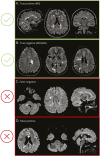
The figure shows examples of MRI, as classified by the DL classifier. (A) For MS, the classifier correctly identified oval-shaped periventricular and deep white matter lesions on axial FLAIR, with Dawson fingers and involvement of the temporal lobe. (B) For MOGAD, the classifier accurately detected large, ill-defined lesions in the juxtacortical/cortical and periventricular regions. (C) An example of MS misclassified as MOGAD revealed a poorly defined hyperintensity in the right cerebellar peduncle, along with more than 6 supratentorial lesions, some of which were round-shaped, and 1 Dawson finger. (D) By contrast, MOGAD misclassified as MS exhibited a well-defined peripheral lesion in the pons, alongside 4 supratentorial confluent lesions, including 2 round-shaped lesions involving the corpus callosum. The integration of clinical and imaging classifiers enabled accurate diagnosis in (C) and (D). DL = deep learning; FLAIR = fluid-attenuated inversion recovery; MNI = Montreal Neurological Institute; MOGAD = myelin oligodendrocyte glycoprotein antibody–associated disease; MS = multiple sclerosis; RRMS = relapsing remitting MS.

References

    1. Marignier R, Hacohen Y, Cobo-Calvo A, et al. Myelin-oligodendrocyte glycoprotein antibody-associated disease. Lancet Neurol. 2021;20(9):762-772. doi: 10.1016/S1474-4422(21)00218-0 - DOI - PubMed
    1. Banwell B, Bennett JL, Marignier R, et al. Diagnosis of myelin oligodendrocyte glycoprotein antibody-associated disease: International MOGAD Panel proposed criteria. Lancet Neurol. 2023;22(3):268-282. doi: 10.1016/s1474-4422(22)00431-8 - DOI - PubMed
    1. Cortese R, Prados Carrasco F, Tur C, et al. Differentiating multiple sclerosis from AQP4-neuromyelitis optica spectrum disorder and MOG-antibody disease with imaging. Neurology. 2023;100(3):E308-E323. doi: 10.1212/WNL.0000000000201465 - DOI - PMC - PubMed
    1. Sechi E, Krecke KN, Messina SA, et al. Comparison of MRI lesion evolution in different central nervous system demyelinating disorders. Neurology. 2021;97(11):e1097-e1109. doi: 10.1212/WNL.0000000000012467 - DOI - PMC - PubMed
    1. Cacciaguerra L, Abdel-Mannan O, Champsas D, et al. Radiologic lag and brain MRI lesion dynamics during attacks in MOG antibody-associated disease. Neurology. 2024;102(10):e209303. doi: 10.1212/WNL.0000000000209303 - DOI - PMC - PubMed

Publication types