. 2025 Sep 16;6(9):102329.
doi: 10.1016/j.xcrm.2025.102329. Epub 2025 Sep 3.

Epigenetic modulation of BARD1 to enhance anti-VEGF therapy

Affiliations

Epigenetic modulation of BARD1 to enhance anti-VEGF therapy

Emine Bayraktar et al. Cell Rep Med. .

Abstract

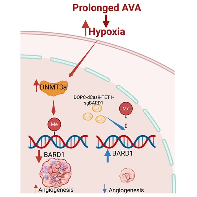
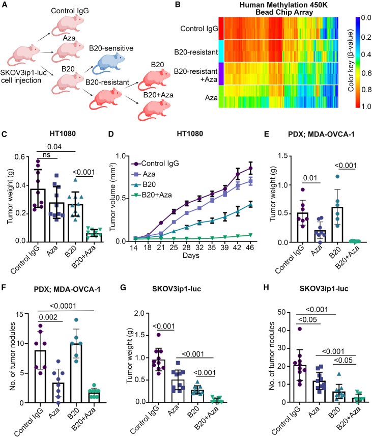
Despite the clinical use of anti-vascular endothelial growth factor (VEGF) antibodies (AVAs) in cancer therapy, resistance frequently develops, leading to disease progression. To address this, we identify a previously unknown role for breast cancer type 1 susceptibility protein (BRCA1)-associated RING domain 1 (BARD1) in modulating AVA sensitivity. Epigenetic modulation-via global and targeted DNA methylation-reveals BARD1 as a key regulator of angiogenesis. Sequential treatment with azacytidine overcomes AVA resistance in vivo. To enable precise epigenetic reactivation, we develop a liposomal CRISPR-deactivated Cas9 (dCas9)-TET1 system guided by BARD1-targeting single-guide RNAs (sgRNAs). This platform achieves CpG-specific demethylation of the BARD1 promoter, restores expression, and enhances AVA response. Additionally, BARD1 restoration, through either dCas9-TET1 or small interfering RNA (siRNA), significantly reduces tumor growth in combination with AVA in ovarian cancer models. These findings uncover a previously unrecognized function of BARD1 in tumor angiogenesis and demonstrate the potential of gene-specific epigenetic targeting to overcome AVA resistance.

Keywords: AVA resistance; BARD1; anti-VEGF antibody therapy; bevacizumab; epigenetic editing; epigenetic therapy; ovarian cancer.


Conflict of interest statement

Declaration of interests A.K.S.: consulting (Onxeo, ImmunoGen, and Kaida), DSMB (Advenchen and Mural Oncology), and research grant (Pfizer). C.I. contributed to the analysis during her time at MDACC; she is currently working at Caris Life Sciences as a data scientist. We declare that we have submitted a patent application related to dCas9-TET1-sgRNA.

Figures

None
Graphical abstract
Figure 1
Figure 1
Addition of Aza restores sensitivity to AVA therapy following resistance development (A) Schematic of treatment groups in the SKOV3ip1-luc mouse model (created in BioRender). (B) Genome-wide methylation profiles across treatment groups (n = 3 mice per group); heatmap contains thousands of probes. (C) Tumor weights in HT1080 xenografts (n = 9 mice for control IgG; n = 10 mice for Aza; n = 10 mice for B20 antibody; n = 9 mice for Aza + B20). ns, not significant. (D) Tumor volumes in HT1080 xenografts (same group sizes as in C). (E) Tumor weights in patient-derived xenograft (PDX; MDA-OVCA-1) model (n = 7 mice for control IgG; n = 8 mice for Aza; n = 6 mice for B20 antibody; and n = 8 mice for Aza + B20). (F) Number of nodules in the MDA-OVCA-1 PDX model (same group sizes as in E). (G) Tumor weights in the SKOV3ip1-luc mouse model (n = 10 mice per group). (H) Number of nodules in the SKOV3ip1-luc mouse model (n = 10 per group). Data are presented as mean ± SD. Statistical analysis was performed using two-tailed Student’s t test (C, E, F, G, and H) and two-way ANOVA with Tukey’s multiple comparisons test (D).
Figure 2
Figure 2
Aza induces transcriptomic and epigenetic changes (A) Heatmap of mRNA expression profiles in the SKOV3ip1-luc model (n = 3 mice per group); includes thousands of probes. (B) Comparison of tumor-specific methylation probes and gene expression between B20 and Aza+B20 groups in the SKOV3ip1-luc mouse model. Some genes appear more than once because the array includes multiple probes per gene. (C) Relative mRNA expression of BARD1 in each treatment group in the SKOV3ip1-luc mouse model. (D) Protein expression of BARD1 in each treatment group in the SKOV3ip1-luc mouse model. (E) Petal diagram of BARD1-associated pathways identified using expression data shown in (B). (F) Relative mRNA expression of BARD1 in normal fallopian tube epithelium (FTE) cells, HIO180 cells, and OC cell lines. (G) Protein expression of BARD1 in normal FTE cells, non-transformed ovarian surface epithelial cells (HIO180), and OC cell lines. Data are presented as mean ± SD.
Figure 3
Figure 3
BARD1 regulates tumor angiogenesis (A) Tube formation assay in RF24 endothelial cells transfected with BARD1 siRNA or control siRNA. Scale bars, 250 μm. (B) Tube formation in RF24 cells cultured with CM from SKOV3ip1-luc cells treated with BARD1 siRNA or control siRNA. Scale bars, 250 μm. (C) Gene Ontology analysis of gene and pathway enrichment in BARD1-silenced SKOV3ip1-luc cells. (D) Hemoglobin content in mice injected with CM from BARD1 siRNA or control siRNA-treated SKOV3ip1 cells. (E) Angiogenesis array results in SKOV3ip1-luc cells treated with BARD1 siRNA or control siRNA. (F) Schematic of the TetOn system: mice fed doxycycline (Dox) chow for BARD1 knockdown and treated with bevacizumab ± Aza. (G) Tumor weights in vivo with or without BARD1 knockdown (n = 10 mice for untreated; n = 10 mice for untreated + Dox; n = 8 mice for Bev antibody; n = 10 mice for Bev antibody + Dox; n = 9 mice for combination; and n = 9 mice for combination + Dox). (H) Relative mRNA expression of BARD1 in vivo across treatment groups ± Dox. Data are presented as mean ± SD. Statistical comparisons were made using two-tailed Student’s t test (A, B, D, F, G, and H).
Figure 4
Figure 4
BARD1 is epigenetically regulated under hypoxic conditions (A and B) Relative mRNA expression of HIF1α and BARD1 in SKOV3ip1 cells grown under normoxic (UT) and hypoxic conditions (1% O2): (A) HIF1α and (B) BARD1. Data are presented as mean ± SD (n = 3 samples per group). (C) Protein expression levels of HIF1α in normoxic (N) and hypoxic (H) conditions. (D) Protein expression of BARD1 under normoxic (N) and hypoxic (H) conditions. (E) Relative BARD1 mRNA levels in SKOV3ip1-luc cells treated with HIFα versus control siRNA. (F) Relative DNMT3A mRNA levels in SKOV3ip1 cells grown under normoxic (UT) or hypoxic conditions (1% O2). (G) Relative DNMT3A mRNA expression levels in SKOV3ip1-luc cells treated with HIF1α siRNA versus control siRNA. (H) Chromatin immunoprecipitation followed by qPCR (ChIP-qPCR) results showing HIF1α enrichment under hypoxic and normoxic conditions (n = 3 samples per group). ∗p < 0.05. (I) HIF1α and DNMT3A enrichment at BARD1 promoter at site 4 under hypoxic and normoxic conditions. Data are presented as mean ± SD (n = 3 samples per group). ∗p < 0.05. (J) Relative TET1 mRNA expression levels under hypoxic and normoxic conditions. (K) Tumor weight in mice treated with DOPC-dCas9-TET1-sgBARD1-3 (sgBARD1-3) versus sgControl. (n = 12 mice per group). (L) Relative BARD1 mRNA expression in mice treated with sgBARD1-3 or sgControl (n = 4 mice per group). (M) Tumor weight in mice treated with sgBARD1-3, alone or in combination with bevacizumab (Bev) (n = 13 mice for untreated, n = 12 mice for DOPC-TET1-sgControl, n = 12 mice for DOPC-TET1-sgBARD1-3, n = 12 mice for DOPC-TET1-sgControl + Bev antibody treatment, and n = 12 mice for DOPC-TET1-sgBARD1-3 + Bev antibody treatment group). (N) Number of nodules in mice treated with sgBARD1-3, alone or in combination with Bev (same group sizes as in M). (O) Tumor weight in mice treated with sgBARD1-1 or control (n = 8 mice per group). Data are presented as mean ± SD (A, B, H, I, and K) or SEM (L–O). Statistical comparisons were made using two-tailed Student’s t test.
Figure 5
Figure 5
Proposed mechanism of BARD1 epigenetic regulation in response to AVA therapy Prolonged AVA treatment induces adaptive resistance characterized by increased tumor hypoxia. Hypoxia stabilizes HIF1α, which enhances DNMT3A expression. DNMT3 then methylates the BARD1 promoter, repressing its transcription. Reduced BARD1 expression in cancer cells is associated with upregulation of proangiogenic factors, promoting tumor angiogenesis and progression. Targeted demethylation of BARD1 restores its expression and suppresses angiogenesis (created in BioRender).

References

    1. Mei C., Gong W., Wang X., Lv Y., Zhang Y., Wu S., Zhu C. Anti-angiogenic therapy in ovarian cancer: Current understandings and prospects of precision medicine. Front. Pharmacol. 2023;14 doi: 10.3389/fphar.2023.1147717. - DOI - PMC - PubMed
    1. Siegel R.L., Miller K.D., Wagle N.S., Jemal A. Cancer statistics, 2023. CA Cancer J. Clin. 2023;73:17–48. doi: 10.3322/caac.21763. - DOI - PubMed
    1. Lengyel E. Ovarian cancer development and metastasis. Am. J. Pathol. 2010;177:1053–1064. doi: 10.2353/ajpath.2010.100105. - DOI - PMC - PubMed
    1. Holash J., Maisonpierre P.C., Compton D., Boland P., Alexander C.R., Zagzag D., Yancopoulos G.D., Wiegand S.J. Vessel cooption, regression, and growth in tumors mediated by angiopoietins and VEGF. Science. 1999;284:1994–1998. doi: 10.1126/science.284.5422.1994. - DOI - PubMed
    1. Yancopoulos G.D., Davis S., Gale N.W., Rudge J.S., Wiegand S.J., Holash J. Vascular-specific growth factors and blood vessel formation. Nature. 2000;407:242–248. doi: 10.1038/35025215. - DOI - PubMed

MeSH terms

LinkOut - more resources