. 2023:3:47.
doi: 10.1038/s43586-023-00231-0. Epub 2023 Jun 22.

Light-based vat-polymerization bioprinting

Affiliations

Light-based vat-polymerization bioprinting

Riccardo Levato et al. Nat Rev Methods Primers. 2023.

Abstract

Light-based vat-polymerization bioprinting enables computer-aided patterning of 3D cell-laden structures in a point-by-point, layer-by-layer or volumetric manner, using vat (vats) filled with photoactivatable bioresin (bioresins). This collection of technologies - divided by their modes of operation into stereolithography, digital light processing and volumetric additive manufacturing - has been extensively developed over the past few decades, leading to broad applications in biomedicine. In this Primer, we illustrate the methodology of light-based vat-polymerization 3D bioprinting from the perspectives of hardware, software and bioresin selections. We follow with discussions on methodological variations of these technologies, including their latest advancements, as well as elaborating on key assessments utilized towards ensuring qualities of the bioprinting procedures and products. We conclude by providing insights into future directions of light-based vat-polymerization methods.


Conflict of interest statement

Competing interests Y.S.Z. consults for Allevi by 3D Systems and sits on the scientific advisory board and holds options of Xellar, both of which, however, did not participate in or bias the work. R.L. is a scientific advisor for Readily3D SA, which did not participate in or bias the work. The other authors declare no competing interests.

Figures
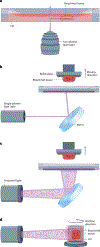
Fig. 1 |
Fig. 1 |. Typical light-based vat-polymerization techniques.
a, Two-photon lithography that raster-scans two-photon lasers to polymerize or deconstruct a bioresin for 3D bioprinting. b, Single-photon stereolithography that raster-scans a single-photon laser for 3D bioprinting. c, Digital light processing that projects a series of light patterns to achieve layer-by-layer 3D bioprinting. The system shown is the bottom-up configuration. d, Tomographic bioprinting that projects a series of intensity-modulated light patterns to achieve rotational 3D bioprinting.
Fig. 2 |
Fig. 2 |. Variations in vat-polymerization techniques, taking digital light processing bioprinting as an example.
a, Digital light processing (DLP) bioprinting in the top-down configuration. b, Multimaterial DLP bioprinting using multiple vats. c, Multimaterial DLP bioprinting using automated bioresin change through a microfluidics-integrated vat. d, Heterogeneous-material DLP bioprinting using multiple wavelengths.
Fig. 3 |
Fig. 3 |. Determining light-dose responses and working curves in light-based vat-polymerization bioprinting.
a, A simple method to establish the single-photon stereolithography or digital light processing working curves consists of projecting an array of disks or squares onto the bioresin vat where each of those is exposed to an increasing light dose. b, After crosslinking, the thicknesses of the bioresin layer are measured and recorded to create a light energy versus thickness plot that can be used to construct the working curves. c, A dose test is performed to identify ideal light-exposure parameters for tomographic bioprinting, by projecting an array of disk-shaped spots within a cuvette containing the bioresin, with each spot corresponding to a varying light intensity and exposure time. Cd, curing depth; VAM, volumetric additive manufacturing.
Fig. 4 |
Fig. 4 |. Resolution assessments in light-based vat-polymerization bioprinting.
a, In point-by-point and layer-by-layer vat polymerization, resolution is assessed by printing diagnostic models with small positive and negative features that range in size at light-exposure parameters in the optimal range identified with the working curve. b, In single-photon stereolithography and digital light processing, the printed structures can display a notable pixelated profile depending on the layer thickness. c, Tomographic bioprinting enables fabrication of objects in a layerless fashion with the resolutions measurable through attainable negative and positive features. d, Measurement of the resolution of negative features can be facilitated by using fluorescent dyes; here a negative cone is filled with a dye, and the maximum attainable negative resolution is determined by measuring the tip dimensions of the cone.
Fig. 5 |
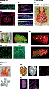
Fig. 5 |. Examples of tissue-engineered constructs.
A, Examples of point-by-point printing. Aa, Point-by-point printing of vascular network by means of two-photon mechanism with human umbilical vein endothelial cells endothelialization. F-actin (green) and nuclei (blue). Scale bar, 100 μm. Ab, Two-photon-based ablation and endothelialization of glomerulus-like vasculature. Scale bar, 100 μm. Ac, Two-photon patterning of growth factors to guide axon outgrowth. Avidin-SAT-F + NGF (blue), BIII-tubulin (red) and F-actin (green). Scale bar, 50 μm. B, Examples of layer-by-layer printing. Ba, Layer-by-layer printing of entangled vasculature networks. Scale bar, 1 mm. Bb, Fast printing of large constructs featuring perfusable channels. Scale bar, 1 cm. Bc, Cellular alignment in FLightbioprinted constructs at day 1 (top) and day 7 (bottom). Filamentous gel (red), normal human dermal fibroblasts (green) and nuclei (blue). Scale bars, 20 μm. Bd, Differentiation of C2C12 muscle cells in bioprinted constructs without (stiff gel, gelatin + hyaluronic acid methacrylate (GH)) or with (soft gel, hyaluronidase (Hase)) enzymatic digestion. Myosin heavy chain (green) and nuclei (blue). Scale bar, 50 μm. C, Examples of volumetric printing. Ca, High-fidelity tomographic printing of mouse pulmonary artery. Scale bars, 5 mm. Cb, Bioprinting of mesenchymal stem cell-laden trabecular bone. Osteogenic medium-primed mesenchymal stem cells (pink). Scale bars, 2 mm and 500 μm. Cc, Bioprinting of C2C12 myoblast-laden complex model. Myosin heavy chain (red) and nuclei (blue). Scale bars, 2 mm and 200 μm. RBC, red blood cell. Part Aa reprinted with permission from ref. , Wiley. Part Ab reprinted with permission from ref. , Wiley. Part Ac reprinted with permission from ref. , Wiley. Part Ba reprinted with permission from ref. , AAAS. Part Bb reprinted with permission from ref. , Wiley. Part Bc reprinted from ref. , CC BY 4.0. Part Bd reprinted from ref. , Springer Nature Limited. Part Ca reprinted from ref. , Springer Nature Limited. Part Cb reprinted with permission from ref. , Wiley. Part Cc reprinted with permission from ref. , Wiley.

References

    1. Groll J et al. Biofabrication: reappraising the definition of an evolving field. Biofabrication 8, 013001 (2016). - PubMed
    1. Levato R et al. From shape to function: the next step in bioprinting. Adv. Mater. 32, 1906423 (2020). - PMC - PubMed
    1. Moroni L et al. Biofabrication: a guide to technology and terminology. Trends Biotechnol. 36, 384–402 (2018). - PubMed
    1. Moroni L et al. Biofabrication strategies for 3D in vitro models and regenerative medicine. Nat. Rev. Mater. 3, 21–37 (2018). - PMC - PubMed
    1. Heinrich MA et al. 3D bioprinting: from benches to translational applications. Small 15, 1805510 (2019). - PMC - PubMed