. 2025 Aug 13;8(5):533-538.
doi: 10.1002/iju5.70086. eCollection 2025 Sep.

Late-Onset Bone Metastasis 46 Years After Initial Surgery for Pheochromocytoma: A Case Report

Affiliations

Late-Onset Bone Metastasis 46 Years After Initial Surgery for Pheochromocytoma: A Case Report

Fumio Ishizaki et al. IJU Case Rep. .

Abstract

Introduction: Pheochromocytoma is a rare catecholamine-producing tumor with metastatic potential. Recurrence after more than 40 years is exceptionally rare.

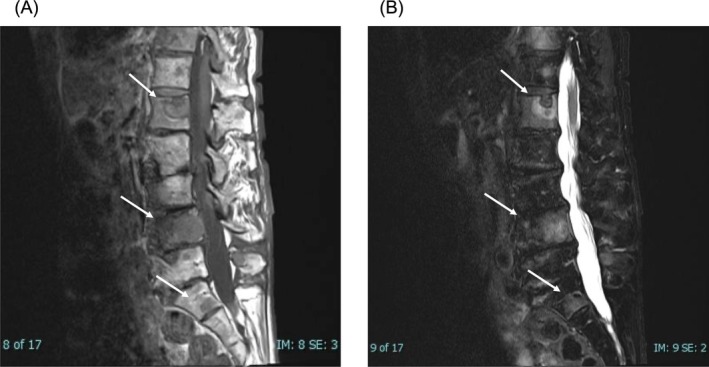
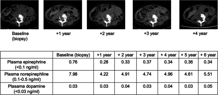
Case presentation: During evaluation for ischemic colitis, a 71-year-old woman was found to have multiple bone metastases, possibly linked to catecholamine excess. She had undergone left adrenalectomy for pheochromocytoma at age 25. Bone biopsy confirmed metastatic pheochromocytoma, and immunohistochemical findings were similar to the original tumor. Urinary metanephrine and normetanephrine were markedly elevated. She declined systemic therapy and has remained clinically stable for 6 years, with her blood pressure well controlled on doxazosin.

Conclusion: This case illustrates a recurrence 46 years after adrenalectomy, potentially representing the longest reported interval to date. It highlights the silent and indolent nature of some metastatic pheochromocytomas and underscores the necessity of lifelong follow-up. The patient's stable course also emphasizes the clinical heterogeneity of metastatic pheochromocytoma and supports the need for individualized follow-up and treatment strategies.

Keywords: bone metastasis; late recurrence; pheochromocytoma.


Conflict of interest statement

The authors declare no conflicts of interest.

Figures

FIGURE 1
FIGURE 1
MRI findings of spinal metastases. (A) Sagittal T1‐weighted MRI showing hypointense lesions in the L1, L4 vertebrae and the sacrum. (B) Sagittal T2‐weighted MRI demonstrating corresponding hyperintense lesions in the same regions, consistent with bone metastases.
FIGURE 2
FIGURE 2
Histological and immunohistochemical findings. (A–E) Primary adrenal pheochromocytoma resected at age 25: Hematoxylin and eosin (HE) staining (A), and immunohistochemical staining for chromogranin A (B), synaptophysin (C), CD56 (D) and SDHB (E). (F–H) Bone metastasis biopsy specimen: HE staining (F) and immunohistochemical staining for chromogranin A (G) and CD56 (H).
FIGURE 3
FIGURE 3
Imaging findings of metastatic pheochromocytoma. (A) 123I‐MIBG scintigraphy showing abnormal tracer uptake in the thoracic and lumbar spine, sacrum, left humerus, and bilateral iliac bones. (B) Axial PET‐CT at the L4 level demonstrating increased FDG uptake in the vertebral lesion. (C) Coronal PET‐CT showing FDG‐avid lesions in multiple vertebrae and pelvic bones, corresponding to the MIBG‐avid regions.
FIGURE 4
FIGURE 4
Follow‐up findings over 6 years after diagnosis of bone metastasis. Serial computed tomography (CT) images showing no significant progression of bone metastases from baseline (biopsy) to year 4. Plasma catecholamine levels remained stably elevated without further increase during the 6‐year observation period.

References

    1. Lam A K.‐y., “Update on Adrenal Tumours in 2017 World Health Organization (WHO) of Endocrine Tumours,” Endocrine Pathology 28 (2017): 213–227. - PubMed
    1. Harari A. and W. B. Inabnet, III , “Malignant Pheochromocytoma: A Review,” American Journal of Surgery 201, no. 5 (2011): 700–708. - PubMed
    1. Ishizaki F., Taguchi T., Murata M., et al., “Long‐Term Outcomes and Prognostic Factors of Metastatic or Recurrent Pheochromocytoma and Paraganglioma: A 20‐Year Review in a Single Institution,” Scientific Reports 14, no. 1 (2024): 26456, 10.1038/s41598-024-75354-9. - DOI - PMC - PubMed
    1. Parisien‐La Salle S., Chbat J., Lacroix A., et al., “Postoperative Recurrences in Patients Operated for Pheochromocytomas and Paragangliomas: New Data Supporting Lifelong Surveillance,” Cancers (Basel) 14, no. 12 (2022): 2942, 10.3390/cancers14122942. - DOI - PMC - PubMed
    1. Plouin P. F., Amar L., Dekkers O. M., et al., “European Society of Endocrinology Clinical Practice Guideline for Long‐Term Follow‐Up of Patients Operated on for a Phaeochromocytoma or a Paraganglioma,” European Journal of Endocrinology 174, no. 5 (2016): G1–G10, 10.1530/EJE-16-0033. - DOI - PubMed

LinkOut - more resources