. 2025 Jul 4:10:330.
doi: 10.12688/wellcomeopenres.24569.1. eCollection 2025.

The genome sequence of the zebra danio, Danio rerio (Hamilton, 1822) (Cypriniformes: Danionidae)

Affiliations

The genome sequence of the zebra danio, Danio rerio (Hamilton, 1822) (Cypriniformes: Danionidae)

Kerstin Howe et al. Wellcome Open Res. .

Abstract

We present a genome assembly from a specimen of Danio rerio (zebra danio; Chordata; Actinopteri; Cypriniformes; Danionidae). The genome sequence has a total length of 1 413.66 megabases. Most of the assembly (99.85%) is scaffolded into 25 chromosomal pseudomolecules. The mitochondrial genome was also assembled, with a length of 16.6 kilobases. Gene annotation of this assembly on Ensembl identified 25 582 protein-coding genes.

Keywords: Cypriniformes; Danio rerio; chromosomal; genome sequence; zebra danio.


Conflict of interest statement

No competing interests were disclosed.

Figures

Figure 1.
Figure 1.. Photograph of Danio rerio by Tohru Murakami.
Figure 2.
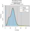
Figure 2.. Frequency distribution of k-mers generated using GenomeScope2.
The plot shows observed and modelled k-mer spectra, providing estimates of genome size, heterozygosity, and repeat content based on unassembled sequencing reads.
Figure 3.
Figure 3.. Hi-C contact map of the Danio rerio genome assembly.
Assembled chromosomes are shown in order of size and labelled along the axes. The plot was generated using PretextSnapshot.
Figure 4.
Figure 4.. Evaluation of k-mer completeness using MerquryFK.
This plot illustrates the recovery of k-mers from the original read data in the final assemblies. The horizontal axis represents k-mer multiplicity, and the vertical axis shows the number of k-mers. The black curve represents k-mers that appear in the reads but are not assembled. The green curve (the homozygous peak) corresponds to k-mers shared by both haplotypes and the red and blue curves (the heterozygous peaks) show k-mers found only in one of the haplotypes.
Figure 5.
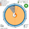
Figure 5.. Assembly metrics for fDanRer4.1.
The BlobToolKit snail plot provides an overview of assembly metrics and BUSCO gene completeness. The circumference represents the length of the whole genome sequence, and the main plot is divided into 1,000 bins around the circumference. The outermost blue tracks display the distribution of GC, AT, and N percentages across the bins. Scaffolds are arranged clockwise from longest to shortest and are depicted in dark grey. The longest scaffold is indicated by the red arc, and the deeper orange and pale orange arcs represent the N50 and N90 lengths. A light grey spiral at the centre shows the cumulative scaffold count on a logarithmic scale. A summary of complete, fragmented, duplicated, and missing BUSCO genes in the set is presented at the top right. An interactive version of this figure can be accessed on the BlobToolKit viewer.
Figure 6.
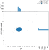
Figure 6.. BlobToolKit GC-coverage plot for fDanRer4.1.
Blob plot showing sequence coverage (vertical axis) and GC content (horizontal axis). The circles represent scaffolds, with the size proportional to scaffold length and the colour representing phylum membership. The histograms along the axes display the total length of sequences distributed across different levels of coverage and GC content. An interactive version of this figure is available on the BlobToolKit viewer.

References

    1. Allio R, Schomaker-Bastos A, Romiguier J, et al. : MitoFinder: efficient automated large-scale extraction of mitogenomic data in target enrichment phylogenomics. Mol Ecol Resour. 2020;20(4):892–905. 10.1111/1755-0998.13160 - DOI - PMC - PubMed
    1. Altschul SF, Gish W, Miller W, et al. : Basic Local Alignment Search Tool. J Mol Biol. 1990;215(3):403–410. 10.1016/S0022-2836(05)80360-2 - DOI - PubMed
    1. Bateman A, Martin MJ, Orchard S, et al. : UniProt: the Universal Protein Knowledgebase in 2023. Nucleic Acids Res. 2023;51(D1):D523–D531. 10.1093/nar/gkac1052 - DOI - PMC - PubMed
    1. Buchfink B, Reuter K, Drost HG: Sensitive protein alignments at Tree-of-Life scale using DIAMOND. Nat Methods. 2021;18(4):366–368. 10.1038/s41592-021-01101-x - DOI - PMC - PubMed
    1. Challis R, Kumar S, Sotero-Caio C, et al. : Genomes on a Tree (GoaT): a versatile, scalable search engine for genomic and sequencing project metadata across the eukaryotic Tree of Life [version 1; peer review: 2 approved]. Wellcome Open Res. 2023;8:24. 10.12688/wellcomeopenres.18658.1 - DOI - PMC - PubMed

LinkOut - more resources