[Preprint]. 2025 Aug 26:2025.08.21.671640.
doi: 10.1101/2025.08.21.671640.

Plasma membrane accessible cholesterol is regulated by ACC1 and lipid droplets

Affiliations

Plasma membrane accessible cholesterol is regulated by ACC1 and lipid droplets

Kalani M Wijesinghe et al. bioRxiv. .

Abstract

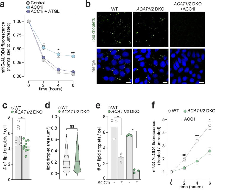
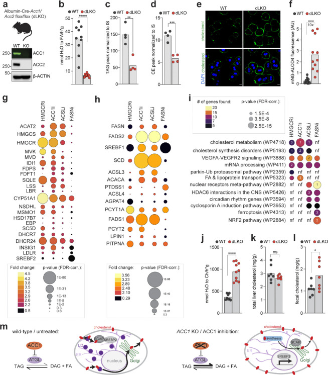
Proper maintenance of plasma membrane (PM) cholesterol is essential for diverse processes ranging from animal development to pathogen evasion. Despite decades of study, the mechanisms governing cellular cholesterol regulation are incomplete. Using genome-wide screens we find that ACC1, the rate-limiting enzyme in fatty acid biosynthesis, regulates PM cholesterol transport. ACC1 loss causes a ~10-fold increase in PM accessible cholesterol in cells and mice. Mechanistically, we find that ACC1 regulates lipid droplet (LD) catabolism, and LDs are intimately tied to PM accessible cholesterol levels since reductions or elevations in their numbers block or promote cholesterol trafficking, respectively. Furthermore, LDs are required for cholesterol trafficking induced by 25-hydroxycholesterol, a modulator of inflammation and an interferon-stimulated second messenger that protects cells from pathogen invasion. This work identifies an unrecognized role for ACC1 and LDs in cholesterol regulation, which has implications for diseases where LD numbers are altered, from metabolic syndromes to neurodegeneration.

Keywords: 25-hydroxycholesterol; ACAT1 and ACAT2; ACC1; AMPK; ATGL; Cholesterol; SREBP2; accessible cholesterol; cholesterol esters; lipid droplet; membrane trafficking; plasma membrane; triacylglycerol.


Conflict of interest statement

Competing Interests: JAO is a member of the scientific advisory board for Vicinitas Therapeutics.

Figures

Extended data Fig. 1
a,b, PCR analysis of GRAMD1A/B/C TKO clones (a) and GRAMD1B KO clones (b). c,d, Plasma membrane (PM) cholesterol analyzed by mNG-ALOD4 staining followed by flow cytometry in wild-type (WT) HAP1 and GRAMD1B KO clones either left untreated (c) or treated with 25HC for the indicated times (d). e, quantitative real-time PCR analysis of Abca1 mRNA levels normalized to Gapdh levels from 3T3 cells treated for 20 hrs with LXRi (1 μM GSK2033). f,g, PM cholesterol analyzed by flow cytometry analysis of mNG-ALOD4 staining in 3T3 cells treated for 20 hrs with or without 1 μM of LXRi alone (f) or followed by treatment with 25-hydroxycholesterol (25HC) for the indicated times (g). h, Domain architecture of ACC1 (top) including the biotin carboxylase domain (BC), biotin transferase domain (BT), biotin carboxyl carrier protein domain (BCCP), central domain (CD), and the carboxyltransferase domain (CT) ,. Residues implicated in catalysis are shown in dark orange. Exon map of ACC1 (bottom) with exon colors matching the domain they encode. i, The number of insertions mapped per intron are normalized to intron length in base pairs (bp). j, Western blot (top) and PCR analysis (bottom) of WT HAP1 and ACC1 KO clonal cell lines. k,l, mNG-ALOD4 flow cytometry analysis of PM cholesterol in WT HAP1 and ACC1 KO cells made with CRISPR sgRNAs targeting the indicated exons either left untreated (k) or treated with 30 μM ACC1i (Firsocostat) for 20 hrs (l). m, Table showing the number of clones isolated from two 96-well dishes and the percentage of those clones that were found to be knockouts with PCR analysis. n, Western blot analysis of WT and NPC1 KO HAP1 cell lines. o,p, Western blot analysis (o) or mNG-ALOD4 flow cytometry analysis of PM cholesterol (p) of Npc1−/− mouse embryonic fibroblasts (MEFs) or Npc1−/− MEFs with NPC1 stably re-introduced using MSCV retrovirus, and then treated with or without ACC1i (30 μM Firsocostat) for 20 hrs. Each data point is the average of three biological replicates (d and g). Cells in k and l were grown in 5% lipoprotein depleted serum for 16 hours prior to analysis. Statistical significance was determined by a Student’s t-test with a Welch’s correction (c-g and l) or an Ordinary one-way ANOVA (k and p). Exact p-values: c, WT vs GRAMD1B KO p=0.0043; d, WT vs GRAMD1B KO (2 hr 25HC) p=0.9961, WT vs GRAMD1B KO (4 hr 25HC) p=0.0847, and WT vs GRAMD1B KO (6 hr 25HC) p<0.0001; e, untreated vs LXRi p<0.0001; f, control vs LXRi p<0.0001; g, control vs LXRi (2 hr 25HC) p=0.0064, control vs LXRi (4 hr 25HC) p=0.0132 and control vs LXRi (6 hr 25HC) p=0.9640; k, WT vs ACC1 KO exon 11 p<0.0001, WT vs ACC1 KO exon 21 p<0.0001, WT vs ACC1 KO exon 31 p<0.0001, WT vs ACC1 KO exon 41 p<0.0001 and WT vs ACC1 KO exon 52 p<0.0001; l, WT untreated vs ACC1i p=0.0076, ACC1 KO exon 11 untreated vs ACC1i p=0.1371 and ACC1 KO exon 52 untreated vs ACC1i p=0.0563; and p, Npc1−/− untreated vs ACC1i p<0.0001 and Npc1−/− vs Npc1−/−:NPC1 p=0.0004.
Extended data Fig. 2
a, Schematic demonstrating that sphingomyelin (SM, purple) binds to cholesterol in membranes. ACC1 loss could elevate cholesterol by 1) increasing cholesterol or by 2) reducing SM levels. b, Mass spectrometry analysis of SM levels in wild-type (WT) HAP1 cells either left untreated or treated with ACC1i (30 μM Firsocostat) for 16 hrs. c, OlyA-EGFP flow cytometry analysis of WT HAP1 cells left untreated or treated with 0.3 mM MꞵCD: cholesterol or 150 mU/mg SMase for 30 minutes, or 30 μM ACC1i for 16 hrs. d, ACC1 KO cells or WT HAP1 cells were left untreated or treated for 16 hrs with 30 μM ACC1i. Cells were then treated with or without 150 mU/mg SMase in 5% FBS for 30 minutes and stained with mNG-ALOD4 and analyzed by flow cytometry. e, Quantification of esterified cholesterol in WT HAP1 cells treated with ACATi (60 μM SZ58–035) for 16 hrs or left untreated, followed by a 6 hr treatment with or without 4 μM 25HC using the Amplex Red assay. f, Flow cytometry analysis of mNG-ALOD4 staining in WT HAP1 cells treated with or without 60 μM ACATi for 16 hrs, followed by 4 μM 25HC for the times indicated. g, h, mNG-ALOD4 flow cytometry analysis of ACC1 KO cells left untreated or treated for 16 hours with ACATi (60 μM SZ58–035) followed by 4 μM 25HC treatment for the indicated times (g) and WT HAP1 cells grown in 5% FBS and treated with HMGCRi (10 μM Lovastatin) for the indicated times (h). Statistical significance was determined by a Student’s t-test with a Welch’s correction (b and d-g) or an Ordinary one-way ANOVA (c and h). Exact p-values: b, untreated vs ACC1i p=0.6634; c, untreated vs cholesterol p<0.0001, untreated vs SMase p=0.0038, untreated vs ACC1i p=0.0036; d, control with vs without SMase p=0.0002, ACC1i with vs without SMase p<0.0001, ACC1 KO with vs without SMase p<0.0001; e, control untreated vs 25HC p=0.0013 and ACATi vs ACATi + 25HC p=0.6469; f, control vs ACATi (3 hr 25HC) p=0.0004, control vs ACATi (4.5 hr 25HC) p=0.0024 and control vs ACATi (6 hr 25HC) p=0.0146; g, ACC1 KO vs ACC1 KO + ACATi (3 hr 25HC) p=0.0134, ACC1 KO vs ACC1 KO + ACATi (4.5 hr 25HC) p=0.0654 and ACC1 KO vs ACC1 KO + ACATi (6 hr 25HC) p=0.0735; h, 0 vs 2 hrs HMGCRi p=0.2028, 0 vs 4 hrs HMGCRi p=0.0137 and 0 vs 6 hrs HMGCRi p=0.0495.
Extended data Fig. 3
a, 3T3 cells were treated with 30 μM ACC1i (Firsocostat) for the indicated times in 5% FBS, then analyzed by flow cytometry after staining with mNG-ALOD4. b, mNG-ALOD4 flow cytometry analysis of WT HAP1 cells cultured in 5% FBS and either left untreated or treated with HMGCRi (10 μM Lovastatin), ACC1i (30 μM Firsocostat) or both for 16 hrs. c, mNG-ALOD4 flow cytometry analysis of WT HAP1 cells and ACC1 KO cells grown in 5% FBS and either left untreated or treated with HMGCRi (10 μM Lovastatin) for 16 hrs. d, Western blot analysis of fractionated LDAH-GFP expressing HAP1 cells with various cellular fractions including whole cell (WC), nucleus (Nuc.; detected by LaminA/C), cytoplasm (Cyt.; detected by GAPDH), plasma membrane (PM; detected by LDLR), and lipid droplets (LD; detected by LDAH-GFP). Cells were either left untreated or treated with ACC1i (30 μM Firsocostat) for 24 hrs. e, Quantification of LD numbers per cell in WT HAP1 cells treated with or without ACC1i (30 μM Firsocostat) for 16 hrs (left) or WT HAP1 cells compared to ACC1 KO HAP1 cells (right). The solid middle line shows the median and dashed lines show the interquartile range. f, Microscopy images of LDs in WT HAP1 cells left untreated or treated with DGAT1 and DGAT2 inhibitors (DGATi) for 16 hrs followed by 150 μM oleic acid (OA) treatment for 6 hrs in 5% FBS stained with Bodipy 493/503. Scale bar is 10 microns. g, Western blot analysis of WT HAP1 cells compared to FASN KO HAP1 clones. h, Microscopy images of LDs in WT HAP1 and FASN KO cells stained with Bodipy 493/503 and DAPI (blue). Scale bar is 10 microns. i,j, Flow cytometry analysis of mNG-ALOD4 PM staining in WT HAP1 and FASN KO clones grown in 5% LDS media for 24 hrs left untreated (i) or then treated with 4 μM 25HC for the indicated times (j). k, mNG-ALOD4 flow cytometry analysis of PM accessible cholesterol (blue curves) and the corresponding fluorescence microscopy quantification of the number of LDs per cell (purple curves) in HAP1 cells grown in 5% FBS and then treated with FASNi (10 μM C75) for the indicated times in 5% LDS. LDs were quantified by taking the mean number of LDs per cell in four fields of view with ~75 cells per field. l, Microscopy images of FASN localization relative to LD staining with Bodipy 493:503. Scale bar is 5 microns. Statistical significance was determined by a Student’s t-test with a Welch’s correction (a-c,e,i and j). Exact p-values: a, 0 vs 15 min ACC1i p=0.0419; b, WT HMGCRi vs HMGCRi + ACC1i p=0.0008 ; c, WT HMGCRi vs ACC1 KO HMGCRi p<0.0001; e, WT untreated vs ACC1i p<0.0001 and WT vs ACC1 KO p<0.0001; i, WT vs FASN KO p<0.0001; j, WT vs FASN KO (2 hr 25HC) p=0.0001, WT vs FASN KO (4 hr 25HC) p<0.0001 and WT vs FASN KO (6 hr 25HC) p=0.0003.
Extended data Fig. 4
a, Flow cytometry analysis of PM mNG-ALOD4 staining. WT HAP1 cells were left untreated, treated with ACC1i (30 μM Firsocostat), or treated with both ACC1i and ATGLi (50 μM NG-497), followed by treatments with 4 μM 25HC for the indicated times. b, Microscopy images of LDs in WT HAP1, untreated ACAT1/2 DKO and ACAT1/2 DKO treated with ACC1i (30 μM Firsocostat) and stained with Bodipy 493/503 and DAPI (blue). Scale bar is 10 microns. c, Quantification of LD numbers per cell in WT and ACAT1/2 DKO HAP1 cells cultured in 5% LDS for 24 hrs. d, LD area for untreated WT and ACAT1/2 DKO HAP1 cells. Each violin plot shows the area for LDs measured in three fields of view. e, LD quantification for WT and ACAT DKO cells, either untreated or treated with ACC1i (30 μM Firsocostat) for 6 hrs. Each data point is the total number of LDs per cell in one field of view. f, Flow cytometry analysis of mNG-ALOD4 staining in WT and ACAT1/2 DKO HAP1 cell lines cultured in 5% LDS for 16 hrs, followed by treatment with ACC1i (30 μM Firsocostat) for the indicated times. Statistical significance was determined by Student’s t-test with Welch’s correction (a and c-f). Exact p-values: a, WT ACC1i vs ACC1i + ATGLi (2 hr 25HC) p=0.0196, WT ACC1i vs ACC1i + ATGLi (4 hr 25HC) p=0.0256, and WT ACC1i vs ACC1i + ATGLi (2 hr 25HC) p=0.0017; c, WT vs ACAT 1/2 DKO p=0.0151; d, WT vs ACAT 1/2 DKO p=0.0588; e, WT untreated vs ACC1i p=0.0186 and ACAT 1/2 DKO untreated vs ACC1i p=0.0146; f, WT vs ACAT1/2 DKO (2 hr ACC1i) p=0.1519, WT vs ACAT1/2 DKO (4 hr ACC1i) p=0.0099 and WT vs ACAT1/2 DKO (6 hr ACC1i) p=0.0160.
Extended data Fig. 5
a,b, Quantification of lipid droplet (LD) numbers per cell (a) or flow cytometry analysis of PM mNG-ALOD4 staining (b) in WT HAP1 cells left untreated, treated with ATGLi (50 μM NG-497), ACC1i (30 μM Firsocostat), or both for 16 hrs. c, Microscopy images of ACC1 localization relative to LD staining (Bodipy 493:503) in cells treated with 100 μM oleic acid for 6 hrs, ATGLi (50 μM NG-497) for 16 hrs, or AMPKi (2.5 μM Dorsomorphin) for 16 hrs. The percentage of LDs with ACC1 co-localization was determined by dividing the number of LDs with adjacent ACC1 puncta by the total number of LDs. n>250 LDs for all conditions. Scale bar is 5 microns. Statistical significance was determined by an Ordinary one-way ANOVA (a and b). Exact p-values: a, untreated vs ATGLi p<0.0001, untreated vs ACC1i p<0.0001 and ACC1i vs ACC1i + ATGLi p=0.0058; and b, untreated vs ATGLi p=0.0002, untreated vs ACC1i p<0.0001 and ACC1i vs ACC1i + ATGLi p<0.0001.
Extended data Fig. 6
a, Quantification of free (unesterified) cholesterol normalized to total protein in WT HAP1 cells treated with ACC1i (30 μM Firsocostat) for the indicated times using the Amplex Red assay. b, Quantification of free (unesterified) cholesterol in WT HAP1 cells and ACC1 KO cells using the Amplex Red assay. Statistical significance was determined by an Ordinary one-way ANOVA (a) or a Student’s t-test with a Welch’s correction (b). Exact p-values: a, untreated vs 2 hrs ACC1i p=0.8245, untreated vs 4 hrs ACC1i p>0.9999, untreated vs 6 hrs ACC1i p=0.5931, untreated vs 24 hrs ACC1i p=0.0004, untreated vs 48 hrs ACC1i p=0.2352 and b, WT vs ACC1 KO p=0.6323.
Fig. 1:
Fig. 1:. Random insertion mutagenesis screens identify new cholesterol regulatory genes.
a-d, Plasma membrane (PM) cholesterol was measured by mNeonGreen-ALOD4 (mNG-ALOD4) staining followed by flow cytometry. Wild type (WT) HAP1 cells are compared to GRAMD1A, GRAMD1B, and GRAMD1C triple knockout HAP1 cells (GRAMD1 TKO) either left untreated (a) or treated with 4 μM 25-hydroxycholesterol (25HC) for the times indicated (b). WT cells were treated with or without LXR inhibitor (LXRi; 1 μM GSK2033) for 16 hrs in 5% FBS and either left untreated (c) or treated with 25HC for the times indicated (d). e, Schematic showing cholesterol trafficking from the PM to the ER membrane where it binds to proteins including SCAP and ACAT1 or ACAT2 (ACAT). ACAT esterifies cholesterol with a fatty acid (FA), enabling it to be stored in lipid droplets (LD). Cholesterol is transported to the PM from the Golgi. f, Schematic of genetic screening pipelines. The mutant cell library was treated with either of NPC1i (1 μM U18666A) for 20 hrs or 4 μM 25HC for 6 hrs and then cells were stained with mNG-ALOD4. The top 10% of mNG-ALOD4 fluorescent cells were isolated for sequencing. g,h, Screen results showing enriched genes with an FDR-corrected p-value of less than 2E-4 for cells treated with NPC1i (g) or 25HC (h). IGTIOB is a measure of the inactivating potential of the mapped insertions, and circle size shows the number of insertions for each gene. i,j, mNG-ALOD4 flow cytometry analysis of PM cholesterol in WT or ACC1 KO HAP1 cells treated with or without 1 μM of NPC1i for 20 hrs (i) or WT and NPC1 KO HAP1 cells treated with or without ACC1i (30 μM Firsocostat) for 16 hrs (j). k-m, Cells were left untreated (k) or treated for 16 hrs with 30 μM ACC1i and then treated with 4 μM 25HC for the times indicated (l and m). The amount of mNG-ALOD4 bound to the PM was measured by flow cytometry. Data is normalized to cells not treated with 25HC for each condition, and each circle is the mean of three biological replicates; error bars are the standard deviation. Statistical tests compare two conditions at the same time point of 25HC treatment (d and k-m). Statistical significance was determined by Student’s t-test with a Welch’s correction (a-d and j-m) or an Ordinary one-way ANOVA (i). Exact p-values are reported in the Methods.
Fig. 2:
Fig. 2:. ACC1 functions independently of ACAT1/2 to maintain plasma membrane cholesterol levels.
a, Fluorescence microscopy image showing wild-type (WT) 3T3 cells left untreated or treated with 0.3 mM MβCD:cholesterol or ACC1i (30 μM Firsocostat) and stained with mNG-ALOD4 and DAPI. Scale bar is 10 microns. b-d, Cell lines were left untreated (b) or treated with 30 μM Firsocostat (c) or 20 μM CP-640186 for 16 hrs (d) and then stained with mNG-ALOD4 and analyzed by flow cytometry. e, Cells were treated with 4 μM 25HC for 4 hrs and then media was replaced with PFO* for 30 minutes at 37°C before cell viability was measured with an XTT assay. f, Schematic of cholesterol ester synthesis. ACAT1 and ACAT2 (ACAT) enzymes transfer a fatty acid from acyl-CoA to cholesterol, forming cholesterol esters. g, Western blot (ACAT1) and PCR (ACAT2) analysis of ACAT1/2 DKO clones. h, WT and ACAT1/2 DKO cell lines were treated with 4 μM 25HC for 6 hrs and the esterified cholesterol was quantified using the Amplex Red assay. i,j, Cells were left untreated (i) or treated for 16 hrs with ACC1i (30 μM Firsocostat) (j); cells were then treated with 4 μM 25HC for the times indicated and then the amount of mNG-ALOD4 bound to the PM was measured by flow cytometry. k, Western blot (ACAT1 and ACC1) or PCR (ACAT2) analysis of ACC1/ACAT1/2 TKO clones. l, Cells were treated with 4 μM 25HC for the indicated times and then mNG-ALOD4 staining was measured by flow cytometry. m, mNG-ALOD4 flow cytometry analysis of PM cholesterol in WT and ACAT1/2 DKO HAP1 cells left untreated or treated with ACC1i (30 μM Firsocostat) for 16 hrs. n, Quantification of total cellular free (unesterified) cholesterol in WT HAP1 cells or ACC1 KO HAP1 cells treated with or without 4 μM 25HC for 8 hrs. Cells (b, d, and m) were grown in lipoprotein depleted serum media for 16 hours prior to treatments. For each condition (i,j and l), data is normalized to cells not treated with 25HC, and each data point represents the average of three biological replicates, with error bars denoting the standard deviation. Statistical significance was determined by a Student’s t-test with a Welch’s correction (b,c,h-j, l and n) or an Ordinary one-way ANOVA (d, and m). Exact p-values are found in the Methods.
Fig. 3:
Fig. 3:. Lipid droplet catabolism increases plasma membrane cholesterol.
a, Plasma membrane (PM) cholesterol was measured by mNG-ALOD4 staining followed by flow cytometry in WT HAP1 cells treated with ACC1i (30 μM Firsocostat) for the indicated times. b,c, Microscopy images of HAP1 cells left untreated or treated with ACC1i (30 μM Firsocostat), and then stained with ACC1 monoclonal antibody (b) or 647-conjugated ACC1 polyclonal antibody (c), Bodipy 493/503 to measure lipid droplets (LDs) and DAPI (blue). Scale bar is 5 microns. d, Schematic showing that enzymes involved in the synthesis of fatty acyl-CoAs, which are then converted into triacylglycerol (TAG), cholesterol esters (CE), sphingolipids (SL) and phospholipids (PL). TAG and CE are the main components of the LD core (green oval). Inhibitors targeting enzymes in this process are shown in gray. e-g mNG-ALOD4 flow cytometry analysis of PM cholesterol (blue curves) and the corresponding fluorescence microscopy quantification of the number of LDs per cell (purple curves) in HAP1 cells grown in 5% LDS (e) or 5% FBS (f,g) and then treated with ACC1i (30 μM Firsocostat) (e,f) or ACSLi (5 μM Triacsin-C) (g). LDs were quantified by taking the mean number of LDs per cell in four fields of view with ~75 cells per field. h,i, mNG-ALOD4 flow cytometry analysis of PM cholesterol in WT HAP1 cells treated with or without ACSLi (5 μM Triacsin-C) for 16 hrs in 5% LDS, followed by treatment with 4 μM 25HC (h) or 30 μM ACC1i (i) for the indicated times. j,k, LD quantification (j) and mNG-ALOD4 flow cytometry analysis of PM cholesterol (k) of WT HAP1 cells treated with or without 30 μM ACC1i or 5 μM ACSLi for 16 hrs, followed by 200 μM oleic acid treatment for 6 hrs in 5% FBS. Each flow cytometry data point (e-i) represents the mean of three biological replicates, with error bars indicating the standard deviation. Values were normalized to the untreated control at the corresponding time point. Statistical significance was determined by Ordinary one-way ANOVA (a) or Student’s t-test with a Welch’s correction (h-k). Exact p-values are found in the Methods.
Fig. 4:
Fig. 4:. ACC1 inhibition triggers lipid droplet catabolism through ATGL.
a, Schematic illustrating the formation of fatty acyl-CoAs from de novo synthesis of fatty acids (top) or from the breakdown of triacylglycerol (TAG) into diacylglycerol and a fatty acid by ATGL (bottom). ATGL localizes at the lipid droplet (LD) surface. b, Western blot analysis of ATGL KO HAP1 cells compared to a wild-type (WT) control. c,d, Quantification of LD numbers per cell (c) or LD area (d) in WT and ATGL KO cells. e-g Microscopy images (e,f) and quantification (g) of LDs in WT (e) and ATGL KO (f) HAP1 cells treated with or without ACC1i (30 μM Firsocostat) for 6 hrs. Cells were stained with Bodipy 493/503 and DAPI (blue). Scale bar is 5 microns. Each data point is the total number of LDs per cell in a single field of view, with ~75 cells per field (c and g). h, i, mNG-ALOD4 flow cytometry analysis of PM cholesterol in WT and ATGL KO cells (h) or WT cells treated with or without ATGLi (50 μM NG-497) (i), followed by treatment with ACC1i (30 μM Firsocostat) for the indicated times. All experiments were carried out in cells pre-cultured for 16 hours in lipoprotein depleted serum. Data points represent the average of three biological replicates, with error bars indicating the standard deviation. All values were normalized to the untreated control (h,i). Statistical significance was determined by a Student’s t-test with a Welch’s correction (c,d and g-i). Exact p-values are found in the Methods.
Fig. 5:
Fig. 5:. Increasing lipid droplet abundance reduces plasma membrane cholesterol.
a-c, Wild-type (WT) HAP1 cells were left untreated or treated with ACSLi (5 μM Triacsin-C), ATGLi (50 μM NG-497) or both for 16 hrs and then analyzed by microscopy to quantify lipid droplet (LD) numbers per cell (a, b) or by mNG-ALOD4 flow cytometry analysis to determine plasma membrane (PM) cholesterol levels (c). d, Schematic demonstrating that AMPK inactivates ACC1 and activates ATGL. Inhibitors for each enzyme are shown in gray. e, Western blot analysis of ACC1 phosphorylation in WT HAP1 cells left untreated or treated with AMPKi (2.5 μM Dorsomorphin), ACC1i (30 μM Firsocostat) or both. f-h WT HAP1 cells were left untreated or treated with 30 μM ACC1i, 2.5 μM AMPKi or both and then analyzed by microscopy to quantify LD numbers per cell (f, g) or by mNG-ALOD4 flow cytometry analysis to determine PM cholesterol levels (h). i,j, LD quantification (i) and flow cytometry analysis of PM cholesterol using mNG-ALOD4 staining (j) in ACAT1/2 DKO cells left untreated or treated with 2.5 μM AMPKi, 30 μM ACC1i or both. All cells were grown in lipoprotein depleted serum media for 16 hours prior to drug treatments. Lipid droplets are visualized with Bodipy 493:503 and nuclei are visualized with DAPI. Scale bars are 5 microns. Statistical significance was determined by an Ordinary one-way ANOVA (b-c and g-j). Each data point is the total number of LD per cell in a single field of view (b,g and i). Exact p-values are found in the Methods.
Fig. 6:
Fig. 6:. ACC1 loss traps cholesterol at the plasma membrane and triggers de novo cholesterol synthesis in a mouse model.
a, Western blot analysis of ACC1 and ACC2 proteins in primary hepatocytes isolated from Acc1/Acc2 flox/flox and Albumin-Cre-Acc1/Acc2 dLKO mice. b-d, Hepatic fatty acid synthesis rates (b), hepatic triacylglycerol (TAG) levels (c), and hepatic cholesterol ester (CE) levels (d) in Acc1/Acc2 flox/flox and Albumin-Cre-Acc1/Acc2 dLKO mice. e, Fluorescence microscopy images of primary hepatocytes stained with mNG-ALOD4 and DAPI. Scale bar is 10 microns. Quantification is shown in (f). g,h, Bubble plot showing differential gene expression from RNA sequencing in WT HAP1 cells. Cells were pre-cultured in lipoprotein depleted serum (LDS) media for 16 hours and then left untreated or treated for 6 hrs with HMGCRi (10 μM Lovastatin), ACC1i (30 μM Firsocostat), ACSLi (5 μM Triacsin-C) or FASNi (10 μM C75). Differential gene expression was computed by comparing each treatment to the untreated (LDS only) control. Bubble size shows the FDR-corrected p-value and bubble color shows the fold change (see legends) for each SREBF2 (g) and SREBP1 (h) target gene. SREBP1 target genes are ranked based on their enrichment in a ChIP-Seq dataset. i, Gene Set Enrichment Analysis from RNA sequencing data using the top 200 genes sorted by the FDR-corrected p-value. “nf” refers to not found. j-l, Hepatic sterol synthesis rate (j), hepatic cholesterol content (k), or fecal cholesterol measurement (l) in chow-fed Acc1/Acc2 flox/flox and Albumin-Cre-Acc1/Acc2 dLKO mice. m, Schematic showing a proposed model: ACC1 inhibits ATGL, blocking the breakdown of TAG to diacylglycerol (DAG) and fatty acids (FA). When ACC1 is inhibited (right), ATGL is activated, leading to TAG hydrolysis. Cell diagrams show that lipid droplets (LD) control cholesterol trafficking from the PM to the ER. When LDs are depleted (right), cholesterol becomes trapped at the PM, and SREBP2 drives the expression of cholesterol biosynthesis genes. Statistical significance was determined by a Student’s t-test with a Welch’s correction (b-d, f and j-l). Exact p-values are found in the Methods.

Similar articles

References

    1. Kumar G. A., Jafurulla M. & Chattopadhyay A. The membrane as the gatekeeper of infection: Cholesterol in host-pathogen interaction. Chem. Phys. Lipids 199, 179–185 (2016). - PubMed
    1. Goluszko P. & Nowicki B. Membrane cholesterol: a crucial molecule affecting interactions of microbial pathogens with mammalian cells. Infect. Immun. 73, 7791–7796 (2005). - PMC - PubMed
    1. Radhakrishnan A., Rohatgi R. & Siebold C. Cholesterol access in cellular membranes controls Hedgehog signaling. Nat. Chem. Biol. 16, 1303–1313 (2020). - PMC - PubMed
    1. Hussain G. et al. Role of cholesterol and sphingolipids in brain development and neurological diseases. Lipids Health Dis. 18, 26 (2019). - PMC - PubMed
    1. Lönnfors M., Doux J. P. F., Killian J. A., Nyholm T. K. M. & Slotte J. P. Sterols have higher affinity for sphingomyelin than for phosphatidylcholine bilayers even at equal acyl-chain order. Biophys. J. 100, 2633–2641 (2011). - PMC - PubMed

Publication types