

This is a preprint.
Identification and Credentialing of Patient Derived Xenograft Models of Invasive Lobular Breast Carcinoma using Multi-omics and Histopathology assessment
- PMID: 40909649
- PMCID: PMC12407743
- DOI: 10.1101/2025.08.21.669685
Identification and Credentialing of Patient Derived Xenograft Models of Invasive Lobular Breast Carcinoma using Multi-omics and Histopathology assessment
Abstract
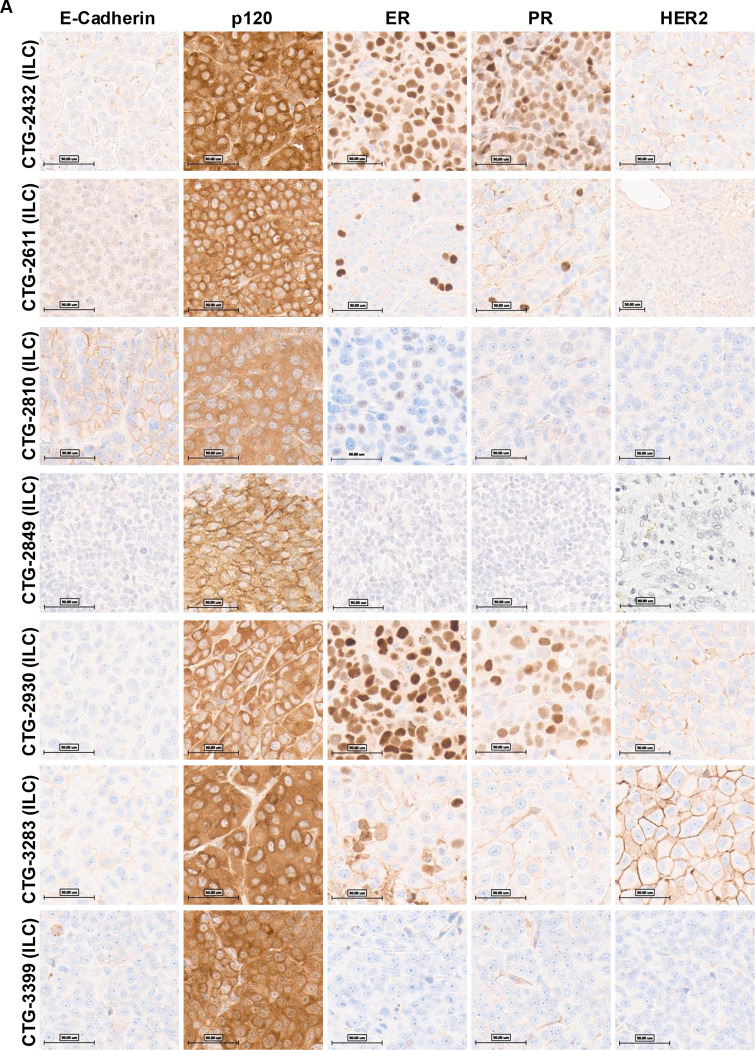
Breast cancer is a heterogeneous disease with numerous histological subtypes. Invasive lobular cancer (ILC) is the most common special subtype, accounting for 10-15% of all breast cancers. The pathognomonic feature of ILC is the loss of E-cadherin (CDH1), which leads to a unique single-file growth pattern of discohesive cells. Although ILCs show better prognostic factors than the most common No Special Type (NST) of breast cancer, patients with ILC have worse long-term outcomes, which is not well understood. In this study, we aimed to identify and characterize Patient-Derived Xenograft (PDX) models of ILC based upon the presence of truncating CDH1 mutations and/or low CDH1 mRNA expression among 128 human breast cancer PDX models. We selected 8 PDX models for validation using Immunohistochemical (IHC) analysis for E-Cadherin, p120, ER, PR, and HER2. We confirmed that seven of these PDX models are indeed ILC while one was identified as mixed NST-ILC PDX. Molecular analysis of the confirmed ILC PDX models showed enrichment of truncating CDH1 mutations, significantly lower levels of CDH1 mRNA expression and predominantly luminal subtypes compared to NST PDX models, in line with the molecular characteristics of human ILC disease. The commonly altered genes in the ILC PDX models included PIK3CA (57%), CDH1 (57%) and TP53 (57%) among others. Our study confirms and characterizes new ILC PDX models, offering valuable tools to advance our understanding of human ILC biology and support the development of innovative treatment strategies.
Keywords: CDH1; Genomic Profiling; Invasive Lobular Breast Cancer; Patient-Derived Xenograft; Targeted Therapy.
Conflict of interest statement
Competing interests The authors declare no competing interests.
Figures


References
-
- Society A.C., Cancer Facts & Figures, in American Cancer Society. 2025.
-
- Christgen M., et al. , ERBB2 mutation frequency in lobular breast cancer with pleomorphic histology or high-risk characteristics by molecular expression profiling. Genes Chromosomes Cancer, 2019. 58(3): p. 175–185. - PubMed
-
- Bozkurt E., et al. , Invasive lobular carcinoma of the breast: clinicopathological features and patient outcomes. Ann Ital Chir, 2021. 92: p. 494–499. - PubMed
Publication types
Grants and funding
LinkOut - more resources
Full Text Sources
Research Materials
Miscellaneous