[Preprint]. 2025 Aug 30:2025.08.28.672867.
doi: 10.1101/2025.08.28.672867.

Catalytic pocket of Clr4 (Suv39h) methyltransferase serves as a substrate receptor for Cullin 4-dependent histone H3 ubiquitination

Affiliations

Catalytic pocket of Clr4 (Suv39h) methyltransferase serves as a substrate receptor for Cullin 4-dependent histone H3 ubiquitination

Katarina Psenakova et al. bioRxiv. .

Abstract

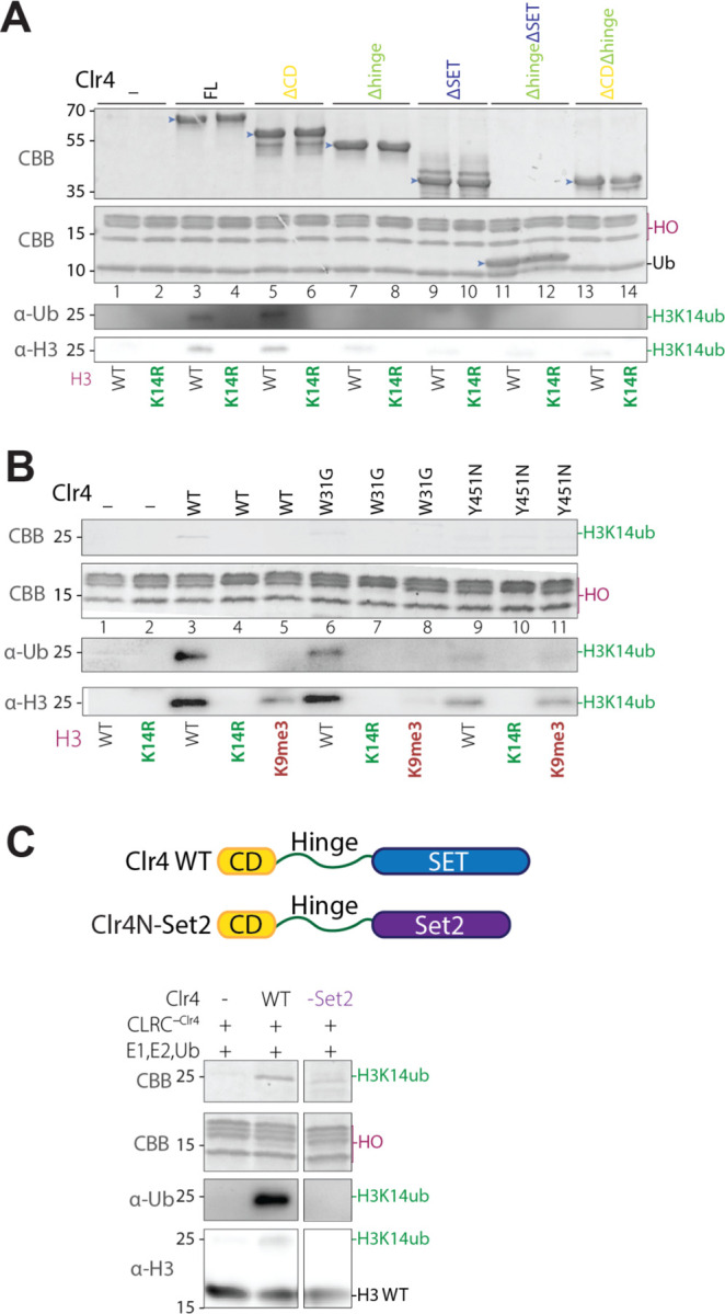
Histone H3 lysine 9 (H3K9) methylation must be regulated to prevent inappropriate heterochromatin formation. Regulation of the conserved fission yeast H3K9 methyltransferase Clr4 (Suv39h) involves an automethylation-induced conformational switch and interaction of its catalytic SET domain with mono-ubiquitinated histone H3 lysine 14 (H3K14ub), a modification catalyzed by the Cul4 subunit of the CLRC complex. Using reconstituted CLRC, we show that Clr4 catalytic pocket serves as a substrate receptor for Cul4-dependent H3K14 ubiquitination. H3K14ub activates Clr4 to catalyze cis methylation of H3K9 on the same histone tail, while Clr4 automethylation enables H3K14ub-bound Clr4 to methylate H3K9 on an unmodified H3 tail in trans. Crosslinking and structural modeling reveal interactions between Clr4 chromo and SET domains, and between the chromodomain and H3K14ub, suggesting that the chromodomain reads H3K9me3 and H3K14ub to allosterically regulate Clr4 activity. H3K14 ubiquitination therefore regulates Clr4 by promoting its recruitment and by positioning H3K9 in the active site.


Conflict of interest statement

Competing interest statement EpiCypher is a commercial developer and supplier of reagents (e.g., semisynthetic nucleosomes) used in this study. EFP, TW, MAC and M.-C.K. own shares in EpiCypher with M.-C.K. also a board member of same. All other authors declare no competing interests.

Figures

Figure 1:
Figure 1:. Reconstitution of CLRC complex-mediated H3K14 ubiquitination.
(A) Strategy for expression and purification of the CLRC E3 complex. The S. pombe CLRC complex consists of Cullin 4 (Cul4), the central E3 ligase, Rik1 (DDB1 homolog), Raf1 (DDB2 homolog, Raf2, and the histone methyltransferase Clr4. (B) Purification of the five-subunit CLRC complex. SDS-PAGE showing the Ni-NTA purified CLRC complex lacking Clr4 (CLRC-Clr4)(left), bacterially expressed and purified Clr4 (middle), and isolation of holo CLRC by size-exclusion chromatography (SEC) on a Superose 6 Increase 3.2/300 column (right). Elution fractions between 1.45–1.55 mL contained all five subunits. *, MBP-Raf1 degradation products. (C) Comparison of AlphaFold-predicted structure of CLRC-Raf2 with the crystal structure of mammalian CRL4 complex. Crystal structure of the mammalian CRL4 complex (PDB 4A0K) (left); AlphaFold3-predicted structure of the S. pombe CLRC-Raf2 complex (right). In a pairwise search for interactions, AF3 predicted an interaction between the N-terminal region of Clr4 and Raf1. Protein components are color-coded based on sequence conservation and homology. (D) Predicted Aligned Error (PAE) Plot of the AlphaFold3 model of the CLRC complex. PAE plot of the AlphaFold3 model showing predicted alignment confidence between residue pairs, where blue indicates low expected positional error (high confidence), white indicates high expected error (low confidence), and red denotes no predicted interaction between regions. (E) Identification of Clr4 domains required for incorporation into CLRC. MBP pulldown assays of CLRC subunits with Clr4 full length (FL) or truncation mutants (light blue arrows). MBP-Raf1 was immobilized on amylose beads and incubated with the indicated Clr4 fragments. After washing and elution, bound proteins were analyzed by SDS-PAGE to identify Clr4 domains that are necessary for its binding to MBP-Raf1. *, MBP-Raf1 degradation products. (F) Specific ubiquitination of H3K14 by CLRC requires Clr4. SDS-PAGE and Western blot analysis of in vitro ubiquitination assays using nucleosome substrates with the indicated histone mutations. Full ubiquitination reactions contained CLRC, E1 (UBE1), E2 (UbcH5c/UBE2D3), ubiquitin (Ub), and ATP. No H3 ubiquitination was observed in the absence of CLRC (lane 1) or without Clr4 (lanes 2–3). Mono-ubiquitination (~25 kDa band) was detected with WT (lane 4) and H3K9M (lane 6) nucleosomes, but not with H3K14R (lanes 3, 5, 7) or H3K9me3 nucleosomes (lane 8). HO, histone octamer.
Figure 2:
Figure 2:. The catalytic pocket of Clr4 acts as a substrate receptor for specific H3K14 ubiquitination.
(A) Clr4 SET domain is required for H3K14 ubiquitination. In vitro ubiquitination assays with full-length (FL) and the indicated Clr4 fragments (light blue arrows). Only Clr4 fragments retaining both the hinge region and the SET domain allowed H3K14 ubiquitination (lanes 3 and 5). HO, histone octamer. (B) Effect of Clr4 mutations and H3K9 methylation on H3K14 ubiquitination. Comparison of WT Clr4 with mutants defective in H3K9me3 binding (W31G, chromodomain) or catalytically dead (Y451N, SET domain). The Y451N mutation markedly impaired H3K14 ubiquitination (lane 9), while W31G caused only a moderate reduction in H3K14 ubiquitination (lane 6). H3K9me3 inhibited H3K14 ubiquitination by both WT and W31G Clr4 proteins. HO, histone octamer. (C) Clr4N-Set2 fusion fails to promote H3K14ub. Ubiquitination assays using WT Clr4 or a chimera consisting of the Clr4 chromodomainhinge fused to the SET domain of S. pombe Set2. WT Clr4 promoted H3K14 ubiquitination (lane 2), whereas the Set2 fusion failed to promote ubiquitination (lane 3), suggesting s specific role for the Clr4 SET domain as a substrate receptor for H3K14 ubiquitination. HO, histone octamer.
Figure 3:
Figure 3:. The relationship between Clr4 automethylation and H3K14 ubiquitination.
(A) H3K14ub activates Clr4 for intranucleosomal methylation independently of its automethylation. Methyltransferase assays with wild-type (WT) and an automethylation-deficient mutant (K455,472R) Clr4 proteins were performed using unmodified and/or H3K14ub-modified nucleosomes. The automethylation mutant showed reduced activity on an unmodified nucleosome (lane 3), but both enzymes displayed similar activity on H3K14ub substrates (lanes 4–7). H3K14ub markedly enhanced Clr4 activity compared to unmodified substrates, despite longer exposure for lanes 2–3. No methylation of unmodified H3 was observed in reactions containing both unmodified and H3K14ub nucleosomes (lanes 4, 6), indicating that under these reaction conditions H3K14ub stimulates intranucleosomal cis H3K9 methylation. HO, histone octamer. (B) Clr4 in vitro methyltransferase assay using radioactively labeled [³H]-SAM. Methyltransferase assays using Clr4 WT and nucleosomes modified with H3K9me3 or double-modified with H3K9me3 and H3K14ub. No H3 methylation signal was observed with H3K9me3 or doubly modified H3K9me3K14ub nucleosomes (lanes 3 and 5), but Clr4 was automethylated in the doubly modified nucleosome (lane 5). HO, histone octamer. (C) H3K14ub promotes Clr4-mediated methylation of K9 on an unmodified H3 tail in an automethylation-dependent manner. In vitro methylation and ubiquitination assays were reconstituted using Clr4 WT and Clr4 K455,472R proteins. Both WT and K455,472R Clr4 proteins ubiquitinated H3 (~25 kDa band detected by Coomassie staining, middle panel, lanes 5 and 6). Both WT Clr4 and Clr4 K455, 472R efficiently methylated a ubiquitinated form of H3, but only WT Clr4 methylated unmodified H3 (autoradiography, lower panel, lanes 5 and 6). HO, histone octamer. (D) Schematic summary based on the results in panels A-C.
Figure 4:
Figure 4:. Structural and functional basis of Clr4 recognition of H3K14ub-modified nucleosomes.
(A) The chromodomain promotes Clr4 binding to H3K14ub nucleosomes. Size-exclusion chromatography (SEC) analysis of complex formation between the indicated full-length (FL) Clr4 or its subfragments and H3K14ub-modified nucleosomes. Full-length Clr4 and a hinge domain-deleted variant (Clr4Δhinge) co-migrated with H3K14ub nucleosomes, suggesting the formation of stable complexes. By contrast, the Clr4-SET, which lacks both the chromodomain and hinge region (ΔCDΔhinge), did not co-migrate with H3K14ub nucleosomes, suggesting that the chromodomain is essential for stable nucleosome binding in the context of H3K14 ubiquitination. HO, histone octamer. (B) Cross-linking mass spectrometry reveals a shared interface between ubiquitin, Clr4 chromodomain, and the autoregulatory loop (ARL). Cross-linking mass spectrometry (XL-MS) was performed on the full-length Clr4-H3K14ub nucleosome complex. Cross-linked lysine residue pairs were mapped onto the linear domain architecture of Clr4, histones, and ubiquitin, and visualized as connecting lines. Intermolecular cross-links between the Clr4 chromodomain and ubiquitin (green lines) or Clr4 autoregulatory loop (ARL, red), and between the chromodomain, hinge region, and ubiquitin (red lines) are highlighted. The histone H3 tail (highlighted in violet) displayed numerous cross-links with both Clr4 chromodomain and ARL. For clarity, only crosslinks involving Clr4 are shown; all crosslinks, including those within the nucleosome, are listed in Table S5. (C) Representative 2D class averages show extranucleosomal density on top of the nucleosomal disk (light blue arrows, scale bar 100 Å). An extended set of 2D classes is shown in Fig S3B. Nucleosome highlighted by dark-pink arrows. (D) XL-MS-guided model of Clr4 in complex with two ubiquitin molecules via ubiquitin interactions site 1 (UBS1, blue) and ubiquitin interaction site 2 (UBS2, yellow). The Clr4-ubiquitin complex model was generated using HADDOCK, with input from an AlphaFold3 model of pairwise Clr4-ubiquitin interactions (see Figure S4), guided by crosslinking mass spectrometry (XL-MS) restraints. The model satisfies 33 crosslinks within a 30 Å threshold, with one mild violation (31–34 Å) in a flexible region and one significant violation (>35 Å). (E) Model of Clr4-H3K14ub nucleosome interaction guided by XL-MS restraints. HADDOCK docking driven by XL-MS distance constraints was used to model the interaction between Clr4 and the H3K14ub nucleosome, using AF3 and XL-MS guided HADDOC model of Clr4 (panel C) and the nucleosome (PDB 1KX5) as input. The resulting complex was refined by molecular dynamics in YASARA. The model suggests that the Clr4 chromodomain (CD, yellow) and autoregulatory loop (ARL, red) interact directly with the nucleosome core and with H3K14 ubiquitinated H3 tail via UBS1 and UBS2; Ub, green, SET domain, blue. (F) CryoEM map of the Clr4-H3K14ub nucleosome complex. The Clr4-H3K14ub nucleosome model was fitted into a low-pass filtered cryo-EM map (purple surface representation, 50% transparency) using COOT (see Figure S4). The density observed at the top of the nucleosome likely corresponds to a composite of the Clr4 chromodomain, one ubiquitin moiety (UBS2), and the ARL region (color coding as in panel E). The dynamic nature of these regions likely contributes to the low-resolution features of the map. The second ubiquitin and the unresolved portions of the SET domain (UBS1, docked from PDB 9ISZ), which could not be confidently fitted due to flexibility, are outlined in gray. (G) Mapping of XL-MS crosslinks onto the Clr4-H3K14ub nucleosome model. Of the 86 crosslinks identified in the XL-MS analysis, the majority were satisfied within expected distance thresholds, with distances ≤30 Å highlighted in green and 31–34 Å in pink. Ten crosslinks exceeded 35 Å (dark red). Six of these longer-distance crosslinks involved the flexible N-terminal linker and hinge regions of Clr4, while the remaining four connected flexible histone tails to Clr4. These outliers likely reflect conformational flexibility and dynamic regions not fully represented in the static model. (H) Methyltransferase activity of full length and truncated Clr4 proteins (light blue arrows) with unmodified and H3K14ub nucleosomes. In vitro methyltransferase assays performed using the indicated full-length or truncated Clr4 proteins and reconstituted nucleosomes containing either unmodified H3 (WT, lanes 5–8) or ubiquitinated H3K14 (H3K14ub, lanes 9–12). Deletion of the N-terminal chromodomain (CD) enhanced Clr4 automethylation (lanes 2 and 6) and H3K9 methylation (lane 6), suggesting that the chromodomain inhibits automethylation and H3 substrate methylation. HO, histone octamer.
Figure 5:
Figure 5:. Cul4-mediated Clr4 ubiquitination.
(A) Overview of the ubiquitinated protein pull-down workflow. Cells were transformed with a plasmid expressing 6×His-tagged ubiquitin (6×His-Ub) and lysed under denaturing conditions to preserve covalent ubiquitin conjugates. Lysates were incubated with Ni-NTA magnetic beads to selectively enrich for ubiquitinated proteins carrying 6×His-Ub. The captured proteins were then subjected to downstream analysis. (B) Schematic diagram of the FLAG-Clr4 protein highlighting the location of lysines in the 1st half and 2nd half of the protein used for mutational analysis. Clr4 was expressed with an N-terminal 3×FLAG epitope tag. A total of 36 lysine residues in Clr4 were divided into two groups: 26 lysines located in the N-terminal half, encompassing the CD and hinge region (1st half lysines) and 10 lysines in the C-terminal half, residing within the SET catalytic domain (2nd half lysines). This classification was used to guide targeted mutagenesis and functional assays. (C) Clr4 ubiquitination in vivo is dependent on Cul4. Anti-FLAG western blot following Ni-NTA pulldown under denaturing conditions from S. pombe cells co-expressing 6×His-tagged ubiquitin (6×His-Ub) and FLAG-Clr4. Ubiquitinated FLAG-Clr4 was detected in wild-type (WT) cells but was greatly diminished in cul4–1 mutants, indicating that Clr4 ubiquitination requires Cul4 in vivo. (D) Clr4 ubiquitination targets lysines in the chromodomain and hinge region. Anti-FLAG western blot following Ni-NTA pulldown under denaturing conditions from S. pombe cells co-expressing 6×His-tagged ubiquitin (6×His-Ub) and the indicated FLAG-tagged Clr4 proteins. Cells expressing Clr4 WT or the 2nd half KtoR mutant (lysines mutated in the SET domain) retained ubiquitination. By contrast, Clr4 1st half KtoR (lysines mutated in the chromodomain and hinge region) and Clr4 allKtoR mutants showed loss of ubiquitination, indicating that ubiquitin conjugation occurs primarily within the N-terminal half of Clr4. *, background band present in untagged cells recognized by the anti-FLAG antibody.
Figure 6:
Figure 6:. Role of Cul4-mediated ubiquitination in Clr4 localization and H3K9 methylation.
(A, B) ChIP-seq genome browser tracks showing localization of FLAG-Clr4 (A) and H3K9me3 (B) modification at the indicated (top) genomic loci in wild-type (cul4+), cul4-K680R, and cul4–1 cells. The locations of centromeric dg, and dh repeats and the mating type (mat) centromere homology (cenH) are highlighted in the bottom. Chromosome coordinates shown on the top, genotypes on left, and reads per million in brackets. (C-N) ChIP-qPCR analysis of FLAG-Clr4, H3K9me2, and H3K9me3 localization at the indicated loci (dg, mat2P, and tlh1) in wild-type, cul4-K680R, cul4–1, H3K14R, H3K14A, 1st half-KtoR (clr4 with 1st half lysines substituted with arginine, see Figure 5B), and clr4 cells. Bars show mean percentage input and error bars show standard deviations of 3 biological replicates.
Figure 7:
Figure 7:. H3K14 ubiquitination regulates both the substrate recognition (“read”) and catalytic activity (“write”) of Clr4.
(A) Clr4 as a dual substrate receptor within the CLRC complex. Left: The catalytic SET domain of Clr4 acts as a substrate receptor for histone H3, enabling Cul4-mediated monoubiquitination of H3K14. Middle: The N-terminus of Clr4 interacts directly with Raf1 that functions as a substrate receptor for Clr4 ubiquitination on multiple surface-exposed lysines, predominantly located within the hinge region of Clr4. Right: Cul4-mediated ubiquitination of Clr4 promotes its dissociation from the CLRC complex, a step required for the spreading of H3K9 methylation. (B) Ubiquitin-binding site 1 (UBS1) drives cis methylation. H3K14 monoubiquitination strongly stimulates Clr4 to methylate H3K9 on the same histone tail in cis. This is mediated by UBS1, an interaction surface between ubiquitin and the SET domain that positions H3K9 in the catalytic site. (C) Ubiquitin-binding site 2 (UBS2) and automethylation enable trans methylation. UBS2, identified in this study, involves interactions between H3K14ub and the chromodomain-ARL interface within Clr4. Together with Clr4 automethylation of the ARL, UBS2 licenses Clr4 to methylate H3K9 on an unmodified histone tail in trans, thereby facilitating methylation spreading.

References

    1. Bühler M. & Moazed D. Transcription and RNAi in heterochromatic gene silencing. Nat Struct Mol Biol 14, 1041–1048 (2007). - PubMed
    1. Allshire R. C. & Madhani H. D. Ten principles of heterochromatin formation and function. Nat Rev Mol Cell Biol 19, 229–244 (2018). - PMC - PubMed
    1. Jia S., Kobayashi R. & Grewal S. I. S. Ubiquitin ligase component Cul4 associates with Clr4 histone methyltransferase to assemble heterochromatin. Nat Cell Biol 7, 1007–1013 (2005). - PubMed
    1. Horn P. J., Bastie J. N. & Peterson C. L. A Rik1-associated, cullin-dependent E3 ubiquitin ligase is essential for heterochromatin formation. Genes Dev 19, 1705–1714 (2005). - PMC - PubMed
    1. Hong E. J. E., Villén J., Gerace E. L., Gygi S. P. & Moazed D. A cullin E3 ubiquitin ligase complex associates with Rik1 and the Clr4 histone H3-K9 methyltransferase and is required for RNAi-mediated heterochromatin formation. RNA Biol 2, 106–111 (2005). - PubMed

Publication types

LinkOut - more resources