[Preprint]. 2025 Aug 27:2025.08.27.672623.
doi: 10.1101/2025.08.27.672623.

Non-polio enteroviruses compromise the electrophysiology of a human iPSC-derived neural network

Affiliations

Non-polio enteroviruses compromise the electrophysiology of a human iPSC-derived neural network

Feline F W Benavides et al. bioRxiv. .

Abstract

The non-polio enteroviruses enterovirus-D68 (EV-D68) and enterovirus-A71 (EV-A71) are highly prevalent and considered pathogens of increasing health concern. While most enterovirus infections are mild and self-limiting, severe complications ranging from meningitis, encephalitis, to acute flaccid paralysis can occur, especially in children and immunocompromised patients. Despite the global burden of neurological complications caused by EV-D68 and EV-A71, the underlying neuropathogenesis remains poorly understood. In particular, the impact of the infection on neural function has not been clearly elucidated. Here, we compare the replication kinetics, cellular tropism, and electrophysiological effects of EV-D68 and EV-A71 infection in a physiologically relevant human pluripotent stem cell-derived neural co-culture model, consisting of excitatory neurons and astrocytes. Inoculation with contemporary circulating EV-D68 strains and an EV-A71 strain resulted in decreased neural activity in the co-cultures, with EV-D68 A2/2018 inducing the most rapid and robust negative effect on neural co-cultures, followed by EV-D68 B3/2019. EV-D68 strains preferentially infected neurons, whereas EV-A71 infection was detected in both cell types to the same extent. Despite the lack of viral release of infectious virus particles of EV-D68 B3/2019 in the supernatant, the infection could spread in the cultures and reduce neurotransmission. Higher viral load and broader tropism of EV-A71 did not result in enhanced impairment of neural function. Our results demonstrate that neurotropic non-polio enteroviruses lead to disruption of spontaneous neural activity in a virus-specific manner, which does not correlate with their replication efficiency.

Keywords: micro-electrode array; neural networks; neurotropism; neurovirulence; non-polio enteroviruses.


Conflict of interest statement

Competing interests The authors report no competing interests.

Figures

Figure 1.
Figure 1.. Ngn2-expressing neurons co-cultured with astrocytes form functional neural networks and respond to stimulation and inhibition.
hPSC-derived Ngn2-expressing neurons were co-cultured with hPSC-derived astrocytes and matured for 21 DIV. (A) Immunofluorescent staining was performed with astrocytic-specific marker GFAP (magenta) and the neuron-specific marker MAP2 (cyan). Cells were counterstained with the nuclear dye Hoechst at DIV 21. (B) Immunofluorescent staining for functional synapses was performed against MAP2 (cyan), synapsin 1 (green, marker for pre-synaptic density), and Homer 1 (red, marker for post-synaptic density) at DIV 22. (C) Brightfield images of neural co-cultures plated on micro-electrode array plates to measure neural activity at day 21. (D) Parameters for neural activity of Ngn2 neural co-cultures between DIV 21 and 31: firing rate, burst frequency, and network burst frequency. Data are depicted as mean ± SEM and are derived from eight independent experiments (n = 48, unless datapoints were excluded based on exclusion criteria, see Material and Methods). (E) Raster plots of neural co-cultures were stimulated with 50 μM FSK and 0.1 μM ROL or inhibited with 10 μM SFX. (F) Firing rate, burst frequency, and network burst frequency measured after stimulation or inhibition are shown. Data are depicted as mean ± SD. Data are derived from two independent experiments (n = 9 per group, unless datapoints were excluded based on exclusion criteria, see Material and Methods). Abbreviations: hPSC = human induced pluripotent stem cells; Ngn2 = Neurogenin-2; DIV = days in vitro; GFAP = glial fibrillary acidic protein; MAP2 = microtubule-associated protein 2; SEM = standard error of the mean; DMSO = dimethylsulfoxide; FSK = forskolin; ROL = rolipram; SFX = (S)-fluoxetine; SD = standard deviation.
Figure 2.
Figure 2.. Enterovirus-D68 and Enterovirus-A71 productively replicate in co-cultures and show similarity in their cell tropism.
(A) Neural co-cultures were infected with different EV-D68 strains and the EV-A71 strain Sep006 from the C4 genotype at a MOI of 0.1. Viral titres were determined in the supernatant at the indicated timepoints by endpoint dilution, with the first data point being the back-titration of the inoculum. Data represent mean ± SEM from three independent differentiation experiments and every growth curve was performed in triplicate. (B) At 24 hpi, the co-cultures were fixed and stained for the presence of EV-D68 structural antigen VP1 (green). MAP2 (cyan) was used as a marker for neurons, and astrocytes were identified by staining for GFAP (magenta). Cells were counterstained with Hoechst (grey) to visualize the nuclei. (C) At 24 hpi, EV-A71 inoculated co-cultures were fixed and stained for the presence of dsRNA, a marker for active EV-A71 replication (green), astrocytes (GFAP, magenta) and neurons (MAP2, cyan). The immunofluorescence data shown are representative examples from three independent experiments for each culture condition. Maximum intensity projections of Z-stacks are displayed. Abbreviations: EV = enterovirus; MOI = multiplicity of infection; SEM = standard error of the main; hpi = hours post inoculation; VP1 = viral protein 1; MAP2 = microtubule-associated protein 2; GFAP = glial fibrillary acidic protein; dsRNA = double-stranded RNA
Figure 3.
Figure 3.. Enterovirus infection does not result in upregulation of cleaved caspase-3.
Neural co-cultures were infected with EV-D68 or EV-A71 at an MOI of 1. At 24 and 72 hpi, the cells were fixed and stained for the presence dsRNA (green/red) as marker for virus infection, for either neural marker MAP2 (cyan) or astrocytic marker GFAP (magenta), and for the apoptosis marker CC-3 (yellow). Data shown are representative examples from two independent experiments. Abbreviations: EV = enterovirus; MOI = multiplicity of infection; hpi = hours post inoculation; VP1 = viral protein 1; dsRNA = double-stranded RNA; MAP2 = microtubule-associated protein 2; GFAP = glial fibrillary acidic protein; CC-3 = cleaved caspase-3
Figure 4.
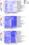
Figure 4.. Heatmap of normalized neural activity data recorded from neural co-cultures inoculated with non-polio enteroviruses.
Neural activity was recorded from neural co-cultures, consisting of Ngn2 neurons and astrocytes, on a MEA platform. Neural co-cultures were mock-inoculated or inoculated with EV-D68 A2/2018, B3/2019, or EV-A71 Sep006 with a MOI of 1. Only data that met inclusion criteria for MEA recordings were used (see Material and Methods). Any experiment that contained Not Available values throughout any of the chosen MEA output variables (Table 3) was excluded for PCA analysis, to ensure biological relevance and integrity of the dataset. Data was normalized against baseline recording obtained before inoculation. Data is shown from one (A), three (B), or 10 dpi (C). Rows represent output variables of MEA data and columns represent experimental data from a single well. All MEA data were z-score normalized for each variable across inoculation groups to enable cross-condition comparisons. Color intensity reflects a positive or negative effect on neural activity, with white to red indicating a more positive effect. Abbreviations: Ngn2 = Neurogenin-2; MEA = micro-electrode array; EV = enterovirus; MOI = multiplicity of infection; dpi = days post inoculation;
Figure 5.
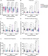
Figure 5.. Enterovirus infection impacts the spontaneous activity of neural co-culture.
Neural co-cultures were inoculated with EV-D68 A2/2018 (pink), B3/2019 (cyan), or EV-A71 Sep006 (purple) with a MOI of 1, and neural activity was measured between one to three dpi. The following parameters were displayed (A) number of active electrodes, where wells were excluded if they contained 5 or less active electrodes at baseline (indicated with a red line); (B) firing rate; (C) burst frequency, (D) number of spikes per burst, (E) network burst frequency and (F) number of spikes per network burst. Data displayed are derived from at least four independent experiments (n = 48 for control and n = 24 per inoculation group, see exclusion criteria in the Material and Methods section). Statistical significance was calculated with a two-way ANOVA with a Šídák’s multiple comparisons post hoc test. Asterisks indicate statistical significance (*P<0.05, **P<0.01, ***P<0.001, ****P<0.0001). Abbreviations: EV = enterovirus; MOI = multiplicity of infection; dpi = days post inoculation.

References

    1. Harvala H, Johannesen CK, Benschop KSM, et al. Enterovirus circulation in the WHO European region, 2015–2022: a comparison of data from WHO’s three core poliovirus surveillance systems and the European Non-Polio Enterovirus Network (ENPEN). Lancet Reg Health Eur. 2025;53:101292. doi: 10.1016/j.lanepe.2025.101292 - DOI - PMC - PubMed
    1. Rankin DA, Spieker AJ, Perez A, et al. Circulation of Rhinoviruses and/or Enteroviruses in Pediatric Patients With Acute Respiratory Illness Before and During the COVID-19 Pandemic in the US. JAMA Netw Open. 2023;6(2):e2254909. doi: 10.1001/jamanetworkopen.2022.54909 - DOI - PMC - PubMed
    1. Nguyen-Tran H, Butler M, Simmons D, Dominguez SR, Messacar K. Return of the Biennial Circulation of Enterovirus D68 in Colorado Children in 2024 Following the Large 2022 Outbreak. Viruses. 2025;17(5):673. doi: 10.3390/v17050673 - DOI - PMC - PubMed
    1. de Schrijver S, Vanhulle E, Ingenbleek A, et al. Epidemiological and clinical insights into enterovirus circulation in Europe, 2018 – 2023: a multi-center retrospective surveillance study. J Infect Dis. Published online April 4, 2025:jiaf179. doi: 10.1093/infdis/jiaf179 - DOI - PMC - PubMed
    1. Chuang YY, Huang YC. Enteroviral infection in neonates. J Microbiol Immunol Infect. 2019;52(6):851–857. doi: 10.1016/j.jmii.2019.08.018 - DOI - PubMed

Publication types

LinkOut - more resources