[Preprint]. 2025 Aug 25:2025.08.21.671520.
doi: 10.1101/2025.08.21.671520.

FSP1 and histone deacetylases suppress cancer persister cell ferroptosis

Affiliations

FSP1 and histone deacetylases suppress cancer persister cell ferroptosis

Masayoshi Higuchi et al. bioRxiv. .

Abstract

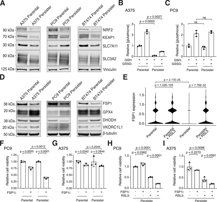
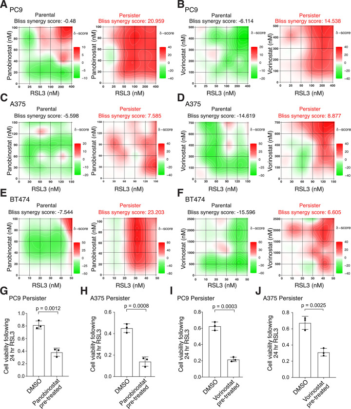
Cancer persister cells populate minimal residual disease and contribute to acquired drug resistance. We previously discovered that persister cells are sensitized to ferroptosis. However, our understanding of this emergent persister cell vulnerability remains limited, impeding ferroptosis drug development efforts. Here, we sought to understand key factors which govern persister cell ferroptosis to inform combinatorial treatment strategies. We found that persister cells can downregulate oxidative phosphorylation, a key source of reactive oxygen species, to avoid death from GPX4 inhibition. However, this can be overcome by pretreatment with clinically available histone deacetylase inhibitors which induce reactive oxygen species in persister cells and synergize with GPX4 inhibition. Furthermore, we found that while levels of iron, glutathione, and antioxidant genes are not universally dysregulated in persister cells, persister cells consistently downregulate alternative ferroptosis suppressor FSP1 and rely upon residual FSP1 to survive GPX4 inhibition. These findings reveal new strategies to eliminate persister cells by combining GPX4 inhibitors with histone deacetylase or FSP1 inhibitors.


Conflict of interest statement

Competing interests: MJH is a co-founder of Ferro Therapeutics, a subsidiary of BridgeBio Pharma, Inc.

Figures

Fig. 1.
Fig. 1.. Oxidative phosphorylation contributes to ferroptosis sensitization in cancer persister cells.
(A) UMAP of PC9 parental and persister cells treated with or without 1 μM RSL3 for 24 hours. (B) Enriched Hallmarks gene sets in persister cells treated with and without RSL3. Positive values indicate enrichment in persister cells treated with RSL3. (C) UMAP of PC9 persister cells treated with and without RSL3. (D) Pseudotime analysis of PC9 persister cells treated with and without RSL3. Solid black line indicates the estimated transitional trajectory across cell states. (E) UMAP of PC9 persister cells treated with and without RSL3 colored by cluster. (F) Ferroptosis-drivers gene set signature score across clusters in (E). (G) Ferroptosis-suppressors gene set signature score across clusters in (E). (F and G) P values calculated with Mann-Whitney test. (H) PC9 persister cells derived from erlotinib plus metformin and then treated with 500 nM RSL3 for 24 hours. n = 3 biological replicates; mean ± s.d. is shown; P values calculated with two-tailed Student’s t-test.
Fig. 2.
Fig. 2.. Persister cells have variable antioxidant deficiencies and depend on FSP1 to survive GPX4 inhibition.
(A) Parental and persister cells analyzed for NRF2, KEAP1, and system xc components SLC7A11 and SLC3A2 expression. (B and C) A375 and PC9 parental and persister cells analyzed for reduced GSH or total GSH (GSSG) levels. (D) Protein expression of ferroptosis suppressor genes in parental and persister cells. (E) FSP1 mRNA expression in PC9 parental and persister cells treated with and without RSL3. P values calculated with the Wilcoxon Rank Sum test with Bonferroni correction. (F and G) PC9 and A375 parental and persister cells treated with and without 5 μM FSP1 inhibitor. (H and I) PC9 and A375 persister cells co-treated with 1 μM FSP1 inhibitor, 50 nM RSL3, or both for 24 hours. (B, C, F to I) n = 3 biological replicates; mean ± s.d. is shown; P values calculated with two-tailed Student’s t-test.
Fig. 3.
Fig. 3.. HDAC inhibition synergizes with GPX4 inhibition to selectively kill persister cells.
(A to F) Heatmaps of synergy between GPX4 inhibitor RSL3 and HDAC inhibitors panobinostat and vorinostat following 24 hour cotreatment. Bliss synergy score calculated with SynergyFinder 3.0. Red color and positive scores indicate synergy, green color and negative scores indicate buffering. (A and B) PC9 parental cells and persister cells derived from 50 nM erlotinib. (C and D) A375 parental cells and persister cells derived from 10 nM dabrafenib with 1 nM trametinib. (E and F) BT474 parental cells and persister cells derived from 2 μM lapatinib. (G to J) Pre-derived PC9 or A375 persister cells were treated for 48 hours with a nontoxic concentration of HDAC inhibitor (see fig. S5), rinsed, and then treated with RSL3 for 24 hours while maintained under targeted therapy treatment. Data normalized to untreated persister cells. Concentration and HDAC inhibitor used: (G) 7.5 nM panobinostat, (H) 5 nM panobinostat, (I) 100 nM vorinostat, (J) 1 μM vorinostat. RSL3 concentrations used: (G) 150 nM, (H) 100 nM, (I) 150 nM, (J) 80 nM. n = 3 biological replicates; mean ± s.d. is shown; P values calculated with two-tailed Student’s t-test.
Fig. 4.
Fig. 4.. HDAC inhibitors induce persister cell oxidative stress to sensitize persister cells to ferroptosis.
(A) UMAP of PC9 parental and persister cells treated with or without HDAC inhibitor panobinostat for 48 hours. (B) Treatment of PC9 persister cells with panobinostat enriches for oxidative phosphorylation genes. (C and D) Treatment with panobinostat does not decrease GSH levels in PC9 or A375 persister cells. 1 mM buthionine sulfoximine (BSO) was used as a positive control for GSH depletion. (E) Ferroptosis sensitization of PC9 persister cells from treatment with panobinostat is not inhibited by glutathione ethyl ester (GSHee, 1 mM). (F) PC9 persister cell treatment with panobinostat decreases rather than increases intracellular iron. (G) Panobinostat treatment of PC9 persister cells increases ROS. (H and I) PC9 persister cells treated with panobinostat are rescued from RSL3 treatment with the antioxidants EUK-134 (10 μM), nordihydroguaiaretic acid (NDGA, 5 μM), decylubiquinone (Dec, 7.5 μM), and α-tocopherol (α-toc, 1.5 mM). Panobinostat concentrations used in PC9 cells: 7.5 nM (A to C and F to G) or 2.5 nM (E and H to I); in A375 cells: 5 nM (D). (C to I) n = 3 biological replicates; mean ± s.d. is shown; P values calculated with two-tailed Student’s t-test.
Fig. 5.
Fig. 5.. Enhancing persister cell ferroptosis with FSP1 and HDAC inhibition.
Cancer persister cells can decrease oxidative stress to survive GPX4 inhibition (GPX4i). However, GPX4i-tolerant persister cells become dependent on the alternative ferroptosis suppressor enzyme FSP1 to survive and addition of FSP1 inhibitor (FSP1i) increases persister cell ferroptotic death. Furthermore, persister cell oxidative stress is increased by nontoxic pre- or co-treatment with clinically available HDAC inhibitors resulting in synergistic persister cell ferroptosis in combination with GPX4 inhibitor (GPX4i). Our findings reveal new approaches to selectively enhance persister cell ferroptosis while potentially sparing other cells.

References

    1. Russo M., Chen M., Mariella E., Peng H., Rehman S. K., Sancho E., Sogari A., Toh T. S., Balaban N. Q., Batlle E., Bernards R., Garnett M. J., Hangauer M., Leucci E., Marine J.-C., O’Brien C. A., Oren Y., Patton E. E., Robert C., Rosenberg S. M., Shen S., Bardelli A., Cancer drug-tolerant persister cells: from biological questions to clinical opportunities. Nat. Rev. Cancer 24, 694–717 (2024). - PubMed
    1. Terai H., Kitajima S., Potter D. S., Matsui Y., Quiceno L. G., Chen T., Kim T., Rusan M., Thai T. C., Piccioni F., Donovan K. A., Kwiatkowski N., Hinohara K., Wei G., Gray N. S., Fischer E. S., Wong K.-K., Shimamura T., Letai A., Hammerman P. S., Barbie D. A., ER Stress Signaling Promotes the Survival of Cancer “Persister Cells” Tolerant to EGFR Tyrosine Kinase Inhibitors. Cancer Res. 78, 1044–1057 (2018). - PMC - PubMed
    1. Hangauer M. J., Viswanathan V. S., Ryan M. J., Bole D., Eaton J. K., Matov A., Galeas J., Dhruv H. D., Berens M. E., Schreiber S. L., McCormick F., McManus M. T., Drug-tolerant persister cancer cells are vulnerable to GPX4 inhibition. Nature 551, 247–250 (2017). - PMC - PubMed
    1. Viswanathan V. S., Ryan M. J., Dhruv H. D., Gill S., Eichhoff O. M., Seashore-Ludlow B., Kaffenberger S. D., Eaton J. K., Shimada K., Aguirre A. J., Viswanathan S. R., Chattopadhyay S., Tamayo P., Yang W. S., Rees M. G., Chen S., Boskovic Z. V., Javaid S., Huang C., Wu X., Tseng Y.-Y., Roider E. M., Gao D., Cleary J. M., Wolpin B. M., Mesirov J. P., Haber D. A., Engelman J. A., Boehm J. S., Kotz J. D., Hon C. S., Chen Y., Hahn W. C., Levesque M. P., Doench J. G., Berens M. E., Shamji A. F., Clemons P. A., Stockwell B. R., Schreiber S. L., Dependency of a therapy-resistant state of cancer cells on a lipid peroxidase pathway. Nature 547, 453–457 (2017). - PMC - PubMed
    1. Sun X., Bieber J. M., Hammerlindl H., Chalkley R. J., Li K. H., Burlingame A. L., Jacobson M. P., Wu L. F., Altschuler S. J., Modulating environmental signals to reveal mechanisms and vulnerabilities of cancer persisters. Sci. Adv. 8, eabi7711 (2022). - PMC - PubMed

Publication types