

Corneal transplantation wound dehiscence after penetrating keratoplasty and deep anterior lamellar keratoplasty
- PMID: 40911254
- PMCID: PMC12413426
- DOI: 10.1007/s10792-025-03708-x
Corneal transplantation wound dehiscence after penetrating keratoplasty and deep anterior lamellar keratoplasty
Abstract
Purpose: To study clinical characteristics and outcomes of penetrating keratoplasty (PK) and deep anterior lamellar keratoplasty (DALK) wound dehiscence.
Methods: This retrospective case series assessed PK and DALK recipients with wound dehiscence at a single institution. We evaluated relationships between dehiscence etiologies, transplant indications, ocular/systemic comorbidities, keratoplasty type, and adverse post-dehiscence outcomes, especially graft failure and visual loss.
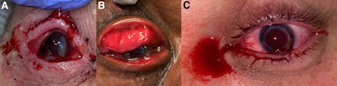
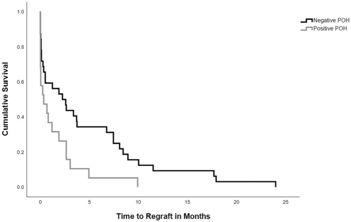
Results: Wound dehiscence occurred in 97/1019 eyes (90/863 PK [10.4%] vs 7/156 DALK [4.5%]; p = 0.002). Median time to dehiscence was 6.6 months (range = 1 day-39.2 years). Primary causes included trauma (44.1%) and ulceration (36.1%). Leading surgical indications associated with dehiscence were microbial keratitis and corneal ectasia. Ocular surface disease, viral keratitis, glaucoma, diabetes, and smoking history were more prevalent in PK eyes. Graft failure post-dehiscence was more frequent after PK than DALK (61% vs 0%; p = 0.002) and more rapid with herpetic keratitis history (Log-Rank p = 0.02). Microbial keratitis-associated dehiscence was the strongest predictor of graft failure (odds ratio = 3.9, 95% CI 1.2-12.9). All 20 enucleations occurred in the PK group. Pre-dehiscence, PK eyes had worse habitually corrected visual acuity (HCVA; p = 0.008). Post-dehiscence, more PK eyes lost ≥ 2 Snellen lines (53.7% vs 14.3%; p = 0.058) and HCVA was worse than 20/200 (55.6% vs 0%; p = 0.005).
Conclusion: Wound dehiscence is a serious keratoplasty complication that may be associated with graft failure and vision loss, especially after PK. Careful selection of transplantation techniques and application of therapeutic strategies tailored for the specific surgical indication and associated comorbidities should be used to mitigate the clinical course.
Keywords: Corneal transplant; Deep anterior lamellar keratoplasty; Penetrating keratoplasty; Wound dehiscence.
© 2025. The Author(s).
Conflict of interest statement
Declarations. Competing interests: The authors have no relevant financial or non-financial interests to disclose. Ethics approval: This retrospective study was approved by the University of Iowa Institutional Review Board, was compliant with the Health Insurance Portability and Accountability Act and adhered to the tenets of the Declaration of Helsinki.
Figures
References
-
- Mathews P, Benbow A, Corcoran K, DeMatteo J, Philippy B, Van Meter W (2023) 2022 eye banking statistical report—executive summary. Eye Bank Corneal Transpl 2(3):e0008. 10.1097/ebct.0000000000000008 - DOI
MeSH terms
LinkOut - more resources
Full Text Sources
Medical
