. 2025 Sep;21(9):e70626.
doi: 10.1002/alz.70626.

Moderating effects of plasma glial fibrillary acidic protein along the Alzheimer's disease continuum

Affiliations

Moderating effects of plasma glial fibrillary acidic protein along the Alzheimer's disease continuum

Shannon Y Lee et al. Alzheimers Dement. 2025 Sep.

Abstract

Introduction: Glial fibrillary acidic protein (GFAP) may contribute to Alzheimer's pathology at early disease stages. GFAP moderation of Alzheimer's disease (AD)-related neurodegeneration and cognition is unclear.

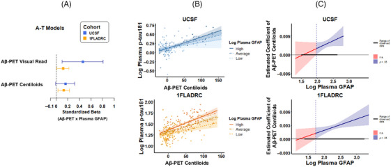
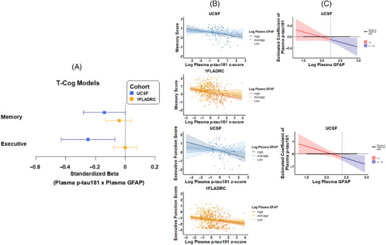
Methods: We examined plasma GFAP moderation of AD biomarkers (amyloid beta [Aβ]-positron emission tomography [PET][A]; plasma phosphorylated tau-181 [p-tau181][T1]), neurodegeneration (plasma NfL[Nplasma]; structural magnetic resonance imaging [MRI][NMRI]), and cognition (Cogmemory; Cogexecutive) in two cohorts: University of California San Francisco (UCSF) (N = 212, 91.0% non-Hispanic/Latino White [NHLW], age = 74.7 [7.6] years, 75.9% cognitively unimpaired [CU]) and 1Florida Alzheimer's Disease Research Centers (1FLADRC; N = 582, 32.8% NHLW, age = 70.7 [8.5] years, 28.9% CU).

Results: Plasma GFAP consistently moderated A-T1 (UCSF: β = 0.46, p = 0.012; 1FLADRC: β = 0.12, p = 0.029). The association between elevated Aβ-PET and increased (p-tau) was strengthened at higher GFAP concentrations. In 1FLADRC, GFAP moderated T1-Nplasma/MRI. In UCSF, GFAP moderated T1-Cogmemory/executive and NMRI-Cogmemory/executive. Higher GFAP consistently related to worse neurodegeneration and cognition (main effects).

Discussion: Across demographically and clinically heterogeneous cohorts, plasma GFAP is a key moderator of AD and may help identify individuals at greatest risk of AD-related neurodegeneration and cognitive decline.

Highlights: AD biomarkers were measured in two demographically and clinically distinct cohorts. Plasma GFAP moderated Aβ-PET to p-tau associations in both UCSF and 1FLADRC. Cohort-dependent, GFAP moderated p-tau to neurodegeneration and cognition associations. All moderations revealed strengthened disease associations with higher plasma GFAP. Plasma GFAP may help identify individuals at greatest risk of AD-related decline.

Keywords: ATN; Alzheimer's disease; GFAP; astrocyte reactivity; cognition; glial fibrillary acidic protein; inflammation; neurodegeneration; neuroinflammation; neuropathology; plasma GFAP; plasma biomarkers.


Conflict of interest statement

Authors report no disclosures relevant to the content of this study. Dr. Armstrong reported grants from the National Institutes of Health (NIH), Florida Department of Health, and Lewy Body Dementia Association Research Center of Excellence; speaker honoraria from the Taiwan International Congress of Parkinson's Disease and Movement Disorders, American Academy of Neurology Annual Meetings, World Congress on Parkinson's Disease and Related Disorders, PRIME CME Program, Dr. Daniel I. Kaufer Lecture Series at the University of Wisconsin Alzheimer's Disease Research Center, Dementia with Lewy Bodies: Filling the Gaps in Translational and Clinical Research NIA‐NINDS Conference, and Michael J. Fox Foundation Neuro‐Impact Workshop; personal compensation for serving as a Data Safety Monitoring Board member with the Alzheimer's Therapeutic Research Institute/ Alzheimer's Clinical Trials Consortium, Alzheimer's Disease Cooperative Study, and an NIH study (R01AG083828); and a non‐compensated relationship as a member of the Scientific Advisory Board Executive Committee for the Lewy Body Dementia Association. Dr. Casaletto reported grants from the National Institute on Aging (NIA), Hillblom Foundation, Alzheimer's Association, and Wellcome Trust, Leap; and non‐compensated relationships as Chair of the International Neuropsychological Society Conflict of Interest Committee and Executive Board member of the Alzheimer's Association ISTAART Cognition Professional Interest Area. Dr. DeKosky reported royalties from UpToDate as the section editor for dementia; consulting fees from Brainstorm Cell Therapeutics, Eisai, and Sanofi; personal fees from Acumen Pharmaceuticals, Biogen, Prevail Pharmaceuticals, Cognition Therapeutics, Vaccinex, Lilly Pharmaceuticals, Nido Biosciences, Neuvivo Pharmaceuticals, Novo Nordisk, and Capricor for serving on the data safety monitoring or medical advisory boards; and honoraria from Neurotherapeutics for serving as an associate editor and UpToDate for serving as a section editor for dementia outside the submitted work. Dr. DeSimone reported consulting fees from Automated Imaging Diagnostics; and being a shareholder for Automated Imaging Diagnostics. Dr. La Joie reported grants from the National Institute on Aging, the Alzheimer's Association, the US Department of Defense; consulting fees from GE Healthcare outside the submitted work; and conference attendance support from the Alzheimer's Association Dr. Rabinovici reported grants from National Institutes of Health during the conduct of the study; consulting fees from C2N, Eli Lilly, Alector, Merck, Roche, and Novo Nordisk; data safety monitoring board fees from Johnson & Johnson; and grants from Avid Radiopharmaceuticals, GE Healthcare, Life Molecular Imaging, and Genentech outside the submitted work. Dr. Rojas reported serving as site principal investigator for clinical trials sponsored by Eli Lilly, Eisai, and Amylyx during the conduct of the study; consulting fees from Ferrer International, AdeptField Solutions, Reach Market Research, and Clarivate; honoraria for lectures through the American Academy of Neurology; conference attendance support from the Alzheimer's Association; and a pending patent. Dr. Paolillo reported a grant from the NIA. Dr. Saloner reported grants from the Alzheimer's Association, New Vision Research Charleston Conference on Alzheimer's Disease, American Academy of Neurology, American Brain Foundation, and Association for Frontotemporal Degeneration. Dr. Smith reported a grant from the National Institute on Aging (NIA). Dr. Staffaroni reported personal fees from ADDF, Alector, Aviado Bio, CervoMed, Passage Bio, Prevail Therapeutics/Eli Lilly, Takeda, and Vesper Bio. Dr. Vaillancourt reported grants from the NIH; Neuropacs Corp shareholder; and licensed patents (11439341; 10758170). Dr. VandeVrede reported grants from the NIH, Alzheimer's Association, and Shenandoah Foundation; consulting fees from Roche, Siemens, and Biogen; personal fees from Peerview CME and Haymarket; payment for expert testimony; meeting attendance/travel support from Biogen, Siems, and Tau Consortium; serving on a monitoring or advisory board for NIO‐SILK; and receipt of materials or services from LabCorp and Quanterix outside the submitted work. Dr. Wiens reported a grant from the NIH. Dr. Wang reported a grant from the NIA. All other authors report no disclosures. The authors have no conflicts of interest. Author disclosures are available in the supporting information.

Figures

FIGURE 1
FIGURE 1
Plasma GFAP moderating effects of Aβ‐PET and plasma p‐tau181 relationships. Aβ‐PET was performed in a subset of participants in each cohort (UCSF: N = 129; 1FLADRC: N = 264). (A) values represent standardized coefficients with bootstrapped 95% confidence intervals for the interaction of Aβ‐PET (visual read or Centiloids) and plasma GFAP on plasma p‐tau181, reported by cohort. (B) Plasma GFAP moderation of Aβ‐PET (Centiloids) and plasma p‐tau181 stratified by high (+1 SD), average (mean), and low (−1 SD) levels of plasma GFAP. (C) The effect size (unstandardized) of Aβ‐PET on plasma p‐tau181 is plotted across plasma GFAP levels. Blue regions represent levels of plasma GFAP at which the relationship between Aβ‐PET (Centiloids) and plasma p‐tau181 is statistically significant. 1FLADRC, 1Florida Alzheimer's Disease Research Center; GFAP, glial fibrillary acidic protein; UCSF, University of California San Francisco.
FIGURE 2
FIGURE 2
Plasma GFAP moderating effects of plasma p‐tau181 and neurodegeneration (AD meta‐ROI and plasma NfL) relationships. (A) values represent standardized coefficients with bootstrapped 95% confidence intervals for the interaction of plasma p‐tau181 and plasma GFAP on AD meta‐ROI or plasma NfL, reported by cohort. (B) Plasma GFAP moderation of plasma ptau‐181 and AD meta‐ROI or plasma NfL, stratified by high (+1 SD), average (mean), and low (−1 SD) levels of plasma GFAP. (C) Effect size (unstandardized) of p‐tau181 on neurodegeneration is plotted across plasma GFAP levels. Blue regions represent levels of plasma GFAP at which the relationship between p‐tau181 and neurodegeneration is statistically significant. AD, Alzheimer's disease; GFAP, glial fibrillary acidic protein; NfL, neurofilament light; ROI, region of interest.
FIGURE 3
FIGURE 3
Plasma GFAP moderating effects of plasma p‐tau181 and cognition (memory and executive function) relationships. (A) Values represent standardized coefficients with bootstrapped 95% confidence intervals for the interaction of plasma p‐tau181 and plasma GFAP on cognition, reported by cohort. (B) plasma GFAP moderation of plasma ptau‐181 and memory or executive function, stratified by high (+1 SD), average (mean), and low (−1 SD) levels of plasma GFAP. (C) The effect size (unstandardized) of p‐tau181 on cognition is plotted across plasma GFAP levels. Blue regions represent levels of plasma GFAP at which the relationship between p‐tau181 and cognition is statistically significant. GFAP, glial fibrillary acidic protein.
FIGURE 4
FIGURE 4
Plasma GFAP moderating effects of neurodegeneration (AD meta‐ROI) and cognition (memory and executive function) relation. (A) Values represent standardized coefficients with bootstrapped 95% confidence intervals for the interaction of AD meta‐ROI and plasma GFAP on cognition, reported by cohort. (B) Plasma GFAP moderation of AD meta‐ROI and memory or executive function, stratified by high (+1 SD), average (mean), and low (−1 SD) levels of plasma GFAP. (C) The effect size (unstandardized) of AD meta‐ROI on cognition is plotted across plasma GFAP levels. Blue regions represent levels of plasma GFAP at which the relationship between neurodegeneration (AD meta‐ROI) and cognition is statistically significant. AD, Alzheimer's disease; GFAP, glial fibrillary acidic protein; ROI, region of interest.

References

    1. Jack CR, Bennett DA, Blennow K, et al. A/T/N: an unbiased descriptive classification scheme for Alzheimer disease biomarkers. Neurology. 2016;87(5):539‐547. doi: 10.1212/WNL.0000000000002923 - DOI - PMC - PubMed
    1. Jack CR, Bennett DA, Blennow K, et al. NIA‐AA Research Framework: toward a biological definition of Alzheimer's disease. Alzheimers Dement. 2018;14(4):535‐562. doi: 10.1016/j.jalz.2018.02.018 - DOI - PMC - PubMed
    1. Jack CR, Andrews JS, Beach TG, et al. Revised criteria for diagnosis and staging of Alzheimer's disease: alzheimer's Association Workgroup. Alzheimers Dement. 2024;20(8):5143‐5169. doi: 10.1002/alz.13859 - DOI - PMC - PubMed
    1. Heneka MT, van der Flier WM, Jessen F, et al. Neuroinflammation in Alzheimer disease. Nat Rev Immunol. 2025;25:321‐352. doi: 10.1038/s41577-024-01104-7 - DOI - PubMed
    1. Heneka MT, Rodríguez JJ, Verkhratsky A. Neuroglia in neurodegeneration. Brain Res Rev. 2010;63(1‐2):189‐211. doi: 10.1016/j.brainresrev.2009.11.004 - DOI - PubMed