. 2025 Sep;26(9):e70149.
doi: 10.1111/mpp.70149.

Mitochondrial Complex Member VdNuo1 Recruits Superoxide Dismutases VdSOD2/4 to Maintain Superoxide Anion Homeostasis During Pathogenesis in Verticillium dahliae

Affiliations

Mitochondrial Complex Member VdNuo1 Recruits Superoxide Dismutases VdSOD2/4 to Maintain Superoxide Anion Homeostasis During Pathogenesis in Verticillium dahliae

Huan Li et al. Mol Plant Pathol. 2025 Sep.

Abstract

During oxidative phosphorylation, the leaked electrons generate superoxide anions to attack the mitochondrial inner membrane and impair mitochondrial activity. Three superoxide dismutases (SODs) are secreted to degrade host superoxide anions in Verticillium dahliae. However, the roles of mitochondrial SODs (mtSODs) in superoxide anion detoxification and in virulence are unknown in this fungus. We had previously shown that complex I VdNuo1 subunit mediates multiple biological functions and mitochondrial morphogenesis in V. dahliae. Here, we demonstrate that among the seven VdSODs of V. dahliae, only VdSOD2 and VdSOD4 were localised in the mitochondria and interact directly with VdNuo1. The VdNuo1 mutants, which exhibited mitochondrial inactivation in response to the superoxide anion inducer menadione, also displayed aberrant VdSODs transcription and SOD activity. VdSOD2 acted as a positive regulator of mitochondrial superoxide anion detoxification, and thus, its overexpression rescued the menadione-sensitive phenotypes of VdNuo1 mutants. In contrast, the increased tolerance of VdSOD2/VdSOD4 double mutants highlights that VdSOD4 negatively affects superoxide anion degradation. Thus, VdSOD2 and VdSOD4 cooperate with VdNuo1 to maintain SOD homeostasis for growth, mitochondrial superoxide anion detoxification, and virulence in V. dahliae. The two active superoxide anion scavengers CgSOD2 and CgSOD4 shared the same localisation and interaction model with CgNuo1 in Colletotrichum gloeosporioides. These results not only demonstrate the roles of mtSODs in V. dahliae and a novel conserved mechanism in which the respiratory chain couples with mtSODs to regulate superoxide anion metabolism in filamentous fungi, but also provide insights for the development of multisite fungicides to control phytopathogenic pathogens.

Keywords: Verticillium dahliae; mitochondrial superoxide dismutase; pathogenicity; respiratory chain complex I; superoxide anion homeostasis.


Conflict of interest statement

The authors declare no conflicts of interest.

Figures

FIGURE 1
FIGURE 1
Mitochondrial complex I subunit VdNuo1 of Verticillium dahliae regulates responses to the superoxide anion generator menadione. (A) Colony morphology of the wild type (WT), VdNuo1 mutants (ΔVdNuo1) and complemented strains (ECΔVdNuo1 ) responding to reactive oxygen species (ROS) inducers. The strains were cultured on Czapek medium supplemented with 15 μM of the superoxide anion generator menadione and 1.5 mM hydrogen peroxide at 25°C in the dark for 7 days. Each strain was inoculated on at least three plates and the experiment was repeated three times independently. (B, C) The inhibition rates of menadione and hydrogen peroxide on the WT, ΔVdNuo1 and ECΔVdNuo1 of V. dahliae. Error bars are standard errors calculated from six plates, and the experiment was repeated three times. Pairwise treatment differences were tested using Student's t test at p < 0.01 (**) . (D) Observation of mitochondrial morphology on the WT, ΔVdNuo1 and ECΔVdNuo1 of V. dahliae treated with menadione by transmission electron microscopy. All strains were grown on Czapek medium supplemented with 15 μM menadione for 5 days. Before observation, the samples were prewashed, fixed, dehydrated, infiltrated, polymerised, sliced and mitochondria stained. The arrows represent mitochondria. Scale bar =500 nm. (E) Staining of active mitochondria the WT, ΔVdNuo1 and ECΔVdNuo1 of V. dahliae before and after menadione treatment. CK, negative control. The conidial suspensions of all strains were collected, diluted and incubated on hydrophobic glass slides in the dark in the presence of moisture at 25°C for 16 h. The germinated hyphae were incubated in 100 μM menadione for 45 min before observation and stained by the mitochondrial probe (CMXRos). Thirty stained hyphae of each strain were observed by fluorescence microscopy using the RFP channel. Scale bar = 10 μm.
FIGURE 2
FIGURE 2
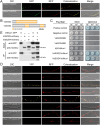
Mitochondria‐localised VdSOD2 and VdSOD4 interact with VdNuo1 in Verticillium dahliae. (A) Subcellular localisation of VdSOD2 and VdSOD4 in V. dahliae. The VdSOD2 and VdSOD4 fused to the mCherry‐encoding fragment were introduced into the VdNuo1::eGFP strain (mitochondrial localisation), and the transformants were incubated on hydrophobic glass slides in the dark in the presence of moisture at 25°C for 16 h. The germinated hyphae were observed by fluorescence microscopy under GFP and RFP channels. Scale bar = 10 μm. (B) The protein sequences of VdSOD2 and VdSOD4 were analysed using pipelines of SMART, InterPro and Pfam. The Mn/Fe‐SOD motifs were labelled by IBS software (Illustrator for Biological Sequences). Scale bar = 50 amino acids. (C) Interaction analyses between VdNuo1 and VdSOD2/4 in yeast two‐hybrid (Y2H) system. The recombinant prey (pGADT7‐VdSOD2/4) and bait (pGBKT7‐VdNuo1) vectors were constructed. The bait vector pGBKT7‐VdNuo1 was cotransformed with the empty pGADT7 or prey vectors into yeast cells to detect self‐activation and interaction, respectively. Yeast cells with 10‐fold serial dilutions were cultured on double dropout (DDO, SD −Leu/Trp) medium and quadruple dropout (QDO, SD −Leu/Trp/His/Ade) medium supplemented with 20 μg/mL X‐α‐Gal and 0.1 μg/mL aureobasidin A (AbA). The pairs of pGADT7‐T coupled with pGBKT7‐53 or pGBKT7‐Lam vectors were used as the positive or negative control interactions, respectively. The interaction phenotypes were photographed at 5 days post‐inoculation. (D) Bimolecular fluorescence complementation assays between VdNuo1 and VdSODs. The recombinant plasmids encoding VdNuo1 or VdSODs fused with N‐ or C‐terminal of YFP‐encoding fragments were paired and cotransformed into the wild‐type (WT) strain protoplasts. The recombinant VdNuo1‐YFPN plasmids cotransformed with empty YFPC vector was used as a negative control. The resistance strains were incubated and stained with mitochondrial probe (CMXRos). The YFP and RFP signals were observed by fluorescence microscopy. Scale bars = 10 μm. (E) Co‐immunoprecipitation analysis between VdNuo1 and VdSOD2 or VdSOD4. The label was detected by analysing total protein of the indicated colocalised, VdNuo1::eGFP and VdSOD2/4::mCherry strains with western blot. The protein of each strain was also incubated with GFP agar beads for detecting the interaction by western blot. The VdNuo1::eGFP and VdSOD2/4::mCherry strains were used as controls.
FIGURE 3
FIGURE 3
VdSOD2 is required for the tolerance of mitochondrial superoxide anions in Verticillium dahliae. (A, B) Analyses of the response of ΔVdNuo1 and ΔVdSOD2 strains of V. dahliae to the superoxide anion generator menadione. All strains were cultured on Czapek medium supplemented with (A) 15 μM (wild‐type [WT], ΔVdNuo1, ΔVdSOD2 and complemented ECΔVdSOD2 strains) or (B) 7.5 μM (ΔVdSOD2 and ΔVdNuo1_VdSOD2 strains) menadione at 25°C in the dark for 7 days. Each strain was inoculated onto at least three plates and the experiment was repeated three times independently. (C, D) The inhibition rates of menadione on the growth of WT, ΔVdNuo1, ΔVdSOD2, ΔVdNuo1_VdSOD2, and ECΔVdSOD2 strains. Colony diameters were determined on medium with 15 μM (C) or 7.5 μM (D) menadione after incubation in the dark at 25°C in the dark for 7 days. Error bars are standard errors calculated from six plates, and the experiment performed three times. Pairwise treatment differences were tested using Student's t test (***p < 0.001). (E, F) The colony morphology and inhibition rate of ΔVdNuo1 and VdNuo1_OE VdSOD2 strains on menadione plates. Compared with ΔVdNuo1, three VdSOD2‐overexpressing strains (different expression levels) were cultured on Czapek medium supplemented with 15 μM menadione at 25°C in the dark for 7 days (E). The inhibition rates on the growth of the respective strains in response to menadione were calculated by colony diameter (F). Error bars are standard errors calculated from six plates, and the experiment was replicated three times. ***p < 0.001 (Student's t test). (G) Staining of active mitochondria of the WT, ΔVdNuo1, ΔVdSOD2, ΔVdNuo1_VdSOD2, ECΔVdSOD2 , and VdNuo1_OE VdSOD2 strains of V. dahliae. The conidial suspensions were incubated on hydrophobic glass slides in the dark with moisture at 25°C for 16 h. The germinated hyphae were treated with 100 μM menadione for 45 min and stained by the mitochondrial probe (CMXRos). The RFP signals were observed by fluorescence microscopy. For each strain, 30 hyphae were observed. Scale bar = 10 μm. (H) Relative expression of VdNuo1 in WT, ΔVdSOD2, and ECΔVdSOD2 strains. The RNA was collected after fungal culturing on Czapek medium at 25°C in the dark for 5 days. The expression level of VdNuo1 in indicated strains with/without menadione treatment was measured by reverse transcription‐quantitative PCR using the 2−ΔΔCt method, with WT as the control. Quantification was calculated from three independent experiments, and there was no significant difference (NS p > 0.05; one‐way ANOVA).
FIGURE 4
FIGURE 4
VdSOD4 negatively regulates the degradation of mitochondrial superoxide anions in Verticillium dahliae. (A, B) The response of VdSOD4 to mitochondrial superoxide anions. The colony morphology of wild‐type (WT), ΔVdNuo1, ΔVdSOD4, ΔVdNuo1_VdSOD4, ECΔVdSOD4 , and VdNuo1_OE VdSOD4 strains (A). All strains were cultured on Czapek medium supplemented with 15 μM menadione at 25°C in the dark for 7 days. The inhibition rates of menadione to these strains (B). Each strain was inoculated on at least three plates and the experiment was conducted three times independently. Pairwise treatment differences were tested using Student's t test (***p < 0.0, NS, p > 0.05) representing no significant difference. (C) Staining of active mitochondria of the WT, ΔVdNuo1, ΔVdSOD4, ΔVdNuo1_VdSOD4, ECΔVdSOD4 , and VdNuo1_OE VdSOD4 strains. Before observation, the germinated hyphae were treated with 100 μM menadione for 45 min and stained by the mitochondrial probe CMXRos. The RFP signals (30 hyphae of each strain) were observed by fluorescence microscopy. Scale bar = 10 μm. (D) Relative expression of VdNuo1 in WT, ΔVdNuo1, ΔVdSOD4, and ECΔVdSOD4 strains. The RNA samples were collected after fungal culturing on Czapek medium with or without menadione treatment at 25°C in the dark for 5 days. The expression levels were measured by reverse transcription‐quantitative PCR with WT expression levels as controls. Quantitative detection was determined from three independent experiments. One‐way analysis of variance was conducted on the data (significant difference ***p < 0.001).
FIGURE 5
FIGURE 5
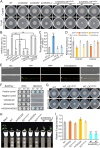
VdSOD2 and VdSOD4 cooperate in mitochondrial superoxide anion homeostasis, growth and virulence of Verticillium dahliae. (A, B) The redundancy analysis of VdSOD2 and VdSOD4 to mitochondrial superoxide anions. The colony morphology of wild‐type (WT), ΔVdSOD2, ΔVdSOD4, ΔVdSOD2_4, ECΔVdSOD4 , VdSOD4_OE VdSOD2 and VdSOD2_OE VdSOD4 strains (A). All strains were cultured on Czapek medium supplemented with 7.5 μM menadione at 25°C in the dark for 7 days. The inhibition rates of menadione to these strains (B) were calculated by colony diameters. Each strain was inoculated onto at least three plates and the experiment was repeated three times independently. Pairwise treatment differences were tested using Student's t test (***p < 0.001,**p < 0.01, NS, p > 0.05). (C, D) Relative expression of VdNuo1, VdSOD2 and VdSOD4 in the indicated strains that were treated (M) or untreated (CK) with menadione. The VdNuo1 expression level in VdSOD2 and WT strains (C). The inter‐regulation of VdSOD2 and VdSOD4 (D). These strains were cultured on Czapek medium with or without menadione treatment at 25°C in the dark. RNA was collected at 5 days post‐inoculation (dpi). The expression levels were measured by reverse transcription‐quantitative PCR. Quantitative detection was calculated from three independent experiments, and the data subjected to one‐way analysis of variance. Significance of mean separation tests were measured (***p < 0.001, **p < 0.01, *p < 0.05). (E, F) The interaction assays between VdSOD2 and VdSOD4 determined by bimolecular fluorescence complementation (E) and by yeast two‐hybrid (F) systems. The recombinant AD/BD and YFPN/YFPC plasmids were obtained. The YFPN/YFPC plasmids were paired and cotransformed into the ΔVdNuo1 strain. The conidia of positive transformants were incubated, and their YFP signals were observed by fluorescence microscopy. Scale bar = 10 μm. The self‐activation of BD‐VdSOD4 was detected and cotransformed into yeast cells with AD‐VdSOD2. The yeast colonies were diluted by 10‐fold and grew on double (DDO) and quadruple (QDO) dropout (added X‐α‐Gal and aureobasidin A [AbA]) media for 5 days. The controls were set in these experiments, and each experiment was repeated three times. (G) Colony morphology of V. dahliae strains overexpressing VdSOD2 and VdSOD4. VdSOD2 and VdSOD4, driven by the TrpC promoter, were transferred to WT strains and positive transformants were grown on Czapek medium with (15 μM) or without menadione treatment in the dark at 25°C for 7 days. Each strain was inoculated onto at least three plates and three independent experiments were carried out. (H) Pathogenicity assay of indicated strains on Shantung maple. Ten 1‐month‐old maple seedlings were inoculated with each strain. The experiment was repeated three times. The water and WT strain treatments served as the negative and positive controls. At 35 dpi, the Verticillium wilt symptoms were photographed. The pathogens were reisolated from the stem and cultured on V8 medium for 4 days in the dark at 25°C. (I) Quantification the fungal biomass in maple stems by quantitative PCR. Stem bases of inoculated seedlings were collected at 35 dpi. The VdEF‐1α of V. dahliae was quantified and the At18S gene of Shantung maple was used as an endogenous control. The biomass was calculated using three independent biological replicates. Error bars represent standard errors of the mean. The data subjected to one‐way analysis of variance. Significance of mean separation tests were measured (***p < 0.001).
FIGURE 6
FIGURE 6
Nuo1, SOD2 and SOD4 homologues share a conserved interaction and functions in Colletotrichum gloeosporioides. (A) Subcellular localisation of CgSOD2 and CgSOD4 in C. gloeosporioides. The CgSOD2‐ and CgSOD4‐encoding genes were fused with a GFP fragment and introduced into the C. gloeosporioides wild‐type (WT) strain. The positive transformants were incubated on hydrophobic glass slides in the dark with moisture at 25°C for 12 h. After mitochondrial staining by the probe (CMXRos), the GFP and RFP signals were observed by fluorescence microscopy. Scale bar = 10 μm. (B) The Mn/Fe‐SOD domain on both CgSOD2 and CgSOD4 were predicted by SMART, InterPro and Pfam. Scale bar = 30 amino acids. (C) Interaction analyses between CgNuo1 and CgSOD2/4 in a yeast two‐hybrid system. The recombinant prey (AD‐CgSOD2/4) and bait (BD‐CgNuo1 tested self‐activation) vectors were constructed, paired and cotransformed into yeast cells. The diluted positive yeast cells were grown on double dropout (DDO) and quadruple dropout (QDO) (+X‐α‐Gal/aureobadisin A) medium. The yeast colonies, including positive and negative controls, were photographed at 5 days post‐inoculation (dpi). (D) Bimolecular fluorescence complementation assays between CgNuo1 and CgSOD2/4. The recombinant CgNuo1‐YFPN plasmid cotransformed with CgSOD2 or CgSOD4‐YFPC plasmids into wild‐type (WT) strain protoplasts. The pair of CgNuo1‐YFPN and empty YFPC were used as controls. Strains were stained with mitochondrial probe CMXRos and YFP and RFP signals were examined by fluorescence microscopy. Scale bars = 10 μm. (E) Colony morphology of Verticillium dahliae strains heterologously overexpressing CgSOD2 and CgSOD4. The recombinant CgSOD2‐GFP and CgSOD4‐GFP plasmids were transferred into the V. dahliae VdSOD2 and VdSOD4 mutants, respectively. The positive transformants were grown in the dark at 25°C for 7 days on Czapek plates only, or those to which 15 μM menadione was added. Each strain was inoculated onto at least three plates and three independent experiments were carried out.
FIGURE 7
FIGURE 7
Working model of VdNuo1 and mtSODs during mitochondrial superoxide anion scavenging in Verticillium dahliae. Among seven superoxide dismutases in V. dahliae, the secreted VdSOD1, VdSOD3 and VdSOD5 are required for superoxide anion scavenging during host invasion, while the function of secreted VdSOD6 and VdSOD7 are unknown. The mitochondria‐localised VdSOD2 and VdSOD4 interact directly with the respiratory chain complex I VdNuo1 subunit to maintain mitochondrial morphological integrity, antioxidant activity, vegetative growth and virulence of V. dahliae by regulating mitochondrial superoxide anion homeostasis.

Similar articles

References

    1. Araiso, Y. , Imai K., and Endo T.. 2022. “Role of the TOM Complex in Protein Import Into Mitochondria: Structural Views.” Annual Review of Biochemistry 91: 679–703. 10.1146/annurev-biochem-032620-104527. - DOI - PubMed
    1. Baschieri, A. , Jin Z., and Amorati R.. 2023. “Hydroperoxyl Radical (HOO˙) as a Reducing Agent: Unexpected Synergy With Antioxidants. A Review.” Free Radical Research 57: 115–129. 10.1080/10715762.2023.2212121. - DOI - PubMed
    1. Broxton, C. N. , and Culotta V. C.. 2016. “SOD Enzymes and Microbial Pathogens: Surviving the Oxidative Storm of Infection.” PLoS Pathogens 12: e1005295. 10.1371/journal.ppat.1005295. - DOI - PMC - PubMed
    1. Camejo, D. , Guzmán‐Cedeño Á., and Moreno A.. 2016. “Reactive Oxygen Species, Essential Molecules, During Plant–Pathogen Interactions.” Plant Physiology and Biochemistry 103: 10–23. 10.1016/j.plaphy.2016.02.035. - DOI - PubMed
    1. Chen, J. Y. , Klosterman S. J., Hu X. P., Dai X. F., and Subbarao K. V.. 2021. “Key Insights and Research Prospects at the Dawn of the Population Genomics Era for Verticillium dahliae .” Annual Review of Phytopathology 59: 31–51. 10.1146/annurev-phyto-020620-121925. - DOI - PubMed

Supplementary concepts

LinkOut - more resources