An Unusual Cause of Massive Per Vaginal Bleeding
- PMID: 40917208
- PMCID: PMC12413294
- DOI: 10.14309/crj.0000000000001496
An Unusual Cause of Massive Per Vaginal Bleeding
Abstract
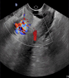
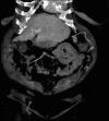
Massive per vaginal bleeding from ectopic pelvic varices is an exceedingly rare presentation in patients with cirrhosis. A 60-year-old postmenopausal woman presented with massive per vaginal (PV) bleeding. Computerized tomography scan showed extensive portosystemic collaterals with a large collateral vessel from the splenic vein to the region of her previous caesarean scar, on a background of liver cirrhosis. The cause of the massive PV bleeding was identified as arising from the uterine varix. She was transferred to a tertiary liver unit where she underwent angiographic embolization of the uterine varix and splenic vein shunt with successful obliteration of the culprit collateral vessel. A high index of suspicion is required in a cirrhotic patient with massive PV bleeding for ectopic variceal bleeding. Once stabilized, prompt consultation should be made to a tertiary center for further assessment and consideration of definitive treatment with obliteration of varices and shunt, as well as transjugular intrahepatic portosystemic shunt, to reduce risk of recurrent bleeding.
Keywords: Cirrhosis; Portal Hypertension; Uterine Varices.
© 2024 The Author(s). Published by Wolters Kluwer Health, Inc. on behalf of The American College of Gastroenterology.
Figures


References
-
- Henry Z, Uppal D, Saad W, Caldwell S. Gastric and ectopic varices. Clin Liver Dis. 2014;18(2):371–88. - PubMed
-
- McHugh PP, Jeon H, Gedaly R, Johnston TD, Depriest PD, Ranjan D. Vaginal varices with massive hemorrhage in a patient with nonalcoholic steatohepatitis and portal hypertension: Successful treatment with liver transplantation. Liver Transpl. 2008;14(10):1538–40. - PubMed
-
- Lee EW, Eghtesad B, Garcia-Tsao G, et al. AASLD Practice Guidance on the use of TIPS, variceal embolization, and retrograde transvenous obliteration in the management of variceal hemorrhage. Hepatology. 2024;79(1):224–50. - PubMed
Publication types
LinkOut - more resources
Full Text Sources

