. 2025 Aug 20:34:102222.
doi: 10.1016/j.mtbio.2025.102222. eCollection 2025 Oct.

The role of 3D printing in skeletal muscle-on-a-chip models: Current applications and future potential

Affiliations

The role of 3D printing in skeletal muscle-on-a-chip models: Current applications and future potential

Wei Bao et al. Mater Today Bio. .

Abstract

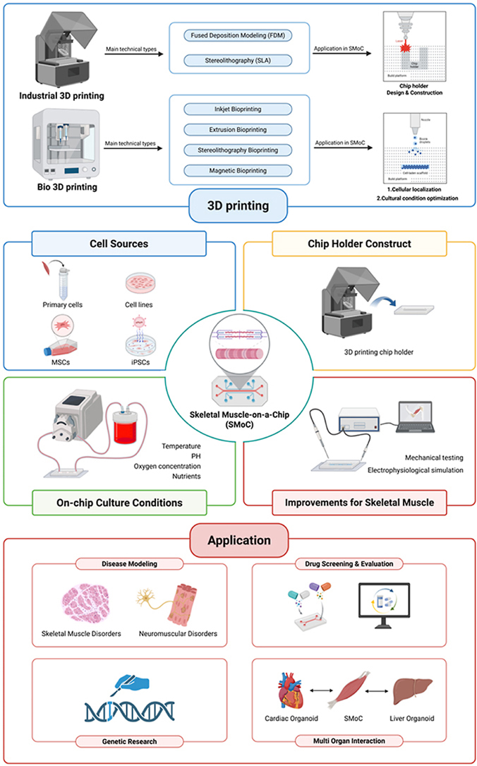
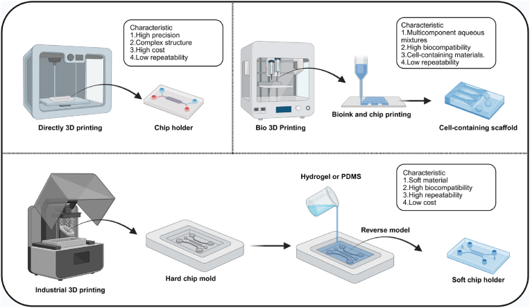
Organ-on-a-chip (OoC) systems can simulate the key functions of human organs, combining microfluidics, cell culture, and biomaterials. 3D printing can be integrated into these technologies to facilitate the construction of OoC models. The high precision and layer-by-layer fabrication process of 3D printing not only enables the creation of complex structures for the microfluidic chip but also improves the cellular microenvironment within the chip by harnessing bioinks for 3D bioprinting. In recent years, OoC models established with 3D printing technology have successfully replicated the functions of various native organs, significantly advancing disease research and drug development. However, due to the complex anatomical structure and unique physiological functions of skeletal muscle, the application of 3D printing in skeletal muscle-on-a-chip (SMoC) models remains relatively limited. Based on existing research on engineered skeletal muscle and OoC, this review discusses the construction of SMoC models by 3D printing to recapitulate the anatomical structure and physiological functions of skeletal muscle. Furthermore, it explores the different applications of 3D printed SMoC models and the future challenges and prospects in this field.

Keywords: 3D printing; Microfluidic technology; Organ-on-a-chip; Skeletal muscle disorders; Skeletal muscle-on-a-chip.


Conflict of interest statement

The authors declare that they have no known competing financial interests or personal relationships that could have appeared to influence the work reported in this paper.

Figures

Image 1
Graphical abstract
Fig. 1
Fig. 1
Anatomical structure of skeletal muscle. A) Different types of cells are present in the skeletal muscle. B) Diagram of the anatomical structure of skeletal muscle and the sliding filament mechanism. C) Schematic representation of muscle ultrastructural changes associated with aging. Notable changes include a reduced number and function of muscle satellite cells, reorganization of motor units, fiber loss and/or atrophy, and increased fibrosis [24]. D) H&E staining of the cross-sections of muscle tissues and mitochondrial states of muscle cells under different conditions [25]. H&E, Hematoxylin and Eosin. HMB, β-hydroxy-β-methyl butyrate, commonly known as HMB, is a widely used supplement in nutrition and sports to help reduce muscle breakdown and promote muscle growth.
Fig. 2
Fig. 2
Construct SMoC from four steps. Step 1: the selection of cell sources. Step 2: the fabrication of the microfluidic device (chip holder). Step 3: establishment of the on-chip culture conditions. Step 4: improve or adapt based on skeletal muscle.
Fig. 3
Fig. 3
Three approaches for constructing OoC microfluidic device using 3D printing technologies. (1) directly using industrial-grade 3D printing to create a chip structure. (2) bioprinting technology to assist in constructing a cell-containing scaffold. (3) industrial-grade 3D printed chip scaffold molds.
Fig. 4
Fig. 4
Examples of constructing skeletal muscle OoC models. A) Model for NVBchip: microfluidic design. a) Design and arrangement of nerve unit, vascular unit, and bone units on the chip. b) Low magnification microscopy tile scan image of the device's full structure showing the long compartments and the medium reservoirs. c, d) Images showing the loading of hydrogel (light blue) to the central channel (t = 60 s) and the addition of aqueous solution (dark blue) to the lateral channel (t = 80 s) Scale bar 500 μm [71]. B) Toward vasculature in SMoC through thermoresponsive sacrificial templates. a) Schematic of the general design of the device. b) Cross-sectional representation of the device. c) Image of the device in the PDMS chambers with the wax template. d) Live/dead staining confocal image of device [89]. C) System design, configuration, and microfabrication of the OoC system. a) Top-view illustration of the sensor glass chip. b) Photograph of the assembled device. c) Schematic cross-sectional view. d) Field-emission scanning electron microscopy image [94]. D) Highly controlled 3D- Self-rolled biosensor array (3D-SR-Bas). a-c) Bright-field optical microscopy images of SR-BAs. d, e) 3D-SR-BAs with varying radii of curvature — simulation and experimental results [101]. E) Schematic of the fabrication process of the neuronemuscle construct and an illustration of the contractile mechanism of the neuronemuscle construct. a) Fabrication of a bundle of free-standing muscle fibers using a PDMS stamp with a striped pattern. b) Formation of the neuronemuscle construct using a muscle fiber bundle and neurospheres. c) Contractile mechanism of the neuronemuscle construct activated by neurotransmitters [105]. (For interpretation of the references to colour in this figure legend, the reader is referred to the Web version of this article.)
Fig. 5
Fig. 5
Types and applications of 3D bioprinting. A. Schematic diagram of 3D bioprinting. B. Major types of 3D bioprinting. C. Applications of 3D bioprinting in tissue engineering repair. D. Applications of 3D bioprinting in disease model construction and drug screening. Examples of the applications of 3D bioprinting can be found in Fig. S1.
Fig. 6
Fig. 6
Engineering skeletal muscle based on 3D printing. A. Construction of engineered skeletal muscle using a combination of electrospinning and 3D printing technology [139]. B. Construction of engineered skeletal muscle based on the electrohydrodynamic direct writing (EHD-DW) process [140]. C. Engineered muscle tissue based on bioink 3D printing guided by electric field and Au nanowires [141]. D. 4D printing for the construction of muscle tissue [142].
Fig. 7
Fig. 7
Examples of SMoC research. A. Construction of skeletal muscle chips and immunofluorescence staining of skeletal muscle on the chips [144]. a) Schematic diagram of the SMoC fabrication process. b) Cross-sectional staining of a representative muscle on the chip. B. Construction of SMoC using SLA technology: z-stack images of the SMoC platform loaded with fluorescent microparticles were obtained using a laser scanning confocal microscope [9]. C. SMoC constructed using 3D printed molds and PDMS molding [145]. a) Schematic diagram of SMoC. b) Immunofluorescence images and scanning electron microscope (SEM) image of musculoskeletal tissue in SMoC. c) Overall experimental workflow. D. Oxygen-controlled microphysiological system based on 3D printed components, used to study functional changes in skeletal muscle tissue under different oxygenation states [146]. a) Fabrication of the MPS for gas-controlled coculture of normoxic and hypoxic SMoC. b) An O2 control well plate insert for compressed gas delivery to SMoC. E. The schematic development process of the MTJ-on-a-chip [148]. MTJ, muscle–tendon junction.
Fig. 8
Fig. 8
Applications of SMoC. A) SMoC applications in disease modeling. B) SMoC applications in drug screening and evaluation. C) SMoC for genetic research. D) SMoC for studying interactions between skeletal muscle and other organs.
Fig. 9
Fig. 9
Examples of SMoC applications. A. SMoC is able to promote stretching-induced human myotube maturation [154]. a) Optical profile of the chip and confocal imaging of the system with the micropatterns (lines in blue), myotubes stained in red with troponin T (TnT), and nuclei stained with DAPI (in blue). b) Confocal immunofluorescence staining of titin and MHC-1 in stretched and unstretched human myotubes. B. Studying gene changes in skeletal muscle under microgravity conditions using SMoC [172]. a) Chip construction and launch preparation. b) RNA-Seq volcano plots. YA flight vs ground; OS flight vs ground; Ground OS vs YA and Flight OS vs YA. C. Neuromuscular junction chip [159]. a) Chip construction schematic. b) Fabrication and use of the chip. D. NMJ chip for disease modeling and drug screening [164]. a) Fabrication of an NMJ chip. b) Formation and detection of NMJ on the chip. E. Multi-organ chip system connected through vascular flow [177]. a) Chip system configurability and modularity. b) Tissue maturity assessment in different organ chips. (For interpretation of the references to colour in this figure legend, the reader is referred to the Web version of this article.)
Fig. 10
Fig. 10
Challenges of 3D printing SMoC. (1) Printing performance: upgrading printing technologies or incorporating multi-nozzle systems can enhance resolution and speed, achieving high-precision SMoC structure construction more efficiently. (2) Material selection: develop materials with enhanced biocompatibility and mechanical properties, better mimicking the skeletal muscle microenvironment while supporting printability. (3) Functional design: integrating dynamic simulation technologies to enable multi-organ interactions, essential for replicating physiological communication between muscle and other systems like nerves and vasculature. (4) Evaluation Systems: introducing more precise detection and analysis techniques enables more effective evaluation of SMoC functionality and provides reliable data support for research.

Similar articles

References

    1. Frontera W.R., Ochala J. Skeletal muscle: a brief review of structure and function. Calcif. Tissue Int. 2015;96(3):183–195. doi: 10.1007/s00223-014-9915-y. - DOI - PubMed
    1. Lipina C., Hundal H.S. Lipid modulation of skeletal muscle mass and function. J. Cachexia Sarcop. Muscle. 2017;8(2):190–201. doi: 10.1002/jcsm.12144. - DOI - PMC - PubMed
    1. Bursac N., Juhas M., Rando T.A. Synergizing engineering and biology to treat and model skeletal muscle injury and disease. Annu. Rev. Biomed. Eng. 2015;17:217–242. doi: 10.1146/annurev-bioeng-071114-040640. - DOI - PMC - PubMed
    1. Saorin G., Caligiuri I., Rizzolio F. Microfluidic organoids-on-a-chip: the future of human models. Semin. Cell Dev. Biol. 2023;144:41–54. doi: 10.1016/j.semcdb.2022.10.001. - DOI - PubMed
    1. Farhang Doost N., Srivastava S.K. A comprehensive review of Organ-on-a-chip technology and its applications. Biosensors (Basel) 2024;14(5) doi: 10.3390/bios14050225. - DOI - PMC - PubMed

LinkOut - more resources