Review
. 2024 Apr 26;5(1):200133.
doi: 10.1016/j.mrl.2024.200133. eCollection 2025 Feb.

Advances in solid-state NMR methods for studying RNA structures and dynamics

Affiliations
Review

Advances in solid-state NMR methods for studying RNA structures and dynamics

Jinhan He et al. Magn Reson Lett. .

Abstract

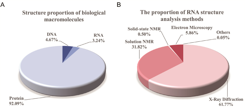
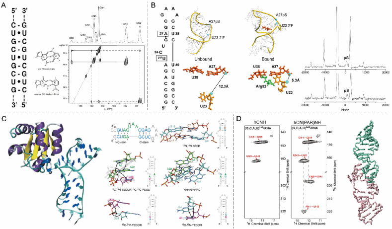
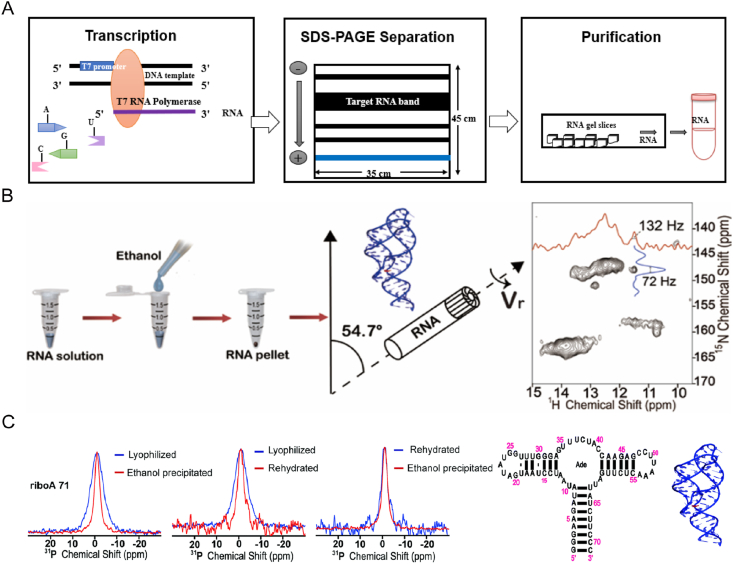
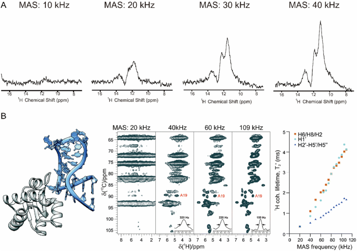
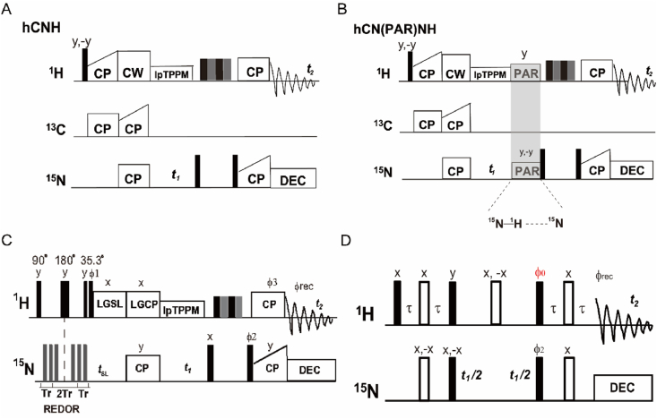
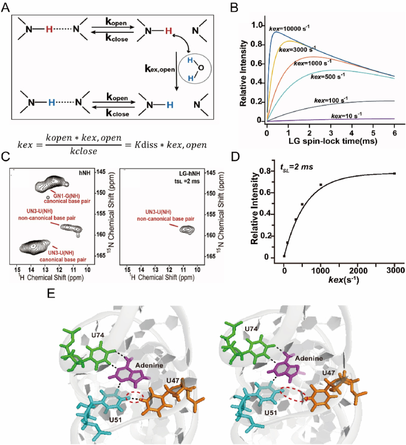
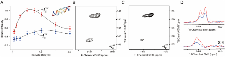
Ribonucleic acid (RNA) structures and dynamics play a crucial role in elucidating RNA functions and facilitating the design of drugs targeting RNA and RNA-protein complexes. However, obtaining RNA structures using conventional biophysical techniques, such as X-ray crystallography and solution nuclear magnetic resonance (NMR), presents challenges due to the inherent flexibility and susceptibility to degradation of RNA. In recent years, solid-state NMR (SSNMR) has rapidly emerged as a promising alternative technique for characterizing RNA structure and dynamics. SSNMR has several distinct advantages, including flexibility in sample states, the ability to capture dynamic features of RNA in solid form, and suitability to character RNAs in various sizes. Recent decade witnessed the growth of 1H-detected SSNMR methods on RNA, which targeted elucidating RNA topology and base pair dynamics in solid state. They have been applied to determine the topology of RNA segment in human immunodeficiency virus (HIV) genome and the base pair dynamics of riboswitch RNA. These advancements have expanded the utility of SSNMR techniques within the RNA research field. This review provides a comprehensive discussion of recent progress in 1H-detected SSNMR investigations into RNA structure and dynamics. We focus on the established 1H-detected SSNMR methods, sample preparation protocols, and the implementation of rapid data acquisition approaches.

Keywords: Dynamics; Pulse sequences; RNA; Solid-state NMR; Structure.


Conflict of interest statement

The authors declare the following financial interests/personal relationships which may be considered as potential competing interests: Shenlin Wang is an editorial board member of Magnetic Resonance Letters but was not involved in the editorial review or the decision to publish this article.

Figures

Image 1
Graphical abstract
Fig. 1
Fig. 1
Statistical analysis of RNA structure quantity and elucidation methods as of January 2024 from PDB. (A) Distribution proportion of solved RNA, DNA, and protein structures. (B) Proportion of biophysical methods for RNA structure determination, including X-ray crystallography, solution NMR, SSNMR, and Cryo-EM.
Fig. 2
Fig. 2
Milestones of SSNMR development towards RNA structures. (A) Schematic diagram illustrating the double-stranded structure of (CUG)97 RNA along with the two-dimensional (2D) 15N–15N SSNMR cross-peak of a lyophilized (CUG)97 sample. Adapted with permission [49]. Copyright 2006, Angewandte Chemie. (B) Representation of the apical 29-mer region of TAR-RNA utilized in SSNMR studies, where the apical circular sequence is substituted by GAAA, 23U is fluorine-labeled, and 26G and 27A are linked by sulfur. Depiction of conformational changes of RNA upon TAT peptide binding. The 1D-31P SSNMR spectra of TAR RNA before and after binding to the TAT peptide are displayed on the right of (B). Adapted with permission [51]. Copyright 2005, Nucleic Acids Research. (C) Structure of 26-mer Box C/D RNA in complex with L7Ae protein. The right panel of (C) illustrates the RNA sequence and the magnetization transfer between the base pairs of Pf Box C/D RNA, including 13C,15N-TEDOR, 13C,13C-PDSD, 13C,31P-TEDOR, 15N,15N-RFDR, NHHN/NHHC, and 13C,15N-TEDOR techniques. Adapted with permission [45,50]. Copyright 2015, Nature Communications. Copyright 2013, Angewandte Chemie International Edition. (D) Depiction of 2D hCNH and 2D hCN(PAR)NH spectra of 15N,13C-labeled RNAs alongside the kissing loop structure of dimeric DIS-HIV-1. Adapted with permission [46]. Copyright 2017, Chemical Communications.
Fig. 3
Fig. 3
Sample preparation methods for RNA in SSNMR studies and spectral resolution comparison. (A) Illustration of the RNA preparation process via the in vitro transcription method. (B) Schematic representation of the ethanol precipitation sample preparation process for riboA71 along with the SSNMR 2D 1H–15N spectrum indicating the 1H and 15N line-width for the selected signal. (C) Comparison of 31P spectra obtained from lyophilized, ethanol-precipitated, and rehydrated lyophilized riboA71 samples. The secondary structure and the crystal structure of riboA71-adenine (PDB: 4TZX) are depicted on the right. Adapted with permission [61]. Copyright 2019, Chemical Communications.
Fig. 4
Fig. 4
1H-detected SSNMR spectra of RNA under fast and ultra-fast MAS rates. (A) SSNMR 1H spectra of DIS-HIV-1 RNA at MAS rates ranging from 10 to 40 kHz. Adapted with permission [46]. Copyright 2017, Chemical Communications. (B) Comparison of 1H–13C spectra of Box C/D RNA and L7Ae protein complex at MAS rates of 20 kHz, 40 kHz, 60 kHz, and 109 kHz. The plot illustrates the overall coherence lifetimes of lower bases H6, H8, and H2, ribose H10, and other protons at different MAS rates. Adapted with permission [66]. Copyright 2018, Chemical Communications.
Fig. 5
Fig. 5
Diagram of pulse sequences of 1H-detected SSNMR for RNA studies. (A–B) hCNH and hCN(PAR)NH experiments for the detection of hydrogen bonds within base pairs of RNA. Adapted with permission [46]. Copyright 2017, Chemical Communications. (C) 2D WaterREXSY scheme for detecting base pairs with low stability. Adapted with permission [60]. Copyright 2022, iScience. (D) Fast acquisition HSQC for RNA detection. Adapted with permission [62]. Copyright 2017, Chemistry -A European Journal.
Fig. 6
Fig. 6
hCNH and hCN(PAR)NH experiments to characterize the topologies of DIS-HIV-1 dimers. (A) 2D 1H–15N hCNH and hCN(PAR)NH spectra of G,C-15N,13C-DIS-HIV-1 with and without dilution of natural abundance RNA. (B) Comparison of 1D15N hCN(PAR)NH spectra of G,C-15N,13C-DIS-HIV-1 (above) and G,C-15N,13C-DIS-HIV-1 mixed with natural abundance DIS-HIV-1 (below). (C) The kissing-loop structure of DIS-HIV-1 (PDB code: 2B8S). Adapted with permission [46]. Copyright 2017, Chemical Communications.
Fig. 7
Fig. 7
Characterization of the dynamic properties of the riboswitch. (A) Schematic diagram illustrating proton exchange with water in the open and closed state of hydrogen bond. (B) Simulation of the exchange rate constant accumulated over time under the 2D WaterREXSY protocol. (C) Comparison of stable and low stability base pairs with 2D WaterREXSY. (D) Subproton intensities of U47 at different tSL times. (E) Closed (left) and open (right) states of U47–U51 base pairs in the U47–U51 (adenine-U74) base tetrad. Adapted with permission [60]. Copyright 2022, iScience.
Fig. 8
Fig. 8
LHSQC experiments of DIS-HIV-1. (A) Experimental sensitivity curves of DIS-HIV-1 for recovery delay. (B and C) Two-dimensional LHSQC spectra in +Izw (B) and −Izw (C) experiments. (D) Comparison of 1D traces of DIS-HIV-1 of LHSQC spectra. The top graph is the signal peak for guanosines with a15N chemical shift of 147 ppm and the bottom graph is the signal peak for uridines with a15N chemical shift of 162 ppm. Adapted with permission [62]. Copyright 2017, Chemistry – A European Journal.

References

    1. Selmer M., Dunham C.M., Murphy F.V., Weixlbaumer A., Petry S., Kelley A.C., Weir J.R., Ramakrishnan V. Structure of the 70S ribosome complexed with mRNA and tRNA. Science. 2006;313:1935–1942. - PubMed
    1. Raj A., van Oudenaarden A. Nature, nurture, or chance: stochastic gene expression and its consequences. Cell. 2008;135:216–226. - PMC - PubMed
    1. Anger A.M., Armache J.-P., Berninghausen O., Habeck M., Subklewe M., Wilson D.N., Beckmann R. Structures of the human and Drosophila 80S ribosome. Nature. 2013;497:80–85. - PubMed
    1. Memczak S., Jens M., Elefsinioti A., Torti F., Krueger J., Rybak A., Maier L., Mackowiak S.D., Gregersen L.H., Munschauer M., Loewer A., Ziebold U., Landthaler M., Kocks C., le Noble F., Rajewsky N. Circular RNAs are a large class of animal RNAs with regulatory potency. Nature. 2013;495:333–338. - PubMed
    1. Bartel D.P. MicroRNAs: target recognition and regulatory functions. Cell. 2009;136:215–233. - PMC - PubMed

LinkOut - more resources