Extrachromosomal DNA-Driven Oncogene Spatial Heterogeneity and Evolution in Glioblastoma
- PMID: 40920091
- PMCID: PMC12498097
- DOI: 10.1158/2159-8290.CD-24-1555
Extrachromosomal DNA-Driven Oncogene Spatial Heterogeneity and Evolution in Glioblastoma
Abstract
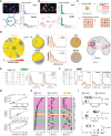
Oncogenes amplified on extrachromosomal DNA (ecDNA) contribute to treatment resistance and poor survival across cancers. Currently, the spatiotemporal evolution of ecDNA remains poorly understood. In this study, we integrate computational modeling with samples from 94 treatment-naive human glioblastomas (GBM) to investigate the spatiotemporal evolution of ecDNA. We observe oncogene-specific patterns of ecDNA spatial heterogeneity, emerging from random ecDNA segregation and differing fitness advantages. Unlike PDGFRA-ecDNAs, EGFR-ecDNAs often accumulate prior to clonal expansions, conferring strong fitness advantages and reaching high abundances. In corroboration, we observe pretumor ecDNA accumulation in vivo in genetically engineered mouse neural stem cells. Variant and wild-type EGFR-ecDNAs often coexist in GBM. Those variant EGFR-ecDNAs, most commonly EGFRvIII-ecDNA, always derive from preexisting wild-type EGFR-ecDNAs, occur early, and reach high abundance. Our results suggest that the ecDNA oncogenic makeup determines unique evolutionary trajectories. New concepts such as ecDNA clonality and heteroplasmy require a refined evolutionary interpretation of genomic data in a large subset of GBMs.
Significance: We study spatial patterns of ecDNA-amplified oncogenes and their evolutionary properties in human GBM, revealing an ecDNA landscape and ecDNA oncogene-specific evolutionary histories. ecDNA accumulation can precede clonal expansion, facilitating the emergence of EGFR oncogenic variants, reframing our interpretation of genomic data in a large subset of GBMs. See related commentary by Korsah et al., p. 1979.
©2025 The Authors; Published by the American Association for Cancer Research.
Conflict of interest statement
M. Haughey reports grants from UK Research and Innovation Future Leaders Fellowship and Cancer Grand Challenges during the conduct of the study. J. Luebeck reports a patent for Methods and Compositions for Detecting ecDNA, licensed and with royalties paid. D. Pradella reports other support from the AIRC Foundation during the conduct of the study. C. Bailey reports personal fees from Bicycle Therapeutics outside the submitted work. C.E. Weeden reports grants from the European Respiratory Society and Marie Skłodowska-Curie Actions during the conduct of the study. M.G. Jones reports personal fees from Tahoe Therapeutics (formerly Vevo) outside the submitted work. K.L. Hung reports patents for Methods for targeted purification and profiling of human ecDNA and DNA element responsive to ecDNA in cancer cells pending. E.J. Norton reports grants from the National Institute for Health and Care Research during the conduct of the study. M. Jamal-Hanjani reports grants from Cancer Research UK (CRUK), Lung Cancer Research Foundation, and Achilles Therapeutics Scientific Advisory Board and Steering Committee; other support from Pfizer, Astex Pharmaceuticals, Oslo Cancer Cluster, Bristol Myers Squibb, Genentech, GenesisCare, and VHIO; and grants from the NCI outside the submitted work and reports that she has received funding from CRUK, NIH National Cancer Institute, IASLC International Lung Cancer Foundation, Lung Cancer Research Foundation, Rosetrees Trust, UKI NETs, and National Institute for Health and Care Research and she is a consultant for Astex Pharmaceuticals, Pfizer, and Achilles Therapeutics and a member of the Achilles Therapeutics Scientific Advisory Board and Steering Committee; has received speaker honoraria from Pfizer, Astex Pharmaceuticals, Oslo Cancer Cluster, Bristol Myers Squibb, Genentech, and GenesisCare; and is listed as a coinventor on a European patent application relating to methods to detect lung cancer PCT/US2017/028013), which has been licensed to commercial entities, and under the terms of employment, she is due a share of any revenue generated from such license(s) and is also listed as a coinventor on the GB priority patent application (GB2400424.4) with the title “Treatment and Prevention of Lung Cancer.” A. Ventura reports grants from the NIH/NCI, Cancer Grand Challenge, ACS, and Mark Foundation for Cancer Research during the conduct of the study. J.A.R. Nicoll reports grants from Brain Tumour Research during the conduct of the study as well as grants from Alzheimer Research UK, Alzheimer Society, and Pathological Society outside the submitted work. D. Boche reports grants from Brain Tumour Research during the conduct of the study as well as grants from Alzheimer’s Society, Alzheimer’s Research UK, Pathological Society, British Neuropathological Society, and UKRI/MRC outside the submitted work. H.Y. Chang reports grants from CRUK, the NIH, and HHMI during the conduct of the study as well as personal fees and other support from Accent Therapeutics, Boundless Bio, Cartography Biosciences, and Orbital Therapeutics; personal fees from Arsenal Bio, nChroma, Vida Ventures, and Amgen; and other support from Spring Science and 10x Genomics outside the submitted work. V. Bafna reports other support from Boundless Bio Inc. and Abterra Inc. outside the submitted work and that he holds equity and is a member of the scientific advisory board for Boundless Bio Inc. and Abterra Inc. P.S. Mischel reports personal fees from Boundless Bio outside the submitted work. C. Swanton reports grants and personal fees from AstraZeneca and Bristol Myers Squibb; grants from Boehringer Ingelheim, Invitae, Ono Pharmaceutical, and Personalis; and personal fees from GRAIL, Bicycle Therapeutics, Genentech, Medixci, China Innovation Centre of Roche, Relay Therapeutics, Saga Diagnostics, Sarah Cannon Research Institute, Novartis, Amgen, GlaxoSmithKline, Illumina, MSD, and Pfizer during the conduct of the study as well as grants and personal fees from AstraZeneca and Bristol Myers Squibb; grants from Invitae, Ono Pharmaceutical, and Personalis; and personal fees from Boehringer Ingelheim, GRAIL, Bicycle Therapeutics, Genentech, Medixci, China Innovation Centre of Roche, Relay Therapeutics, Saga Diagnostics, Sarah Cannon Research Institute, Novartis, Amgen, GlaxoSmithKline, Illumina, MSD, and Pfizer outside the submitted work and that he has patents for PCT/EP2022/077987, PCT/EP2022/070694, PCT/GB2020/050221, and PCT/EP2023/059039 issued; patents for PCT/EP2016/071471, PCT/GB2017/053289, and PCT/US2017/028013 licensed; and patents for PCT/GB2018/051912, PCT/EP2021/059989, PCT/GB2019/051028, PCT/US2010/033755, and PCT/EP2023/065074 pending and he is the chief investigator of the MeRmaiD 1 and 2 clinical trials funded by AstraZeneca and also the co-chief investigator of the NHS Galleri Trial funded by GRAIL and is a cofounder and holds stock/options in Achilles Therapeutics and stock/options in the following: Bicycle Therapeutics, Saga Diagnostics, and Relay Therapeutics, and previously held stock/options with GRAIL until June 2022. B. Werner reports grants from UKRI, Cancer Grand Challenge, and Barts Charity during the conduct of the study. No disclosures were reported by the other authors.
Figures


Update of
-
Extrachromosomal DNA driven oncogene spatial heterogeneity and evolution in glioblastoma.bioRxiv [Preprint]. 2024 Oct 25:2024.10.22.619657. doi: 10.1101/2024.10.22.619657. bioRxiv. 2024. Update in: Cancer Discov. 2025 Oct 6;15(10):2078-2095. doi: 10.1158/2159-8290.CD-24-1555. PMID: 39484416 Free PMC article. Updated. Preprint.
References
-
- Qazi MA, Vora P, Venugopal C, Sidhu SS, Moffat J, Swanton C, et al. Intratumoral heterogeneity: pathways to treatment resistance and relapse in human glioblastoma. Ann Oncol 2017;28:1448–56. - PubMed
MeSH terms
Substances
Grants and funding
- C11496/A17786/Cancer Research UK (CRUK)
- OT2CA278635/National Cancer Institute (NCI)
- U24 CA264379/CA/NCI NIH HHS/United States
- K00 CA274692/CA/NCI NIH HHS/United States
- 21-029-ASP/Mark Foundation For Cancer Research (The Mark Foundation for Cancer Research)
- U24-CA264379/National Cancer Institute (NCI)
- R01-GM114362/National Cancer Institute (NCI)
- NIH K99CA286968/National Cancer Institute (NCI)
- OT2 CA278635/CA/NCI NIH HHS/United States
- R01 GM114362/GM/NIGMS NIH HHS/United States
- 835297/HORIZON EUROPE European Research Council (ERC)
- FC001169/WT_/Wellcome Trust/United Kingdom
- R01 CA282913/CA/NCI NIH HHS/United States
- MGU045/Barts Charity
- K99 CA286968/CA/NCI NIH HHS/United States
- C416/A21999/Cancer Research UK (CRUK)
- MR/V02342X/1/UK Research and Innovation (UKRI)
- CGCATF-2021/100025/Cancer Research UK (CRUK)
- P30 CA008748/CA/NCI NIH HHS/United States
- NIH K00CA274692/National Cancer Institute (NCI)
- ID16584/Novo Nordisk Foundation Center for Basic Metabolic Research (NovoNordisk Foundation Center for Basic Metabolic Research)
- OT2 CA278688/CA/NCI NIH HHS/United States
- RP/EA/180007/Royal Society (The Royal Society)
- OT2CA278688/National Cancer Institute (NCI)
- CGCATF-2021/100012/Cancer Research UK (CRUK)
- C11496/A30025/Cancer Research UK (CRUK)
LinkOut - more resources
Full Text Sources
Medical
Research Materials
Miscellaneous

