. 2025 Sep 2;66(12):22.
doi: 10.1167/iovs.66.12.22.

Genome-Wide Insights Into the Genes and Pathways Shaping Human Foveal Development: Redefining the Genetic Landscape of Foveal Hypoplasia

Affiliations

Genome-Wide Insights Into the Genes and Pathways Shaping Human Foveal Development: Redefining the Genetic Landscape of Foveal Hypoplasia

Callum Hunt et al. Invest Ophthalmol Vis Sci. .

Abstract

Purpose: To define the genetic architecture of foveal morphology and explore its relevance to foveal hypoplasia (FH), a hallmark of developmental macular disorders.

Methods: We applied deep-learning algorithms to quantify foveal pit depth from central optical coherence tomography (OCT) B-scans in 61,269 UK Biobank participants. A genome-wide association study (GWAS) was conducted using REGENIE, adjusting for age, sex, height, and ancestry. Rare coding variants (frequency <1%) were analyzed in an exome-wide rare-variant association study (RVAS). Candidate genes were prioritized using integrative mapping; pathway, cross-ancestry, and genetic-correlation analyses were exploratory.

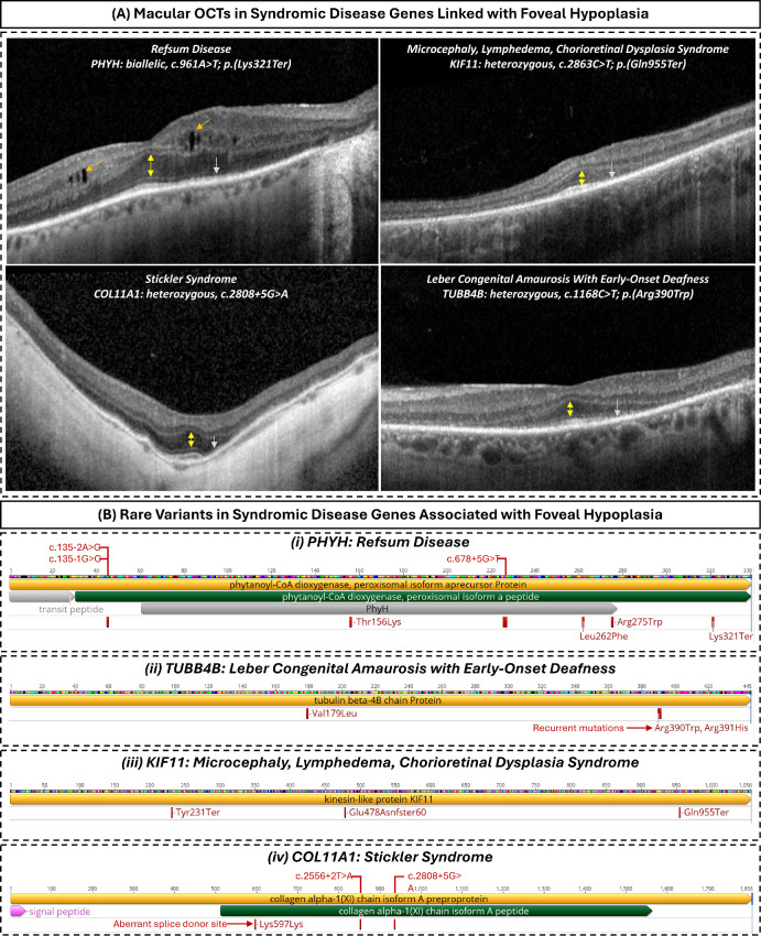
Results: GWAS identified 126 sentinel variants, including 47 novel associations. Integrative mapping prioritized 129 putative causal genes, with 64 not previously implicated in foveal biology. Enriched pathways included retinoic acid metabolism (e.g., CYP26A1), photoreceptor differentiation (e.g., VSX2), extracellular matrix organization, and pigmentation. RVAS identified missense variants in ACTN3 and ESYT3 (P < 5 × 10-⁹) associated with FH features. Polygenic scores were predictive across African and South Asian ancestries. Overlap was observed with monogenic FH genes (TYR, OCA2, PAX6, AHR) and with genes underlying systemic diseases (COL11A1, KIF11, TUBB4B, PHYH). Re-examination of OCTs in affected individuals confirmed FH in select cases, including those with recurrent TUBB4B p.(Arg390Trp) variants.

Conclusions: This is the first GWAS of human foveal morphology. Our findings redefine the genetic and biological framework underlying normal foveal development and foveal hypoplasia (FH). By linking common variation to rare monogenic disease, we establish a continuum model of FH with implications for future mechanistic and clinical investigation.


Conflict of interest statement

Disclosure: C. Hunt, None; H.-J. Yoon, None; A. Lirio, None; K. Coley, None; J. Wang, None; N. Shrine, None; J. Shao, None; G.D.E. Maconachie, None; Z. Tu, None; J.H. Zippin, None; P.G. Hysi, None; C.J. Hammond, None; O.A. Mahroo, None; M. Moosajee, None; M. Michaelides, None; A.R. Webster, None; A. Moshiri, None; R. Chen, M.D. Tobin, Orion Pharma (R), GlaxoSmithKline (S); C. Batini, None; M.G. Thomas, None

Figures

Figure 1.
Figure 1.
Manhattan and quantile-quantile (QQ) plots for genome-wide association analysis of foveal pit depth. The Manhattan plot displays genome-wide association results, with each point representing a genetic variant. The x-axis indicates chromosomal position, and the y-axis shows the −log10 (P value). The red dotted lines represent the genome-wide significance threshold (P < 5 × 10⁻⁸). The QQ plot compares observed versus expected −log10 (P values) under the null hypothesis, with the genomic inflation factor (λGC) provided as a measure of population stratification or confounding.
Figure 2.
Figure 2.
Summary of the variant-to-gene evidence supporting putative causal genes. Evidence for novel gene associations with foveal pit depth is highlighted in blue, whereas genes previously reported in GWAS of the macular region or associated with known foveal diseases are shaded in gray. Columns are ordered by the number of genes implicated by each line of evidence.
Figure 3.
Figure 3.
Schematic representation of the retina and foveal region within the human eye. Putative causal genes relevant to prioritized functional groups involved in foveal development are displayed alongside their respective functional categories. Genes highlighted in bold are novel gene associations not reported in previous GWAS of the macular region or as foveal disease genes.
Figure 4.
Figure 4.
Abnormal foveal morphology in individuals carrying rare genetic variants. (A) Heatmaps showing average retinal thickness across the central macula, measured from the internal limiting membrane (ILM) to Bruch's membrane. The left panel shows the mean retinal thickness map for the broader study cohort of European ancestry. The middle and right panels show retinal thickness maps from individuals heterozygous for rare variants in ACTN3 (11:66560171-T) and ESYT3 (3:138472579-A), respectively. Compared to the average map, both variants are associated with a shallower and more flattened foveal pit contour. (B) Violin plots comparing foveal pit depth in the general cohort (black) versus individuals carrying the ACTN3 (blue) and ESYT3 (red) rare variants. Both variants are associated with a significant reduction in pit depth, consistent with abnormal foveal development.
Figure 5.
Figure 5.
(A) Macular OCT scans from individuals with molecularly confirmed systemic or retinal syndromes demonstrate foveal architectural disruption and outer retinal abnormalities. In Refsum disease due to a PHYH nonsense mutation, the scan shows a shallow foveal pit, cystic changes in the inner retina (orange arrows), and loss of the inner segment ellipsoid (ISe) band (green arrow). In KIF11- and COL11A1-associated syndromes, the foveal pit is absent, with accompanying ISe loss (gray arrows). In the TUBB4B-associated case, a shallow indentation is present at the fovea. Across all examples, widening of the outer nuclear layer at the foveal center (yellow arrows) helps localize the fovea, despite the absent or shallow pit. The intrusion of inner retinal layers at the foveola, combined with a shallow or absent pit, is a hallmark of FH, although outer retinal abnormalities are also present in all cases. (B) Schematic protein models showing locations of rare variants observed in our cohort for PHYH, TUBB4B, KIF11, and COL11A1. Variants are mapped along the canonical protein sequence with relevant annotations. Predicted splice-site variants are shown above each protein track, positioned according to their anticipated impact on coding sequence, while missense, nonsense, and frameshift variants are shown below. TUBB4B shows a notable recurrent missense variant p.(Arg390Trp) in multiple individuals with FH. The COL11A1 synonymous variant (Lys597Lys) activates an aberrant splice donor site. The mapping illustrates diversity of variants and location of recurrent variants.

Update of

References

    1. Thomas MG, Papageorgiou E, Kuht HJ, Gottlob I.. Normal and abnormal foveal development. Br J Ophthalmol. 2022; 106: 593–599. - PubMed
    1. Vajzovic L, Hendrickson AE, O'Connell RV, et al.. Maturation of the human fovea: correlation of spectral-domain optical coherence tomography findings with histology. Am J Ophthalmol. 2012; 154: 779–789.e2. - PMC - PubMed
    1. Hendrickson A, Possin D, Vajzovic L, Toth CA.. Histologic development of the human fovea from midgestation to maturity. Am J Ophthalmol. 2012; 154: 767–778.e2. - PMC - PubMed
    1. Hendrickson AE, Yuodelis C.. The morphological development of the human fovea. Ophthalmology. 1984; 91: 603–612. - PubMed
    1. Thomas MG, Kumar A, Mohammad S, et al.. Structural grading of foveal hypoplasia using spectral-domain optical coherence tomography a predictor of visual acuity? Ophthalmology. 2011; 118: 1653–1660. - PMC - PubMed

Supplementary concepts

LinkOut - more resources