. 2025 Sep 2;66(12):20.
doi: 10.1167/iovs.66.12.20.

Novel Grm6 Variant in a no b-wave (nob) Mouse Model: Phenotype Characterization and Gene Therapy

Affiliations

Novel Grm6 Variant in a no b-wave (nob) Mouse Model: Phenotype Characterization and Gene Therapy

Pei-Hsuan Lin et al. Invest Ophthalmol Vis Sci. .

Abstract

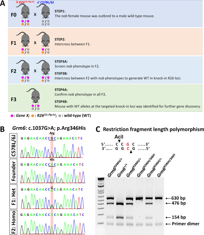
Purpose: To characterize a no b-wave (nob) mouse model of congenital stationary night blindness (CSNB) caused by a Grm6 variant that disrupts photoreceptor-to-bipolar cell signaling. Additionally, we aim to evaluate the efficacy of gene therapy in restoring visual function.

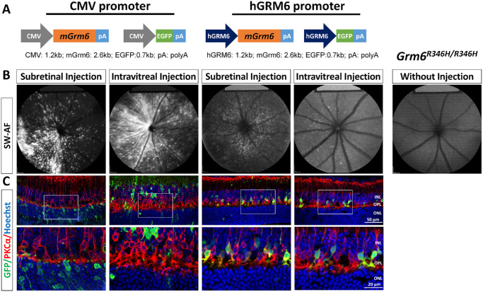
Methods: The nob mouse was generated through selective breeding to regenerate the nob phenotype. Adeno-associated viruses encoding Grm6 and GFP, driven by two promoters (hGRM6 and CMV), were administered to nob mice at postnatal days 5 (P5) and 30 (P30), respectively. Electroretinography and spectral domain optical coherence tomography (SD-OCT) were conducted three months after gene therapy.

Results: The nob phenotype was successfully regenerated, and a homozygous missense variant c.1037G>A (p.Arg346His) in Grm6 was identified as the causal variant. Scotopic b waves were absent, whereas a waves remained normal, indicating intact rod function but impaired bipolar cell function. SD-OCT revealed thinning of the retinal nerve fiber layer and outer plexiform layer (OPL) in affected mice. Immunofluorescence and immunoblotting revealed decreased mGluR6 levels and associated signaling proteins. Gene therapy restored mGluR6 expression and reestablished synaptic protein localization in the OPL, although improvements in b/a ratios and OPL thickness were modest. Notably, the hGRM6 promoter at P5 was more effective at restoring OPL.

Conclusions: We identified a new nob mouse model that mimics the CSNB phenotype in human patients. Whereas gene therapy successfully restored mGluR6 expression, functional improvements were limited. Early treatment using a specific promoter is critical, and increasing transduction efficiency may improve gene therapy strategies.


Conflict of interest statement

Disclosure: P.-H. Lin, None; E.Y.-C. Kang, None; N. Makrides, None; W. Lee, None; Y.-J. Tseng, None; P.-L. Wu, None; J. Peregrin, None; E. Sherman, None; J.H. Wang, None; T.C. Swayne, None; T.-D. Li, None; M.A. Agosto, None; J.R. Sparrow, None; X. Zhang, None; S.H. Tsang, None; N.-K. Wang, None

Figures

Figure 1.
Figure 1.
ERGs recorded in the founder mouse. Representative scotopic, photopic, and flicker ERG recordings comparing R26LSL-Pgc1α no b-wave (nob) founder mouse with C57BL/6J mice reveal: (A) no b-waves in scotopic serial intensities in R26LSL-Pgc1α nob founder mouse, (B) significantly reduced b waves in photopic serial intensities in R26LSL-Pgc1α nob founder mouse, and (C) decreased amplitudes in a flicker series in R26LSL-Pgc1α nob founder mouse. The a and b waves are labeled in normal C57BL/6J mouse.
Figure 2.
Figure 2.
Breeding scheme, genetic analysis, and variant segregation in the nob phenotype. (A) Breeding scheme and strategy for gene discovery. (B) Sanger sequencing revealed segregation with the nob phenotype. Mice with the nob phenotype were homozygous for the variant, whereas those without it were either WT or heterozygous. (C) AciI digestion products of Grm6+/+, Grm6R346H/+, and Grm6R346H/+ samples.
Figure 3.
Figure 3.
ERG of C57BL/6J, Grm6R346H/+, and Grm6R346H/R346H mice. Representative scotopic (A) and photopic (B) ERG recordings of C57BL/6J, Grm6R346H/+, and Grm6R346H/R346H mice. The scotopic and photopic ERG a- and b-wave amplitudes for the three groups are plotted at the bottom of the figure. n = 4, 5, and 4 mice for C57BL/6J, Grm6R346H/+, and Grm6R346H/R346H mice, respectively. (C) Flicker ERG recordings of C57BL/6J, Grm6R346H/+, and Grm6R346H/R346H mice. n = 4, 5, and 8 mice for C57BL/6J, Grm6R346H/+, and Grm6R346H/R346H mice, respectively. The graphs are presented as mean ± SEM. *P < 0.05; **P < 0.01; ***P < 0.001.
Figure 4.
Figure 4.
SD-OCT and Histology. (A) Left, representative SD-OCT images with automatic segmentation. Right, although no obvious structural loss was observed, the RNFL and OPL were significantly thinner in Grm6R346H/R346H mice than in C57BL/6J mice. n = 18 and 18 mice for C57BL/6J and Grm6R346H/R346H mice, respectively. (B) Left, representative retinal histology of C57BL/6J and Grm6R346H/R346H mice. The results show a significant reduction in the thickness of the OPL. n = 4 and 5 mice for C57BL/6J and Grm6R346H/R346H mice, respectively. The graphs are presented as mean ± SEM. *P < 0.05; **P < 0.01; ***P < 0.001. RFNL, retinal nerve fiber layer.
Figure 5.
Figure 5.
IF and Western blotting. (A) Representative IF using antibodies against CtBP2, Pikachurin, mGluR6, TRPM1, Bassoon proteins at synaptic terminals of C57BL/6J and Grm6R346H/R346H mice. The images at the bottom are enlarged views of the white-boxed areas from the images above. (B) Representative IF with antibodies against CtBP2, PKCα, PSD95, and Goα on C57BL/6J and Grm6R346H/R346H mice. (C) Western blots demonstrate reduced levels of proteins mGluR6, GRIK1, CtBP2, PKCα, and PSD95 in Grm6R346H/R346H mice compared with those in C57BL/6J mice. n = 7 and 7 mice for C57BL/6J and Grm6R346H/R346H mice, respectively. The graphs are presented as mean ± SEM. **P < 0.01; ***P < 0.001; ****P < 0.0001.
Figure 6.
Figure 6.
AAV constructs and their transduction efficiency. (A) Schematic diagram of AAVs used. (B) SW-AF in vivo for detection of GFP fluorescence after SRI or IVI of AAV-CMV-GFP or AAV-hGRM6-GFP. Representative SW-AF of a Grm6R346H/R346H mouse without injection is shown on the right. (C) Retinal cryosections were stained with anti-GFP and anti-PKCα antibodies using immunofluorescence. The images at the bottom are enlarged views of the white-boxed areas from the images above.
Figure 7.
Figure 7.
The mGluR6 restored and synaptic protein localization re-established after treatment. (A) Representative confocal images of cross-sections centered on the OPL of C57BL/6J and Grm6R346H/R346H mice. Grm6R346H/R346H mice received IVI or SRI of AAV vectors with the CMV or hGRM6 promoter. Sections were stained with antibodies against mGluR6 (red) and GFP (green). The images at the bottom are enlarged views of the white-boxed areas from the above images. (B) Representative IF using antibodies against mGluR6, TRPM1, Pikachurin, CtBP2 and Bassoon proteins at synaptic terminals of C57BL/6J and Grm6R346H/R346H mice. GFP*, intrinsic green fluorescent protein.
Figure 8.
Figure 8.
ERG and SD-OCT recordings after treatment. (A) OPL thickness measurements comparing C57BL/6J, control (untreated Grm6R346H/R346H mice), and Grm6R346H/R346H mice after treatment by IVI or SRI of the CMV or hGRM6 promoter. n = 18, 18, 11, 10, 5, and 5 mice for C57BL/6J, Control, IVI CMV, IVI hGRM6, SRI CMV, and SRI hGRM6 group, respectively. (B) ERG b/a ratio measurements comparing control (untreated Grm6R346H/R346H mice) and treated Grm6R346H/R346H mice. n = 7, 18, 22, 6, and 12 mice for Control, IVI CMV, IVI hGRM6, SRI CMV, and SRI hGRM6 group, respectively. (C) Recovery of the scotopic b-wave in one mouse from the IVI with the hGRM6-treated group across a range of light intensities, from −4.0 log cd · s/m² to 1.0 log cd · s/m². Red lines, injected eye; black lines, uninjected fellow eye from the same mouse; gray lines: ERG from a C57BL/6J mouse. The graphs are presented as mean ± SEM. *P < 0.05; ***P < 0.001; ****P < 0.0001.

References

    1. Zeitz C, Robson AG, Audo I.. Congenital stationary night blindness: an analysis and update of genotype-phenotype correlations and pathogenic mechanisms. Prog Retin Eye Res. 2015; 45: 58–110. - PubMed
    1. Riggs LA. Electroretinography in cases of night blindness. Am J Ophthalmol. 1954; 38(1:2): 70–78. - PubMed
    1. Schubert G, Bornschein H.. Analysis of the human electroretinogram. Ophthalmologica. 1952; 123: 396–413. - PubMed
    1. Jo A, Deniz S, Xu J, Duvoisin RM, DeVries SH, Zhu Y.. A sign-inverted receptive field of inhibitory interneurons provides a pathway for ON-OFF interactions in the retina. Nat Commun. 2023; 14: 5937. - PMC - PubMed
    1. Scalabrino ML, Boye SL, Fransen KM, et al.. Intravitreal delivery of a novel AAV vector targets ON bipolar cells and restores visual function in a mouse model of complete congenital stationary night blindness. Hum Mol Genet. 2015; 24: 6229–6239. - PMC - PubMed

MeSH terms

Substances

Supplementary concepts