. 2025 Sep 5;53(17):gkaf852.
doi: 10.1093/nar/gkaf852.

Nuclear compression-mediated DNA damage drives ATR-dependent Lamin expression and mouse ESC differentiation

Affiliations

Nuclear compression-mediated DNA damage drives ATR-dependent Lamin expression and mouse ESC differentiation

Tanusri Roy et al. Nucleic Acids Res. .

Abstract

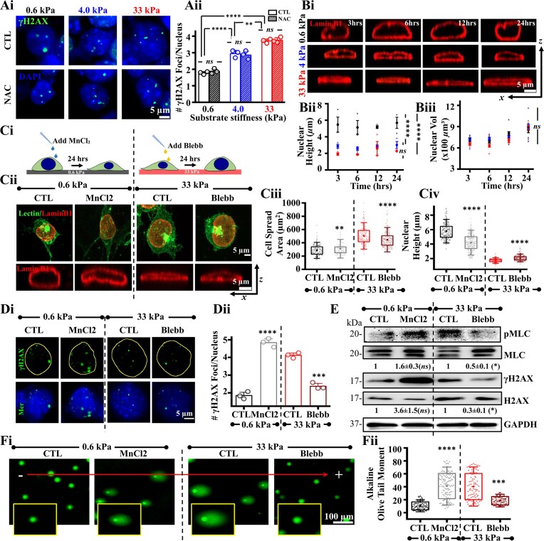
Embryonic stem cells (ESCs), which are susceptible to DNA damage, depend on a robust and highly efficient DNA damage response (DDR) mechanism for their survival. However, the implications of physical force-mediated DNA damage on ESC fate remain unclear. We show that stiffness-dependent spreading of mouse ESCs (mESCs) induces DNA damage through nuclear compression, with DNA damage causing differentiation through Lamin A/C. Interestingly, differentiation is associated with DNA damage and activation of the DDR factors such as ATR and CHK1. While ATR is typically known to play roles in DDR pathway, its role during stiffness-mediated nuclear compression and mESC differentiation is unknown. While our results show activation of CHK1 pathway and nuclear enrichment of activated ATR on stiff substrates, inhibiting ATR and CHK1 both result in reduction of Lamin A/C expression by different mechanisms. Overall, we demonstrate that mESC differentiation is driven by nuclear compression-mediated DNA damage and involves ATR-dependent modulation of Lamin A/C.


Conflict of interest statement

None declared.

Figures

Graphical Abstract
Graphical Abstract
Figure 1.
Figure 1.
Association of mechanoadaptation and DNA damage in mESCs. (Ai, ii) Representative lectin-stained images of mESCs on fibronectin (FN)-coated polyacrylamide (PA) gels of varying stiffness post 3, 6, 12, 24, and 72 h of seeding and quantification of cell spreading (formula image cells per condition across N = 3 independent experiments). Scale Bar = 10 μm. Error bars represent SD. Two-way ANOVA with Tukey’s test was used for comparing means (**formula image-value formula image0.01, **** formula image-value formula image0.0001). (Bi, ii) Assessment of pMLC expression in mESCs cultured on PA gels for 24 h based on immunofluorescence and western blotting showing mean ± SEM of pMLC/MLC (N = 3 independent experiments). Two-way ANOVA with Tukey’s test was used for comparing means with respect to 0.6 kPa conditions (**** formula image-value formula image0.0001). Scale Bar = 10 μm.(Ci-iii) Measurement of mESC cortical stiffness using atomic force microscopy (AFM) after 24 h in culture. Cells were probed with a soft pyramidal probe and first 500 nm of the raw force-indentation curves were fitted with Hertz model to estimate cortical stiffness (formula image cells per condition across N = 3 independent experiments). One-way ANOVA with Tukey’s test was used for comparing means (**** formula image-value formula image0.0001). (Di-iii) Quantification of DNA damage in mESCs cultured on PA gels for 24 h. DNA damage was assessed by co-staining cells with DAPI and γH2AX and counting γH2AX foci per nucleus (formula image nuclei per condition across N = 3 independent experiments) and western blotting of whole cell lysates showing mean ± SEM of γH2AX/H2AX (N = 3 independent experiments). Two-way ANOVA with Tukey’s test was used for comparing means with respect to 0.6 kPa conditions (*formula image-value formula image0.05). Error bars represent SEM. One-way ANOVA with Tukey’s test was used for comparing means (**** formula image-value formula image0.0001). (Ei, ii) Representative images of alkaline and neutral comet assay of mESCs cultured on PA gels (Insets show single comets) and quantitative analysis of olive tail moment (formula image cells per condition across N = 3 independent experiments). One-way ANOVA with Tukey’s test was used for comparing means (*formula image-value formula image0.05, ** formula image-value formula image0.01, ns = non-significant P-valueformula image0.05). For all blots, GAPDH served as loading control. See also Supplementary Fig. S1A–C.
Figure 2.
Figure 2.
Mechanoadaptation mediated nuclear compression induces DNA damage in mESCs: (Ai, Aii) Representative images of mESC nuclei co-stained with γH2AX on PA gels of varying stiffnesses after 24 h in culture in presence and absence of the ROS inhibitor, NAC and quantification of DNA damage (formula image cells per condition across N = 3 independent experiments). Scale Bar = 5 μm. Error bars represent SEM. Two-way ANOVA followed by Tukey’s post-hoc test was used to compare means (**formula image-value formula image0.01, **** formula image-valueformula image 0.0001, ns = non-significant formula image-valueformula image0.05). (Bi-Biii) Representative images showing transverse sections of mESC nuclei immunostained with Lamin B1 on PA gels of varying stiffness post 3, 6, 12, and 24 h of seeding and quantification of nuclear height and nuclear volume (formula image cells per condition across N = 3 independent experiments). Scale Bar = 5 μm. Error bars represent SD. Two-way ANOVA with Tukey’s test was used for comparing means (**** formula image-value formula image0.0001, ns = non-significant P-valueformula image0.05). (Ci-Civ) Schematic workflow, representative lectin-stained images and transverse Lamin B1 stained nuclear sections of mESCs on PA gels of 0.6 kPa ± MnCl2 and 33 kPa ± Blebbistatin and quantification of cell spreading and nuclear height (formula image cells per condition across N = 3 independent experiments). Scale Bar = 5 μm. Error bars represent SD. Unpaired Student’s t-test was used for comparing means between control and treated samples on each PA gel (**formula image-value formula image0.01, **** formula image-valueformula image 0.0001). (Di, Dii) γH2AX and DAPI immunostained images of mESCs on PA gels of 0.6 kPa ± MnCl2 and 33 kPa ± Blebbistatin. Quantification of γH2AX foci (formula image cells per condition across N = 3 independent experiments). Scale Bar = 5 μm. Error bars represent SEM. Unpaired student t-test was used for comparing means between control and treated samples on each PA gel (***formula image-value formula image0.001, **** formula image-valueformula image 0.0001). (E) Representative immunoblots and quantification of mean ± SEM of pMLC/MLC and γH2AX/H2AX levels in mESCs cultured on 0.6 kPa gels ± MnCl2 and 33 kPa gels ± Blebbistatin (N = 3 independent experiments). Unpaired Student t-test was used for comparing means with respect to untreated controls of each PA gel (*formula image-value formula image0.05, ns = non-significant formula image-valueformula image0.05). (Fi, Fii) Representative images of alkaline comet assay of mESCs cultured on 0.6 kPa ± MnCl2 and 33 kPa ± Blebbistatin (Insets show single comets), and quantitative analysis of olive tail moment (formula image cells per condition across N = 3 independent experiments). Scale Bar = 100 μm. Error bars represent SD. Unpaired Student’s t-test was used for comparing means with respect to untreated controls of each PA gel (***formula image-value formula image0.001, **** formula image-value formula image0.0001). For all blots, GAPDH served as loading control.
Figure 3.
Figure 3.
DNA damage induces Lamin A/C expression: (Ai, Aii) Representative images of mESC nuclei immunostained with Lamin A/C on PA gels of varying stiffnesses post 24 h of culture. Scale Bar = 5 μm. Representative immunoblots and quantification of mean ± SEM of Lamin A/C expression and pluripotency (Oct3/4 and Nanog) of mESCs on varying stiffnesses post 24 h culture (N = 3 independent experiments). Two- way ANOVA with Tukey’s test was used for comparing means with respect to 0.6 kPa conditions (*formula image-value formula image0.05, ns = non-significant formula image-valueformula image0.05). (Bi, Bii, Biii) Experimental setup and representative images of Lamin A/C expression and DNA damage in presence of 2 μM etoposide (ETO) post 24 h treatment on mESCs by immunofluorescence on 0.6 kPa gels. Scale Bar = 5 μm. Representative immunoblots and quantification of mean ± SEM of Lamin A/C expression, pluripotency levels and γH2AX/H2AX of whole cell mESC lysates in presence and absence of ETO post 24 h treatment on 0.6 kPa gels (formula image independent experiments). Unpaired student’s t-test was used for comparing means with respect to untreated control. (*formula image-value formula image0.05, **formula image-value formula image0.01). (Ci, Cii, Ciii) Experimental setup for studying temporal correlation between DNA damage and Lamin A/C induction. Cells were allowed to attach onto 0.6 kPa gels for 6 h prior to ETO addition, and then cultured for upto 3 h. Representative immunoblots and quantification of mean ± SEM showing temporal evolution of Lamin A/C, Oct3/4, Nanog, γH2AX/H2AX (formula image independent experiments). One- way ANOVA with Tukey’s test was used for comparing means with respect to untreated controls (*formula image-value formula image0.05, **formula image-value formula image0.01, ns = non-significant P-valueformula image0.05). (Di, Dii, Diii) Schematic workflow and representative immunofluorescence images of Lamin A/C and γH2AX on mESCs post 3 and 24 h of retinoic acid (RA) and ascorbic acid (AA) treatment on 0.6 kPa PA gels. Scale Bar = 5 μm. Representative immunoblots and quantification of mean ± SEM of Lamin A/C, Lamin B1, Oct3/4, Nanog and γH2AX/H2AX of whole cell mESC lysates post treatment of RA and AA on 0.6 kPa PA gels (formula image independent experiments). Two-way ANOVA with Tukey’s test was used for comparing means with respect to 3 h CTL conditions (*formula image-value formula image0.05, **formula image-value formula image0.01, ns = non-significant P-valueformula image0.05). For all blots, GAPDH served as loading control. See also Supplementary Fig. S1 Di, ii and Supplementary Fig. S2.
Figure 4.
Figure 4.
Germ layer differentiation is associated with loss in Lamin A/C: (A) 3-D PCA plot representing clusters of mESCs in + LIF condition, Etoposide (ETO), and varying stiffness post 3, 6, 24, and 72 of culture based on RNAseq profiles. (B) Heatmaps showing temporal evolution of mESC RNAseq profiles of genes involved in pluripotency, endodermal, mesodermal, and ectodermal lineage across different stiffnesses and in ETO-treated cells on 0.6 kPa gels. Normalization was carried out with respect to + LIF condition. (Ci, Cii) Representative immunoblots and quantification of Oct3/4 and Nanog in mESCs cultured in the presence of LIF and in the absence of LIF on PA gels across different time-points (formula image independent experiments; error bars represent formula imageSEM). Two-way ANOVA with Tukey’s test was used for comparing means with respect to + LIF controls (*formula image-value formula image0.05, **formula image-value formula image0.01, ***formula image-value formula image0.001, ns = non-significant P-valueformula image0.05). (Di) Heatmaps showing RNAseq profiles of Lamin genes (LMNA, LMNB1 and LMNB2) in mESCs cultured on PA gels across different time-points as well as in ETO treated cells on 0.6 kPa gels. Normalization was carried out with respect to + LIF condition. (Dii, Diii) Representative immunoblots and quantification of Lamin A/C and Lamin B1 in mESCs cultured on PA gels across different time-points (formula image independent experiments; error bars represent ± SEM). Quantification of Lamin A/C and Lamin B1 levels were normalized with respect to 0.6 kPa at 3 h at different time-points on the gels. Statistical significance obtained by Two-way ANOVA followed by Tukey's tests (*formula image-value formula image0.05, **formula image-value formula image0.01, ****formula image-value formula image0.0001, ns = non-significant P-value formula image0.05). (Ei-Eiii) Representative immunofluorescence images of Lamin A/C co-stained with DAPI in mESCs cultured on PA gels across different time-points showing heterogenous expression of Lamin A/C (red arrowhead: high expression; yellow arrowhead: low expression) (formula image cells per condition across N = 3 independent experiments). Scale Bar = 20 μm. Lamin A/C heterogeneity was captured by plotting the normalized Lamin A/C distribution across different time-points. Normalization was done wrt the maximum intensity observed across all conditions and time-points (formula image cells per condition across N = 3 independent experiments). Assessment of percentage of low (Lo), moderate (Mo) and high (Hi) Lamin A/C expression across different conditions (see Materials & Methods for details). For all blots, GAPDH served as loading control. See also Supplementary Figs S3–S5.
Figure 5.
Figure 5.
Differentiation involves DNA repair through ATR activation and nuclear enrichment: (Ai, Aii) Representative images of temporal evolution of mESCs immunostained for γH2AX co-stained with DAPI on varying stiffness and quantification of γH2AX foci count per nucleus (formula image cells per condition across N = 3 independent experiments). Scale Bar = 5 μm. Error bars represent SD. Two- way ANOVA with Tukey’s test was used for comparing means (**formula image-value formula image0.01, **** formula image-valueformula image 0.0001). (Bi, Bii) Representative images of temporal evolution of mESCs immunostained for DDR factor, 53BP1, co-stained with DAPI on varying stiffness and quantification of 53BP1 foci count per nucleus (formula image cells per condition across N = 3 independent experiments). Scale Bar = 5 μm. Error bars represent SD. Two-way ANOVA with Tukey’s test was used for comparing means (*** P-valueformula image0.001, **** formula image-valueformula image0.0001, ns = non-significant formula image-valueformula image0.05). (C) Immunoblots representing temporal expression of DDR factors, γH2AX/H2AX and pRPA2 of mESCs on varying stiffness. (D) Heatmaps showing temporal evolution of mESC RNAseq profiles of genes involved in DDR factors and DNA repair across different stiffnesses and in ETO-treated cells on 0.6 kPa gels. Normalization was carried out with respect to + LIF condition. (Ei, Eii) Immunoblots representing temporal expression of DDR factors (pATM/ATM, pATR/ATR) and DNA repair factor (RAD 51) in the presence of LIF and in the absence of LIF on 33 kPa PA gels. Quantification of pATR/ATR, ATR, ATM, and RAD51 levels were done with respect to + LIF condition at different time-points across N = 3 independent experiments. Error bars represent ± SEM. One-way ANOVA with Tukey’s test was used for comparing means (*formula image-value formula image0.05). (Fi, ii) Representative pATM-Ser1981/pATR-Thr1989/DAPI stained images of mESCs across different time-points on 33 kPa gels. Scale Bar = 5 μm. Representative intensity profiles along white dotted lines depict nuclear/cytoplasmic localization of pATM and pATR. (Fiii) Quantification of nuclear to cytoplasmic (N/C) ratio of pATM and pATR (formula image cells per condition across N = 3 independent experiments). Error bars represent SD. One-way ANOVA with Tukey’s test was used for comparing means (ns = non-significant formula image-valueformula image0.05). For all blots, GAPDH served as loading control. See also Supplementary Fig. S6 and Supplementary Movies S1–S3.
Figure 6.
Figure 6.
ATR regulates Lamin expression: (Ai, Aii) Immunoblots representing temporal evolution of pATR/ATR, pLamin A/C/Lamin A/C, pCHK1/CHK1, γH2AX/H2AX in the presence of LIF and in the absence of LIF on 33 kPa PA gels upto day 7 (168 h). Quantification of mean ± SEM of pATR/ATR, pCHK1/CHK1, γH2AX/H2AX, Lamin A/C and pLamin A/C/Lamin A/C normalized with respect to + LIF condition (formula image independent experiments). One- way ANOVA with Tukey’s test was used for comparing means with respect to + LIF controls (*formula image-value formula image0.05, **formula image-value formula image0.01, ***formula image -value formula image0.001, ****formula image -value formula image0.0001). (Bi, ii, iii) Cells were allowed to attach onto 0.6 kPa gels for 6 h prior to ETO addition, and then cultured for upto 3 h. Representative immunoblots showing temporal evolution of pATM/ATM, pATR/ATR, and pCHK1/CHK1, and its quantification of mean formula imageSEM (formula image independent experiments). One-way ANOVA with Tukey’s test was used for comparing means with respect to untreated controls (*formula image-value formula image0.05, **formula image-value formula image0.01, ***formula image-value formula image0.001). (Ci-iv) Experimental setup for studying effect of KU-55933 (KU) and VE-821 (VE) on Lamin A/C. Cells were cultured on 33 kPa gels for 24 h. Representative immunostained images of γH2AX co-stained with DAPI and Lamin A/C on mESCs cultured in the presence and absence of KU and VE. Bar plots show quantification of γH2AX foci per nucleus in these conditions (formula image cells per condition across N = 3 independent experiments). Scale Bar = 5 μm. Error bars represent SEM. Unpaired student t-test was used for comparing means between control and treated samples (**formula image-value formula image0.01, ****formula image-value formula image0.0001). Representative immunoblots showing quantification of mean ± SEM of γH2AX/H2AX (N = 3 independent experiments). One- way ANOVA with Tukey’s test was used for comparing means with respect to untreated control (***formula image -value formula image0.001, ns = non-significant formula image-valueformula image0.05). (D) Representative immunoblots of mESCs on 33 kPa gels in the presence and absence of KU and VE and quantification of mean ± SEM showing pLamin A/C/Lamin A/C, Lamin A/C and Lamin B1 expression (formula image independent experimentsformula image. One- way ANOVA with Tukey’s test was used for comparing means with respect to untreated controls (*P-value formula image0.05, **formula image-value formula image0.01, ***formula image-value formula image0.001, ns = non-significant). (E) Representative immunoblots of mESCs on 33 kPa gels in the presence and absence of KU and VE and quantification of mean ± SEM showing Oct3/4, Nanog, GATA4, Brachyury, NeuN and GATA3 expression (formula image independent experiments). One- way ANOVA with Tukey’s test was used for comparing means with respect to untreated control (*P-value formula image0.05, **formula image-value formula image0.01, ****formula image-value formula image0.0001, ns = non-significant). For all blots, GAPDH served as loading control. See also Supplementary Figs S7 and S8.
Figure 7.
Figure 7.
Schematic representation depicting stiffness-dependent differentiation in mouse ESCs. Stiffness-dependent mechanoadaptation induces nuclear compression leading to DNA damage (SSBs), activation of ATR and ATR-mediated lamin A/C modulation. Differentiation involves ATR-mediated lamin A/C stabilization by inhibition of Lamin A/C phosphorylation at early time-points followed by gradual loss of Lamin A/C and cell death. ATR activation in surviving cells at later time-points restores Lamin A/C.

References

    1. Martin GR Isolation of a pluripotent cell line from early mouse embryos cultured in medium conditioned by teratocarcinoma stem cells. Proc Natl Acad Sci USA. 1981; 78:7634–8. 10.1073/pnas.78.12.7634. - DOI - PMC - PubMed
    1. Mahla RS Stem Cells Applications in Regenerative Medicine and Disease Therapeutics. Int J Cell Biol. 2016; 2016:6940283. 10.1155/2016/6940283. - DOI - PMC - PubMed
    1. Kapinas K, Grandy R, Ghule P et al. The abbreviated pluripotent cell cycle. J Cell Physiol. 2013; 228:9–20. 10.1002/jcp.24104. - DOI - PMC - PubMed
    1. Ahuja AK, Jodkowska K, Teloni F et al. A short G1 phase imposes constitutive replication stress and fork remodelling in mouse embryonic stem cells. Nat Commun. 2016; 7:10660. 10.1038/ncomms10660. - DOI - PMC - PubMed
    1. Vitale I, Manic G, De Maria R et al. DNA damage in stem cells. Mol Cell. 2017; 66:306–19. 10.1016/j.molcel.2017.04.006. - DOI - PubMed

MeSH terms

LinkOut - more resources