Case Reports
. 2025 Oct:135:111892.
doi: 10.1016/j.ijscr.2025.111892. Epub 2025 Sep 6.

Delayed diagnosis of post-traumatic bronchial transection in a pediatric patient: A case report

Affiliations
Case Reports

Delayed diagnosis of post-traumatic bronchial transection in a pediatric patient: A case report

Mohammad Alaa Aldakak et al. Int J Surg Case Rep. 2025 Oct.

Abstract

Background: Tracheobronchial injuries (TBI) are rare yet potentially fatal complications of blunt chest trauma, often underdiagnosed due to nonspecific clinical manifestations.

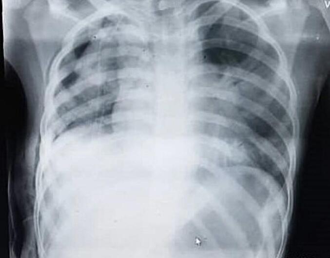
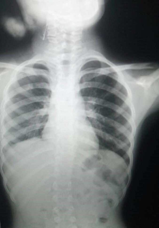
Case presentation: We report the case of an 11-year-old Arab girl who developed progressive dyspnea two months after a motor vehicle accident initially managed conservatively. Imaging revealed complete atelectasis of the right lung and obstruction of the right main bronchus by granulation tissue. Bronchoscopy confirmed complete bronchial occlusion, and surgical intervention revealed a delayed bronchial transection. Successful end-to-end anastomosis restored full lung expansion and respiratory function.

Clinical discussion: This case highlights the diagnostic challenge of TBIs in children, particularly when symptoms present late. Anatomical vulnerability of the right bronchus, delayed symptom onset, and nonspecific radiologic signs may obscure early recognition. Granulation-induced bronchial obstruction, a known complication of undiagnosed TBIs, was confirmed intraoperatively.

Conclusion: Delayed presentation of tracheobronchial injury may lead to progressive airway obstruction due to granulation tissue. High suspicion, timely bronchoscopy, and early surgical intervention are essential to prevent irreversible lung damage and restore pulmonary function.

Keywords: Bronchial transection; Bronchoscopy; Granulation tissue; Pediatric trauma; Surgical repair; Tracheobronchial injury.


Conflict of interest statement

Conflict of interest statement The authors declared no potential conflicts of interest concerning the research, authorship, and/or publication of this article.

Figures

Fig. 1
Fig. 1
Frontal chest X-ray showing near-complete opacification of the right hemithorax due to hypoventilation and collapse. Small air lucencies are visible within the opacity, suggestive of pulmonary contusion. A moderate right-sided pleural effusion and mild mediastinal shift to the right are also noted.
Fig. 2
Fig. 2
Axial contrast-enhanced chest CT scan showing complete absence of aeration in the right lung, right-sided pleural effusion, and mediastinal shift toward the right side. A soft tissue density is noted in the right main bronchus, suggestive of an obstructing lesion, consistent with granulation tissue formation following traumatic bronchial transection.
Fig. 3
Fig. 3
Intraoperative view showing complete transection of the right main bronchus.
Fig. 4
Fig. 4
Intraoperative view following completion of end-to-end anastomosis of the right main bronchus. The right lung is seen fully re-expanded, indicating restoration of airway continuity and adequate ventilation.
Fig. 5
Fig. 5
Postoperative chest X-ray demonstrating significant re-expansion of the right lung with improved aeration and resolution of the previously noted pleural effusion. The mediastinum is centrally positioned, and lung fields appear well inflated bilaterally.
Fig. 6
Fig. 6
Follow-up chest X-ray obtained two months postoperatively showing sustained re-expansion and aeration of the right lung. No evidence of pleural effusion, atelectasis, or other complications is observed. The mediastinum remains centrally aligned, and lung fields appear clear bilaterally.

References

    1. Altinok T., Can A. Management of tracheobronchial injuries. Eurasian J. Med. 2014;46(3):209–215. doi: 10.5152/eajm.2014.42. - DOI - PMC - PubMed
    1. Dominguez E., De La Torre C., Sánchez A.V., Hernandez F., Ortiz R., Moreno A.M., et al. Severe tracheobronchial injuries: our experience. Eur. J. Pediatr. Surg. 2015;25(1):71–76. doi: 10.1055/s-0034-1386642. - DOI - PubMed
    1. Prokakis C., Koletsis E.N., Dedeilias P., Fligou F., Filos K., Dougenis D. Airway trauma: a review on epidemiology, mechanisms of injury, diagnosis and treatment. J. Cardiothorac. Surg. 2014;9:117. doi: 10.1186/1749-8090-9-117. - DOI - PMC - PubMed
    1. Iwasaki M., Kaga K., Ogawa J., Inoue H., Shohtsu A. Bronchoscopy findings and early treatment of patients with blunt tracheo-bronchial trauma. J. Cardiovasc. Surg. 1994;35(3):269–271. - PubMed
    1. Chesterman J.T., Satsangi P.N. Rupture of the trachea and bronchi by closed injury. Thorax. 1966;21(1):21–27. doi: 10.1136/thx.21.1.21. - DOI - PMC - PubMed

Publication types

LinkOut - more resources