. 2025 Aug 31;34(4):156-167.
doi: 10.5607/en25013.

Differences in Learning Strategy Selection and Object Location Memory Impairments in APP/PS1 Mice

Affiliations

Differences in Learning Strategy Selection and Object Location Memory Impairments in APP/PS1 Mice

Yoon-Sun Jang et al. Exp Neurobiol. .

Abstract

This study investigated the learning strategy preferences of 11-month-old APP/PS1 double transgenic (Tg) mice, a well-established murine model of Alzheimer's disease (AD). APP/PS1 Tg and non-Tg control mice were serially trained in visual and hidden platform tasks in the Morris water maze. APP/PS1 Tg mice performed poorly in visual platform training compared with non-Tg mice but performed as well as non-Tg mice in hidden platform training. Further analysis of their search paths for locating a hidden platform revealed that APP/PS1 Tg mice used more cued/response search patterns than place/spatial search patterns compared with non-Tg mice. Three months later, the object/location recognition memory of APP/PS1 Tg mice was assessed. Although their object recognition memory was intact, their object location memory was impaired. Neuropathological AD features of APP/PS1 transgenic mice were observed in the medial prefrontal cortex, retrosplenial cortex, and hippocampus, key brain regions involved in learning strategy shifts and spatial cognition. These results indicate that distinct search patterns and spatial memory deficits in APP/PS1 Tg mice are key features of AD animal models.

Keywords: APP/PS1; Alzheimer’s disease; Amyloid beta; Cued/response learning; Learning strategy; Object location memory.


Figures
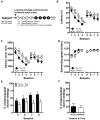
Fig. 1
Fig. 1
Performance of 11-month-old APP/PS1 and non-transgenic (Tg) mice in the visual and hidden platform training in the Morris water maze task. (A) Overview of the behavioral experimental schedule. (B, C) Significant differences in latency and search error were observed between non-Tg and APP/PS1 mice during visual platform training but not during hidden platform training. (D) Non-Tg and APP/PS1 mice showed equivalent swimming speed. (E) In the hidden platform training, APP/PS1 mice employed a place/spatial search strategy less frequently than non-Tg mice. (F) No difference in the percentage of place or spatial search strategy was found between the two groups on the competition test. Non-Tg (n=12), APP/PS1 (n=12), Data are expressed as mean±SEM. *p<0.05, **p<0.01.
Fig. 2
Fig. 2
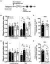
Performance of the 14-month-old non-Tg and APP/PS1 mice in the novel object/location recognition task. (A) Overview of the experimental procedure. All test phases (T) were conducted 24 h after the familiarization phase (F). (B) In the novel object recognition task, non-Tg and APP/PS1 mice equally explored the two identical objects placed on the left (L) and right sides (R) of the open-field box in the familiarization phase. (C) Both non-Tg and APP/PS1 mice explored the novel object (N) significantly more than the familiar object (F). No difference was found between the two. (D) In the novel object location task, non-Tg and APP/PS1 mice equally explored the two identical objects on the L and R sides of the open-field box. During the test phase, non-Tg mice preferred the object at a new location (N) over that at a familiar location (F), whereas APP/PS1 mice did not. (E) A significant difference was found in the preference for the novel location between the two. Data are expressed as mean±SEM. *p<0.05, **p<0.01, #p<0.05 (one-tail); Non-Tg (n=11), APP/PS1 (n=12).
Fig. 3
Fig. 3
Increases in amyloid-β, microglial activation, and astroglial activation in the medial prefrontal cortex of 14-month-old APP/PS1 mice. (A) Representative image showing immunofluorescence staining of the medial prefrontal cortex of the non-Tg and APP/PS1 mice. (B~E) Quantification of 4G8-, Iba-1-, GFAP-, and NeuN-positive signals in the medial prefrontal cortex of the non-Tg and APP/PS1 mice. Intensities of 4G8-, Iba-1-, and GFAP-positive signals in the medial prefrontal cortex of the APP/PS1 mice were higher than those of non-Tg mice. Non-Tg mice (n=5); APP/PS1 mice (n=4). Data are expressed as mean±SEM. Scale bar=200 μm; Scale bar in the lower rectangle=100 μm. **p<0.01, ***p<0.001.
Fig. 4
Fig. 4
Increases in amyloid-β, microglial activation, and astroglial activation in the retrosplenial cortex of 14-month-old APP/PS1 mice. (A) Representative images showing immunofluorescence staining of the retrosplenial cortex of non-Tg and APP/PS1 mice. (B~E) Quantification of 4G8-, Iba-1-, GFAP-, and NeuN-positive signals in the retrosplenial cortex of the non-Tg and APP/PS1 mice. Intensities of 4G8-, Iba-1-, and GFAP-positive signals in the retrosplenial cortex of the APP/PS1 mice were higher than those of non-Tg mice. Non-Tg (n=5); APP/PS1 (n=4). Data are expressed as mean±SEM. Scale bar=200 μm; Scale bar in the lower rectangle=100 μm. p<0.05, **p<0.01, #p<0.05 (one-tail).
Fig. 5
Fig. 5
Increases in amyloid-β, microglial activation, and astroglial activation in the hippocampus of 14-month-old APP/PS1 mice. (A) Representative images showing immunofluorescence staining of the hippocampus of non-Tg and APP/PS1 mice. (B~E) Quantification of 4G8-, Iba-1-, GFAP-, and NeuN-positive signals in the hippocampus of non-Tg and APP/PS1 mice. Intensities of 4G8-, Iba-1-, and GFAP-positive signals in the hippocampus of APP/PS1 mice were higher than those of non-Tg mice. Non-Tg (n=5); APP/PS1 (n=4). Data are expressed as mean±SEM. Scale bar=200 μm; Scale bar in the lower rectangle=100 μm. p<0.05, **p<0.01, #p<0.05 (one-tail).

References

    1. Ashe KH. Learning and memory in transgenic mice modeling Alzheimer's disease. Learn Mem. 2001;8:301–308. doi: 10.1101/lm.43701. - DOI - PubMed
    1. Hsiao K, Chapman P, Nilsen S, Eckman C, Harigaya Y, Younkin S, Yang F, Cole G. Correlative memory deficits, Abeta elevation, and amyloid plaques in transgenic mice. Science. 1996;274:99–102. doi: 10.1126/science.274.5284.99. - DOI - PubMed
    1. Cho WH, Park JC, Chung C, Jeon WK, Han JS. Learning strategy preference of 5XFAD transgenic mice depends on the sequence of place/spatial and cued training in the water maze task. Behav Brain Res. 2014;273:116–122. doi: 10.1016/j.bbr.2014.07.033. - DOI - PubMed
    1. Hall AM, Roberson ED. Mouse models of Alzheimer's disease. Brain Res Bull. 2012;88:3–12. doi: 10.1016/j.brainresbull.2011.11.017. - DOI - PMC - PubMed
    1. Kim DH, Jang YS, Jeon WK, Han JS. Assessment of cognitive phenotyping in inbred, genetically modified mice, and transgenic mouse models of Alzheimer's disease. Exp Neurobiol. 2019;28:146–157. doi: 10.5607/en.2019.28.2.146. - DOI - PMC - PubMed