. 2025 May-Jun;40(3):127-135.
doi: 10.4103/ijnm.ijnm_13_25. Epub 2025 Aug 7.

Utility of SPECT/CT in Non-Traumatic Osteonecrosis: Revisited with New Insights from a Prospective Diagnostic Study

Affiliations

Utility of SPECT/CT in Non-Traumatic Osteonecrosis: Revisited with New Insights from a Prospective Diagnostic Study

Mangu Srinivas Bharadwaj et al. Indian J Nucl Med. 2025 May-Jun.

Abstract

Objectives: Bone scintigraphy is a sensitive imaging method to evaluate patients with suspected osteonecrosis. We assessed the diagnostic performance of combined bone single-photon emission computed tomography/computed tomography (SPECT/CT) (CBS) in patients with known rheumatic disease or other connective tissue disorders and clinical suspicion of osteonecrosis compared to magnetic resonance imaging (MRI).

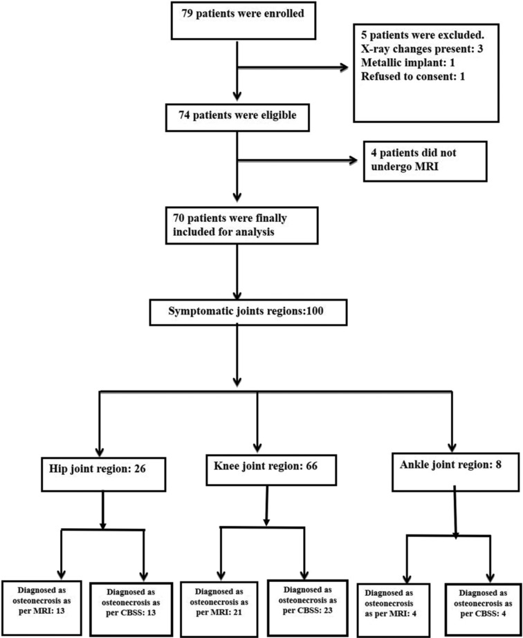
Methods: This prospective diagnostic accuracy study included 70 patients with clinical suspicion of osteonecrosis in any bone who underwent a planar triple-phase bone scan along with a regional SPECT/CT (CBS) and regional MRI. MRI was considered the standard for diagnosing the sensitivity, specificity, predictive values, and accuracy of CBS. Cohen's kappa statistic of the agreement was also calculated.

Results: The distribution of the patients based on the joint regions suspected to have osteonecrosis is as follows: 21 hip, 43 knee, and six ankle. MRI detected osteonecrosis in 30/70 patients. CBS had a sensitivity of 100% (30/70 were detected) and a specificity of 97% (2/40 were false positive). Overall, there was good agreement between the two scans regarding the diagnosis of osteonecrosis (Cohen's kappa statistic = 0.94). In addition to the suspected sites, CBS detected osteonecrosis in 19 additional asymptomatic sites in 13 patients.

Conclusion: The study has demonstrated that CBS, which includes whole-body imaging and SPECT/CT, is highly sensitive in detecting osteonecrosis with accuracy comparable to regional MRI. Its inherent whole-body imaging technique enabled the detection of multifocal osteonecrosis. It can be used as an early investigating modality after routine plain radiography to establish the diagnosis.

Keywords: Magnetic resonance imaging; nontraumatic osteonecrosis; rheumatic and autoimmune diseases; single-photon emission computed tomography/computed tomography; triple-phase bone scan.


Conflict of interest statement

There are no conflicts of interest.

Figures

Figure 1
Figure 1
STARD flow diagram
Figure 2
Figure 2
A 30-year-old female with systemic lupus erythematosus on corticosteroids presented with bilateral hip pain. Flow (a) and pool phase (b) images were normal. Bone scan delayed images (c) show increased uptake in bilateral hips with central photopenia, suggesting osteonecrosis. Single-photon emission computed tomography/computed tomography (d) reveals subchondral cysts and sclerosis, consistent with osteonecrosis. Magnetic resonance imaging T2 turbo rapid acquisition (TRA) turbo spin echo (TSE) sequences images (e) confirm geographic T2 hyperintensities, establishing the diagnosis
Figure 3
Figure 3
An 18-year-old female with systemic lupus erythematosus presented with bilateral knee pain. Perfusion (a) and tissue phase (b) images show mildly increased tracer perfusion and pooling in bilateral knees. Delayed phase (c) images reveal diffuse tracer uptake in the distal femur and proximal tibia, indicating osteonecrosis. Single-photon emission computed tomography/computed tomography (d and e) confirms increased uptake with sclerotic marrow changes. Magnetic resonance imaging T2 repetition time- independent multislice imaging (TRIM) coronal sequences (f) show serpiginous marrow lesions with peripheral hyperintensities, consistent with bone infarcts
Figure 4
Figure 4
A 30-year-old female with systemic lupus erythematosus presented with left ankle pain. Perfusion (a) and tissue phase (b) images show increased flow in the left ankle. Delayed phase (c) images reveal increased uptake in bilateral knees and the left ankle. Single-photon emission computed tomography/computed tomography (d) confirms osteonecrosis in the left distal tibia and knees. Magnetic resonance imaging images, T2W short tau inversion recovery (STIR) (e) and proton density (PD) turbo spin echo (TSE) fat saturation (FS) (f) of the left distal tibia and knee show irregular geographic lesions, consistent with osteonecrosis
Figure 5
Figure 5
A 33-year-old female with systemic lupus erythematosus on corticosteroids presented with bilateral knee pain. Perfusion (a) and tissue phase (b) images were normal. Delayed phase (c and d) images reveal increased uptake in lateral condyles of both femurs. Single-photon emission computed tomography/computed tomography (e) shows focal tracer uptake in the right lateral femoral condyle (red arrow), indicating osteonecrosis. Magnetic resonance imaging T2 TSE axial images (f) demonstrate subarticular irregularity and bone marrow edema in the right lateral femoral condyle (yellow arrow)
Figure 6
Figure 6
A 42-year-old female with systemic lupus erythematosus presented with left knee pain. Perfusion (a) and tissue phase (b) images were normal. Delayed phase (c) showed increased tracer uptake in bilateral knees. Single-photon emission computed tomography/computed tomography (d) indicated abnormal uptake in the left tibial condyle (red arrow), suggesting osteonecrosis. Magnetic resonance imaging (MRI) T2 Repetition time- independent multislice imaging (TIRM) coronal sequences (e) performed 25 days later showed normal signal intensity with joint effusion. The discrepancy may reflect transient osteonecrosis, resolving before the MRI

References

    1. Mankin HJ. Nontraumatic necrosis of bone (osteonecrosis) N Engl J Med. 1992;326:1473–9. - PubMed
    1. Assouline-Dayan Y, Chang C, Greenspan A, Shoenfeld Y, Gershwin ME. Pathogenesis and natural history of osteonecrosis. Semin Arthritis Rheum. 2002;32:94–124. - PubMed
    1. Weiner ES, Abeles M. Aseptic necrosis and glucocorticosteroids in systemic lupus erythematosus: A reevaluation. J Rheumatol. 1989;16:604–8. - PubMed
    1. Gurion R, Tangpricha V, Yow E, Schanberg LE, McComsey GA, Robinson AB, et al. Avascular necrosis in pediatric systemic lupus erythematosus: A brief report and review of the literature. Pediatr Rheumatol Online J. 2015;13:13. - PMC - PubMed
    1. Yang Y, Kumar S, Lim LS, Silverman ED, Levy DM. Risk factors for symptomatic avascular necrosis in childhood-onset systemic lupus erythematosus. J Rheumatol. 2015;42:2304–9. - PubMed

LinkOut - more resources