. 2025 Sep 8:8:0854.
doi: 10.34133/research.0854. eCollection 2025.

PRMT1-Mediated PARP1 Methylation Drives Lung Metastasis and Chemoresistance via P65 Activation in Triple-Negative Breast Cancer

Affiliations

PRMT1-Mediated PARP1 Methylation Drives Lung Metastasis and Chemoresistance via P65 Activation in Triple-Negative Breast Cancer

Jinhui Zhang et al. Research (Wash D C). .

Abstract

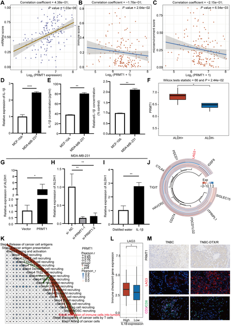
Triple-negative breast cancer (TNBC) is the most aggressive breast cancer subtype, characterized by a high propensity for metastasis, poor prognosis, and limited treatment options. Research has demonstrated a substantial correlation between the expression of protein arginine N-methyltransferase 1 (PRMT1) and enhanced proliferation, metastasis, and poor outcomes in TNBC. However, the specific role of PRMT1 in lung metastasis and chemoresistance remains unclear. Single-cell RNA sequencing coupled with bioinformatics analysis was employed to identify pertinent genes within metastatic TNBC samples. Functional assays, including cell cycle, apoptosis, wound healing, Transwell migration, colony formation, and Cell Counting Kit-8 Assay (CCK-8), were conducted to evaluate the role of PRMT1. The interaction between PRMT1 and PARP1 was validated by mass spectrometry (MS) and immunoprecipitation. Downstream signaling pathways were explored, with a focus on P65 activation. Enzyme-linked immunosorbent assay was used to quantify the effect of PRMT1 on interleukin-1β secretion. Our study identified a significant association between elevated PRMT1 expression and both lung metastasis and chemoresistance in TNBC. PRMT1 boosts TNBC cell growth, invasion, and lung metastasis. Additionally, high PRMT1 expression contributed to increased resistance to docetaxel in TNBC. Mechanistically, PRMT1 methylates PARP1. On the one hand, this methylation promotes the DNA damage repair ability of PAPA1. On the other hand, it in turn modulates the NF-κB signaling pathway. This modulation enhances the stemness of tumor cells and induces immune suppression within the tumor microenvironment, thereby exacerbating chemoresistance in TNBC. PRMT1 drives lung metastasis and chemoresistance in TNBC through PARP1 methylation and P65 activation. These findings position PRMT1 as a promising biomarker and therapeutic target to overcome resistance and limit metastatic progression in TNBC.


Conflict of interest statement

Competing interests: The authors declare that they have no competing interests.

Figures

Fig. 1.
Fig. 1.
Elevated PRMT1 levels are linked to lung metastasis and chemoresistance in TNBC. (A) Differential analysis was performed using the GSE110153 dataset. (B) Survival analysis was conducted by utilizing the GSE7390 dataset. (C) The Venn diagram demonstrates the intersection of up-regulated genes in resistant breast cancer tissues with genes identified as risk factors. (D) Representative images display PRMT1 immunohistochemical staining in breast cancer tumor tissues, corresponding normal tissues, and lung metastatic tissues. (E) A box plot illustrates the differences in PRMT1 expression between metastatic and nonmetastatic MDA-MB-231 cells in the GSE54465 dataset. (F) Multivariate Cox analyses provide the P values, hazard ratios (HR), and confidence intervals (CI) for PRMT1 expression and clinical characteristics. (G) The t-distributed stochastic neighbor embedding (t-SNE) plot depicts PRMT1 expression levels within the breast cancer single-cell RNA sequencing cohort BRCAGSE143423. (H) Differential expression analysis of PRMT1 was performed using TCGA database. (I) A box plot compares PRMT1 expression levels in the GSE76250 dataset. (J) A Spearman correlation analysis plot was conducted to examine the role of PRMT1 in the G2/M checkpoint.
Fig. 2.
Fig. 2.
PRMT1 promotes the proliferation and migration of TNBC. (A) Within the TCGA database, single-sample gene set enrichment analysis (ssGSEA) was performed to assess the expression levels of PRMT1 and its association with DNA replication. (B) Within the TCGA database, ssGSEA was performed to assess the expression levels of PRMT1 and its association with apoptosis. (C) Flow cytometry was utilized to analyze the cell cycle distribution of MDA-MB-231 cells following the knockdown and overexpression of PRMT1. (D) Flow cytometry was utilized to examine the apoptosis of MDA-MB-231 cells subsequent to the knockdown of PRMT1. (E) The CCK-8 assay was employed to determine the growth curve of MDA-MB-231 cells following the knockdown and overexpression of PRMT1. (F) The wound healing assay was employed to assess the migratory capacity of MDA-MB-231 cells following the knockdown and overexpression of PRMT1. (G) The Transwell migration assay was conducted to evaluate the invasive capacity of MDA-MB-231 cells subsequent to the knockdown and overexpression of PRMT1. (H) The colony formation assay was conducted to evaluate the colony-forming potential of MDA-MB-231 and BT-549 cells, subsequent to the knockdown and overexpression of PRMT1.
Fig. 3.
Fig. 3.
PRMT1 contributes to docetaxel resistance in TNBC, while TC-E 5003 can inhibit TNBC cell growth and migration. (A) Evaluate the IC50 values of MDA-MB-231 cells transfected with either a PRMT1 overexpression plasmid or siRNA. (B) Treat MDA-MB-231 cells transfected with PRMT1 siRNA with either docetaxel or dimethyl sulfoxide, and subsequently quantify the apoptosis rate via flow cytometry. (C) The colony formation ability of MDA-MB-231 cells with PRMT1 knockdown and overexpression was evaluated by a colony formation assay after treatment with docetaxel. (D) Analyze the 3D structure of the interaction between the PRMT1 protein and the small-molecule TC-E 5003. (E) Ascertain the IC50 value of TC-E 5003 in MDA-MB-231 cells. (F) Investigate the expression levels of PRMT1 in MDA-MB-231 cells following treatment with varying concentrations and time intervals of TC-E 5003. (G) The colony formation assay was employed to assess the effects of combined treatment with TC-E 5003 and docetaxel on the MDA-MB-231 cells. (H) Utilize a Transwell migration assay to assess the invasive potential of MDA-MB-231 cells after exposure to TC-E 5003.
Fig. 4.
Fig. 4.
Arginine methylation mediated by PRMT1 enhances the activation of PARP1. (A) MS analysis indicated that PARP1 functions as a downstream target of PRMT1. (B) The molecular docking of PRMT1 and PARP1 was used to observe the binding. (C and D) Immunoprecipitation experiment and silver staining assay were employed to investigate the interaction between PRMT1 and PARP1. (E and F) The methylation sites of PARP1 were identified using MS. (G) Expression correlation between PRMT1 and PARP1 in MDA-MB-231 cells. (H) The methylation sites of PARP1 were verified by immunoprecipitation. (I) Gene–gene correlation between PRMT1 and PARP1 was conducted using data from the TCGA database and the GSE76250 dataset. (J) Immunofluorescence staining was utilized to evaluate the expression levels of PRMT1 and PARP1 in MDA-MB-231 and BT-549 cell. (K) The expression of PARP1 in chemoresistant TNBC tissues was assessed using the GSE28784 dataset. (L) Immunofluorescence was used to compare the expression of PRMT1 and PARP1 in docetaxel-resistant breast cancer tissues relative to TNBC tissues.
Fig. 5.
Fig. 5.
PRMT1 facilitates the proliferation and lung metastasis of TNBC in vivo. (A) A Spearman correlation analysis plot was conducted to investigate the relationship between PRMT1 and tumor progression. (B and C) A subcutaneous breast cancer injection model was developed to assess the carcinogenic potential of PRMT1. Tumor volume was measured weekly, and growth curves were plotted for each experimental group. Subsequently, tumor weight was recorded. (D and E) A TNBC lung metastasis model was established to evaluate the role of PRMT1 in the lung metastasis of TNBC. The impact of PRMT1 on the metastatic potential of TNBC was analyzed using bioluminescence imaging. (F) H&E staining was employed to evaluate tumor tissue metastasis in the lung metastasis model groups. (G) Western blot was used to determine the expression levels of PRMT1 and PARP1 in sh-PRMT1 cells. (H and I) A wound healing assay and a colony formation assay were conducted to assess functional rescue.
Fig. 6.
Fig. 6.
PRMT1 facilitates the activation of the NF-κB signaling pathway through the regulation of PARP1. (A) Perform KEGG pathway enrichment analysis on the proteins identified by MS. (B) Perform pathway enrichment analysis of PRMT1 using the TCGA database. (C and D) Conduct gene–gene correlation of PARP1 and P65 in the TCGA database, and validate the findings through immunoprecipitation experiments. (E and F) Analyze the expression levels of P65 and IL-1β of MDA-MB-231 cells after PRMT1 overexpression using Western blot analysis and ELISA assays. (G and H) Analyze the expression levels of P65 and IL-1β of MDA-MB-231 cells after PRMT1 knockdown using Western blot analysis and ELISA assays. (I and J) The expression levels of P65 and IL-1β of MDA-MB-231 cells after TC-E 5003 treatment were analyzed using Western blot analysis and ELISA assays. (K) A Transwell migration assay was used to analyze the invasive ability of MDA-MB-231 cells after treatment with recombinant IL-1β protein. (L) The correlation between PRMT1 and IL1B was verified in the TCGA database. (M and N) A prognostic model was constructed through LASSO Cox regression analysis of the PRMT1, PARP1, P65, and IL1B genes and was validated in the dataset GSE6532. (O) The expression levels of PRMT1, PARP1, and P65 in TNBC lung metastasis tissue and normal breast tissue were determined using multiplex immunofluorescence.
Fig. 7.
Fig. 7.
PRMT1 enhances tumor stemness and immune suppression in TNBC, sustaining chemoresistance. (A) The “stemness” score of PRMT1 was assessed. (B and C) The XCELL immune scores were employed to investigate the association between PRMT1 and the tumor microenvironment in TNBC. (D and E) The expression levels of IL-1β in MDA-MB-231 and MCF-10A cell lines were quantified through RT-qPCR and ELISA assays. (F) The expression of PRMT1 in ALDH+ and ALDH cells was compared in the dataset GSE136287. (G and H) RT-qPCR was conducted to evaluate the expression of ALDH1 following the knockdown and overexpression of PRMT1 in MDA-MB-231 cells. (I) The expression of ALDH1 in MDA-MB-231 cells was assessed via RT-qPCR subsequent to the administration of recombinant IL-1β protein. (J) A heatmap analysis was conducted to explore the relationship between PRMT1 and immune checkpoint. (K) The tumor immunophenotype analysis was performed to evaluate the proportion of tumor-infiltrating immune cells within the cancer immune cycle in relation to PRMT1 expression. (L) A boxplot analysis was executed to investigate the association between IL1B and LAG3. (M) Immunohistochemistry and immunofluorescence assay were used to examine the expression of PRMT1, LAG3 CD3, and CD8 in DTX/R or control TNBC tissues.
Fig. 8.
Fig. 8.
PRMT1-mediated PARP1 methylation drives lung metastasis and chemoresistance via P65 activation in TNBC.

References

    1. Siegel RL, Giaquinto AN, Jemal A. Cancer statistics, 2024. CA Cancer J Clin. 2024;74(1):12–49. - PubMed
    1. Bray F, Laversanne M, Sung H, Ferlay J, Siegel RL, Soerjomataram I, Jemal A. Global cancer statistics 2022: GLOBOCAN estimates of incidence and mortality worldwide for 36 cancers in 185 countries. CA Cancer J Clin. 2024;74(3):229–263. - PubMed
    1. Chen W, Zheng R, Baade PD, Zhang S, Zeng H, Bray F, Jemal A, Yu XQ, He J. Cancer statistics in China, 2015. CA Cancer J Clin. 2016;66(2):115–132. - PubMed
    1. DeSantis CE, Ma J, Gaudet MM, Newman LA, Miller KD, Goding Sauer A, Jemal A, Siegel RL. Breast cancer statistics, 2019. CA Cancer J Clin. 2019;69(6):438–451. - PubMed
    1. Tang Y, Tian W, Zheng S, Zou Y, Xie J, Zhang J, Li X, Sun Y, Lan J, Li N, et al. Dissection of FOXO1-induced LYPLAL1-DT impeding triple-negative breast cancer progression via mediating hnRNPK/β-catenin complex. Research. 2023;6:0289. - PMC - PubMed

LinkOut - more resources