. 2025 Sep 8:120:e240266.
doi: 10.1590/0074-02760240266. eCollection 2025.

Concomitant use of anti-leishmanial therapy and antibacterial prophylaxis reduces plasma LPS levels and improves several aspects of experimental Leishmania infantum infection in golden hamsters

Affiliations

Concomitant use of anti-leishmanial therapy and antibacterial prophylaxis reduces plasma LPS levels and improves several aspects of experimental Leishmania infantum infection in golden hamsters

Joanna Reis Santos-Oliveira et al. Mem Inst Oswaldo Cruz. .

Abstract

Background: Parasite antigens and plasma lipopolysaccharide (LPS) levels from luminal origin in visceral leishmaniasis (VL) patients are correlated with cellular activation and low CD4+T cell counts.

Objectives: Our aim was to verify whether Leishmania infantum infection damages the intestinal barrier and whether combination antimonial/antibiotic contributes to the reduction of LPS levels and immune activation.

Methods: Golden hamsters were grouped in: G1-uninfected; G2-infected with L. infantum; and G3/G4 and G5-infected, treated with antimonial, antibiotic or both drugs, respectively. The treatment initiated 45 days post infection (dpi), daily by 10 days.

Findings: G2, G3, and G4 animals showed a significant increase in spleen weight compared to G1. An elevated parasite load was observed in G2, unlike the G3, G4, and especially, G5, whose decrease was significant at 120 dpi. Intestinal mucosal alterations and elevated LPS levels were observed in G2 group. However, G3, G4 and G5 animals showed lower LPS levels than G2. Moreover, G4 and G5 presented higher CD4+T-cell percentages and lower activation levels than G2 and G3, either at 60 or 101-120 dpi.

Main conclusions: Our results showed evidence of bacterial translocation in experimental VL and that the concomitant use of antimonial and antibiotic may reduce LPS levels, along with an improvement of the immunosuppression and reduction of lymphocyte activation.


Conflict of interest statement

The authors declare that they have no competing interests.

Figures

Fig. 1:
Fig. 1:. study flowchart. Experimental design of infection, monitoring of disease development and treatment of hamsters infected or not with Leishmania infantum. Golden hamsters were infected with 2 × 10⁷ parasites/mL at day 0 and at 45 days post infection (dpi) were treated with meglumine antimoniate, amikacin sulphate or both drugs during 10 days. G1, animals without infection; G2, infected, untreated animals; G3, infected animals treated with anti-Leishmania therapy (antimonial); G4, infected animals treated with antibiotic (amikacin); and G5, infected animals treated with both drugs.
Fig. 2:
Fig. 2:. immunofluorescence detection of Leishmania infantum amastigotes in colonic tissue from experimentally infected hamsters. (A) Negative control showing absence of fluorescence in colonic tissue from an infected animal processed without the primary antibody. Presence of mucosal and crypt epithelial cells interspersed with a dense eosinophilic infiltrate. (B) Cluster of amastigotes within a perivascular region, surrounded by connective tissue and colonic crypts. Staining was performed using in-house mouse serum from L. infantum-infected animals, followed by an Alexa Fluor® 488-conjugated secondary antibody. Host cell nuclei were counterstained with DAPI (blue) and Evans blue (red). Scale bars: as indicated.
Fig. 3:
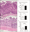
Fig. 3:. histological section of the duodenum stained with Haematoxylin and Eosin. (A) Duodenum of uninfected hamster (G1) showing the mucosa, submucosa, and muscular layer. (B) Infected golden hamster with Leishmania infantum (G2) showing the damaged mucosal surface (black arrow), inflammatory cell infiltrate in the submucosa (black point), and a thickened muscle layer. (C) Mucosal morphometry; (D) Submucosal morphometry; (E) Morphometry of the muscular layer. Representative image of the duodenum of an uninfected (G1, n = 2) and an infected animal (G2, n = 4). Mu: mucosa; Sm: submucosa; M: muscular layer; PP: peyer’s patches; ●: inflammatory infiltrate. Scale bar: 50µm.
Fig. 4:
Fig. 4:. histologic evaluation of the colon stained with Haematoxylin and Eosin. (A) Colon of uninfected hamster (G1), showing intact mucosa, submucosa and muscular layer. (B) Colon of infected hamster golden hamster with Leishmania infantum (G2), exhibiting histological variations in shape and length of the crypts, damage to the striated border of the colon mucosa, a narrow submucosa, and a thick muscular layer. (C) Mucosal morphometry; (D) Submucosal morphometry; (E) Morphometry of the muscular layer. Representative image of the colon of an uninfected (G1, n = 2) and an infected animal (G2, n = 4). Mu: mucosa; Sm: submucosa; M: muscular layer. Scale bar: 50µm.
Fig. 5:
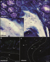
Fig. 5:. evidence of bacterial translocation in large intestine of golden hamsters infected with Leishmania infantum. (A) Histological sections of large intestine (colon) stained with Giemsa and (B and C) Fluorescence microscopy using DAPI and fluorescence in situ hybridisation (FISH) for analysis of bacterial invasion in the submucosa layer. (A) Infected-hamster tissue with L. infantum showing mucosa, submucosa, and muscular layer, with bacteria in the submucosa. Detail of the showing cocci-shaped and bacilli bacteria in the submucosa. (B and C) Fluorescence microscopy showing bacteria (green) and host tissue (DAPI). (B) Sections of colon from uninfected hamster with absent bacteria. (C) Leishmania-infected tissue showing bacteria in the mucosa, and invasion of bacteria in the submucosa. Mu: mucosa; Sm: submucosa; M: muscularis; arrow, bacteria. Scale bar: A - 20µm; detail of A - 10µm; B and C - 50µm.
Fig. 6:
Fig. 6:. prospective evaluation of plasma lipopolysaccharide (LPS) levels in golden hamsters infected with Leishmania infantum. (A) Assessment of LPS levels in uninfected hamsters (G1) and infected-untreated animals (G2) that were followed over 101-120 days post-infection (dpi) and (B) in all experimental groups followed at 60 and 101-120 days post-infection. At 45 days post-infection, the treatment of the animals was started with the respective drugs of each group. G1: uninfected animal (red circle), G2: infected and untreated (green square), G3, G4 and G5: infected and treated with antimonial (purple triangle), amikacin (blue inverted triangle) or both drugs (orange diamond), respectively. Each point in the figure represents an animal, and the horizontal bar represents the median of the values. *p < 0.05 [t test, non-parametric, Mann-Whitney test or analysis of variance (ANOVA), non-parametric, Kruskal-Wallis test ].
Fig. 7:
Fig. 7:. prospective evaluation of total IgG anti-Leishmania levels in golden hamsters infected with Leishmania infantum. (A) IgG levels were measured by enzyme-linked immunosorbent assay (ELISA) and performed in unifected (G1) and infected-untreated animals (G2) that were followed over 101-120 days post-infection (dpi) and (B) in all experimental groups followed at 60 and 101-120 days post-infection. At 45 days post-infection, the treatment of the animals was started with the respective drugs of each group. G1 (red cicle), G2: infected and untreated (green square), G3, G4 and G5: infected and treated with antimonial (purple triangle), amikacin (blue inverted triangle) or both drugs (orange diamond), respectively. Each point in the figure represents an animal, and the horizontal bar represents the median of the values. *p < 0.05 [Analysis of variance (ANOVA), non-parametric, Kruskal-Wallis test ].
Fig. 8:
Fig. 8:. prospective evaluation of the CD4 T-cell levels (A and C) and cellular activation degree (B and D) in golden hamsters infected with Leishmania (L.) infantum. The percentages of CD4+ in PBMC (A and C) and CD25+ on T CD4+ lymphocytes (B and D) was obtained by flow cytometry and were evaluated in unifected (G1) and Leishmania infected-untreated animals (G2) that were followed of 72 hours up to 101-120 days post-infection (dpi) (A-B). The same evaluation was performed in all experimental groups that followed at 60 and 101-120 dpi (C-D). At 45 dpi, the treatment of the animals was started with the respective drugs of each group. G1: uninfected (red circle), G2: infected and untreated (green square), G3, G4 and G5: infected and treated with antimonial (purple triangle), amikacin (blue inverted triangle) or both drugs (orange diamond), respectively. Each point in the figure represents an animal, and the horizontal bar represents the median of the values. *p < 0.05 [t test, non-parametric, Mann-Whitney test or analysis of variance (ANOVA), non-parametric, Kruskal-Wallis test ]. Sample acquisition was performed using the FACSCalibur cytometer and analysis was performed using the CellQuest™ software.
Fig. 9:
Fig. 9:. schematic representation of the bacterial translocation in experimental visceral leishmaniasis (VL) and its systemic consequences for the immunopathogenesis of the disease and the impact of combined therapy. The figure represents an animal infected with Leishmania infantum presenting intestinal damage (duodenum and colon) resulting from the parasitism and inflammatory response itself. The increase in intestinal permeability culminates in translocation of bacterial products from intestinal lumen to the bloodstream, such as LPS from Gram (-) bacteria, resulting in systemic disturbances such as increased cellular activation, increased levels of anti-Leishmania IgG and decreased CD4+ T lymphocyte counts. Our hypothesis is that this scenario can be alleviated by combined anti-Leishmania and antibiotic therapy, leading to blocking the clinical progression of VL.

References

    1. van Griensven J, Diro E. Visceral leishmaniasis. Infect Dis Clin North Am. 2012;26(2):309–322. doi: 10.1016/j.idc.2012.03.005. - DOI - PubMed
    1. Alvar J, Vélez ID, Bern C, Herrero M, Desjeux P, Cano J. Leishmaniasis worldwide and global estimates of its incidence. PLoS One. 2012;7(5):e35671. doi: 10.1371/journal.pone.0035671. - DOI - PMC - PubMed
    1. MS Situação epidemiológica da leishmaniose visceral. Ministério da Saúde. 2022 https://www.gov.br/saude/pt-br/assuntos/saude-de-a-a-z/l/leishmaniose-vi...
    1. Costa CH, Werneck GL, Costa DL, Holanda TA, Aguiar GB, Carvalho AS. Is severe visceral leishmaniasis a systemic inflammatory response syndrome A case control study. Rev Soc Bras Med Trop. 2010;43(4):386–392. doi: 10.1590/s0037-86822010000400010. - DOI - PubMed
    1. Colmenares M, Kar S, Goldsmith-Pestana K, McMahon-Pratt D. Mechanisms of pathogenesis: differences amongst Leishmania species. Trans R Soc Trop Med Hyg. 2002 - PubMed

MeSH terms