. 2025 Sep 9;7(1):100514.
doi: 10.1016/j.xhgg.2025.100514. Online ahead of print.

PATJ deficiency leads to cystic kidney disease and related ciliopathies

Affiliations

PATJ deficiency leads to cystic kidney disease and related ciliopathies

Daniel Epting et al. HGG Adv. .

Abstract

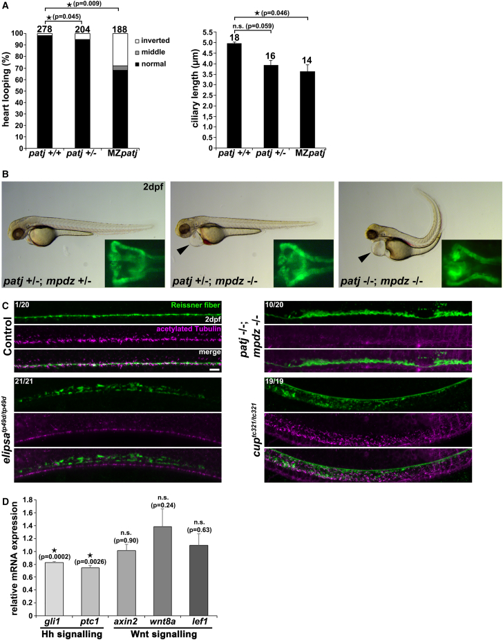
Cystic kidney disease and related ciliopathies are caused by pathogenic variants in genes that commonly result in ciliary dysfunction. For a substantial number of individuals affected by those cilia-related diseases, the causative gene remains unknown. Using massively parallel sequencing, we here identified a pathogenic bi-allelic variant in the gene encoding PALS1-associated tight junction protein ([PATJ] also known as inactivation-no-afterpotential D-like, INADL) in an individual with ciliopathy. The affected fetus carried the homozygous truncating PATJ nonsense variant c.830delC (p.Pro277fsX), and presented with a syndromic phenotype mainly characterized by polycystic kidney disease and hydrocephalus. Using zebrafish (Danio rerio) as a vertebrate in vivo model organism, we could validate our patient findings and demonstrated a ciliopathy phenotype. In addition, we were able to address a hitherto not described role of Patj for cilia formation and function. Taken together, with the Crumbs cell polarity complex member PATJ, we add a new member to the large family of ciliopathy-related human disease proteins that is different from the classical ciliopathy protein classes, and may offer new perspectives for drug development.

Keywords: MPDZ; MUPP1; Reissner fiber; cilia; ciliopathy; hydrocephalus; massively parallel sequencing; morpholino; polycystic kidney disease; zebrafish.


Conflict of interest statement

Declaration of interests The authors declare no competing interests.

Figures

Figure 1
Figure 1
Identification of bi-allelic PATJ variants in an affected individual (A) Pedigree for family with PATJ genetic variant c.830delC (p.Pro277fsX). Healthy parents and affected individual are shown in white and black boxes, respectively. PATJ genetic changes are shown below the symbol of the affected individual. The affected pregnancy was prenatally terminated (black symbol with transverse line); an autopsy was not performed. (B) Schematic of human PATJ comprising 43 exons. Human PATJ encodes for a 1,801 amino acid protein with a single L27 domain and 10 PDZ domains (GenPept: NP_795352.3). The site of human PATJ variant identified is located in the second PDZ domain.
Figure 2
Figure 2
Expression analyses reveal specific expression of patj during embryogenesis and in adult organs in zebrafish (A and B) Analyses of patj expression via semi-quantitative RT-PCR on cDNA of different embryonic developmental stages (A) or adult organs (B) in zebrafish, respectively. H2O served as negative control and ef1α as loading control. PD, primer dimer. (C and D) Analyses of patj expression via WISH at 1 and 2dpf; pronephric tubules (black arrowheads), neuronal cell populations in the spinal cord (white arrowheads), otic vesicle (white arrow), hindbrain (black arrow), and notochord (n). Embryos are shown from lateral with anterior to the left.
Figure 3
Figure 3
Knockdown of Patj results in ciliopathy-associated phenotypes in zebrafish (A) Representative bright-field images of Co-MO- and patj-MO1-injected embryos at 2dpf; hydrocephalus (black arrowhead). Embryos are shown from lateral with anterior to the left. Quantification of hydrocephalus formation, ventral body curvature, and altered heart looping (analyzed as normal, middle (unlooped), and inverted) of Co-MO- and patj-MO1-injected embryos at 2dpf. (B) Quantification of WISH-analyzed Co-MO- and patj-MO1-injected embryos at 18 somites (S) using southpaw (spaw) as LR asymmetry marker. Spaw expression was analyzed in respect to its localization in the embryo as normal (on the left side, black arrowhead), bilateral (on both sides, black arrowheads), inverted (on the right side, black arrowhead), and absent (no expression). Embryos are shown from dorsal with anterior to the top. (C) Quantification of WISH-analyzed Co-MO- and patj-MO1-injected embryos at 2dpf using foxa3 as LR asymmetry marker. Foxa3 expression was analyzed in respect to its localization in the embryo as normal (liver [black arrowhead] on the left side, pancreas [white arrowhead] on the right side and normal looping of the intestine [black arrow]), bilateral (liver, pancreas, and intestine in the middle of embryonic axis), and inverted (liver [black arrowhead] on the right side, pancreas [white arrowhead] on the left side, and reversed looping of the intestine [black arrow]). Embryos are shown from dorsal with anterior to the left. (D) Representative confocal images of the Kupffer’s vesicle of Co-MO- and patj-MO1-injected embryos at the stage of 8S immunostained with anti-acetylated tubulin as a ciliary marker. Scale bar, 10 μm. Quantification of the ciliary length in the Kupffer’s vesicle of Co-MO- and patj-MO1-injected embryos at 8S. Number of embryos used for analyses are shown above respective bar. Data were analyzed by Student’s t test (2-sided, unpaired); error bars represent the standard error of the mean (SEM). A p value of <0.05 was considered statistically significant.
Figure 4
Figure 4
CRISPR-Cas9-induced patj and patj;mpdz zebrafish mutants display ciliopathy-associated phenotypes (A) Quantification of altered heart looping (analyzed as normal, middle (unlooped) and inverted), and of the ciliary length in the Kupffer’s vesicle of wild-type (patj +/+), heterozygous patj mutants (patj +/−), and MZpatj mutants at 2dpf and 8S, respectively. Number of embryos used for analyses are shown above respective bars. Data were analyzed by Student’s ttest (2-sided, unpaired); error bars represent the SEM. A p value of <0.05 was considered statistically significant. (B) Representative bright-field images of a homozygous mpdz mutant (patj +/−; mpdz −/−) displaying pericardial edema (black arrowhead) and of a double homozygous patj;mpdz mutant (patj −/−; mpdz −/−) displaying pericardial edema (black arrowhead) and dorsal body curvature compared with a respective control clutch embryo (patj +/−; mpdz +/−) at 2dpf. Quantitative analysis from randomly selected control clutch embryos (without phenotype) revealed following genotypes: 3x (patj +/+; mpdz +/+), 3x (patj +/+; mpdz +/−), 5x (patj +/−; mpdz +/+), 5x (patj +/−; mpdz +/−), 2x (patj −/−; mpdz +/+) and 6x (patj −/−; mpdz +/−). Quantitative analysis from randomly selected embryos displaying pericardial edema revealed following genotypes: 9x (patj +/+; mpdz −/−) and 15x (patj +/−; mpdz −/−). Quantitative analysis from randomly selected embryos displaying pericardial edema and dorsal body curvature revealed following genotypes: 24x (patj −/−; mpdz −/−). The respective inserts show a fluorescent image of EGFP expression of the same embryo (dorsal view) indicating no detectable pronephric cyst formation for all three different genotypes. Embryos are shown from lateral with anterior to the left. (C) Representative confocal images of cuptc321/tc321, elipsatp49d/tp49d, patj −/−; mpdz −/− mutant embryos and respective control sibling embryos at 2dpf immunostained with anti-RF and anti-acetylated tubulin as a ciliary marker. Numbers represent embryos displaying RF disorganization and embryos that have been analyzed in total. Scale bar, 10 μm. (D) Quantitative RT-PCR analyses reveal unaltered expression of Wnt signaling components axin2, wnt8a, and lef1 while Hh signaling components gli1 and ptc1 were significantly downregulated in double homozygous patj;mpdz mutant embryos compared with control sibling embryos at 2dpf (E−ΔΔCT, normalized to control samples for all genes). Data were analyzed with Graphpad Prism software and one sample t test; error bars represent the SEM.

References

    1. Reiter J.F., Leroux M.R. Genes and molecular pathways underpinning ciliopathies. Nat. Rev. Mol. Cell Biol. 2017;18:533–547. doi: 10.1038/nrm.2017.60. - DOI - PMC - PubMed
    1. Bazellieres E., Aksenova V., Barthelemy-Requin M., Massey-Harroche D., Le Bivic A. Role of the Crumbs proteins in ciliogenesis, cell migration and actin organization. Semin. Cell Dev. Biol. 2018;81:13–20. doi: 10.1016/j.semcdb.2017.10.018. - DOI - PubMed
    1. Omori Y., Malicki J. oko meduzy and related crumbs genes are determinants of apical cell features in the vertebrate embryo. Curr. Biol. 2006;16:945–957. doi: 10.1016/j.cub.2006.03.058. - DOI - PubMed
    1. Hazime K., Malicki J.J. Apico-basal Polarity Determinants Encoded by crumbs Genes Affect Ciliary Shaft Protein Composition, IFT Movement Dynamics, and Cilia Length. Genetics. 2017;207:1041–1051. doi: 10.1534/genetics.117.300260. - DOI - PMC - PubMed
    1. Whiteman E.L., Fan S., Harder J.L., Walton K.D., Liu C.J., Soofi A., Fogg V.C., Hershenson M.B., Dressler G.R., Deutsch G.H., et al. Crumbs3 is essential for proper epithelial development and viability. Mol. Cell Biol. 2014;34:43–56. doi: 10.1128/MCB.00999-13. - DOI - PMC - PubMed

LinkOut - more resources