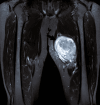
BCOR::CCNB3 Sarcoma of the Thigh: A Rare Case
- PMID: 40932977
- PMCID: PMC12417219
- DOI: 10.7759/cureus.89654
BCOR::CCNB3 Sarcoma of the Thigh: A Rare Case
Abstract
BCOR-rearranged sarcoma is a rare mesenchymal tumor and a recognized subtype of undifferentiated small round-cell sarcoma. It shares morphological similarities with other round-cell sarcomas but is distinguished by a unique molecular hallmark that differentiates it from Ewing sarcoma. These tumors primarily arise in bones and soft tissues. This case report details the case of an 11-year-old female who developed swelling on the medial aspect of her right distal thigh. There was no history of trauma, surgery, or fracture. Clinically, the presentation initially suggested osteosarcoma. However, histopathological evaluation confirmed a diagnosis of BCOR-rearranged sarcoma. This case highlights the importance of considering BCOR-rearranged sarcoma in the differential diagnosis of pediatric bone and soft tissue tumors, as its clinical and radiological features can mimic more common malignancies like osteosarcoma. Early recognition and accurate molecular diagnosis are crucial for guiding appropriate treatment.
Keywords: bcor-ccnb3; clinical pathology; ewing-like sarcoma; immunohistochemistry(ihc); sarcoma soft tissue.
Copyright © 2025, Khan et al.
Conflict of interest statement
Human subjects: Informed consent for treatment and open access publication was obtained or waived by all participants in this study. Conflicts of interest: In compliance with the ICMJE uniform disclosure form, all authors declare the following: Payment/services info: All authors have declared that no financial support was received from any organization for the submitted work. Financial relationships: All authors have declared that they have no financial relationships at present or within the previous three years with any organizations that might have an interest in the submitted work. Other relationships: All authors have declared that there are no other relationships or activities that could appear to have influenced the submitted work.
Figures


References
-
- BCOR-CCNB3 rearranged sarcoma arising in neck misdiagnosed as thyroid cancer: a case report. Tan LC, Yu PC, Wang J, Zhou XY, Ji QH, Wang YL. Oral Oncol. 2021;120:105290. - PubMed
-
- Diagnostic classification of soft tissue malignancies: a review and update from a surgical pathology perspective. Cloutier JM, Charville GW. Curr Probl Cancer. 2019;43:250–272. - PubMed
-
- Updates on WHO classification for small round cell tumors: Ewing sarcoma vs. everything else. Dehner CA, Lazar AJ, Chrisinger JS. Hum Pathol. 2024;147:101–113. - PubMed
Publication types
LinkOut - more resources
Full Text Sources
