. 2025 Sep 14;31(34):109825.
doi: 10.3748/wjg.v31.i34.109825.

B cell CLL/lymphoma 10 promotes colorectal cancer cell proliferation and regulates cuproptosis sensitivity through the NF-κB signaling pathway

Affiliations

B cell CLL/lymphoma 10 promotes colorectal cancer cell proliferation and regulates cuproptosis sensitivity through the NF-κB signaling pathway

Peng-Tuo Xiao et al. World J Gastroenterol. .

Abstract

Background: Colorectal cancer (CRC) is a major global health burden. B cell CLL/lymphoma 10 (BCL10), a key component of the caspase recruitment domain protein-BCL10-mucosa-associated lymphoid tissue lymphoma paracaspase complexes, is upregulated in CRC and associated with poor patient prognosis, suggesting its potential role in CRC development and progression. Cuproptosis, a novel form of programmed cell death, has emerged as a promising therapeutic strategy for cancer.

Aim: To explore the role of BCL10 in regulating the sensitivity of CRC cells to cuproptosis.

Methods: A series of in vitro and in vivo experiments were conducted using CRC cell lines and CRC mouse models to evaluate the effects of BCL10 on CRC cell proliferation, migration, invasion, and sensitivity to copper-induced cell death. Mechanistic studies were performed to elucidate the underlying molecular pathways.

Results: BCL10 promoted CRC cell proliferation, migration, and invasion, while its knockdown had the opposite effects. BCL10 also influenced the sensitivity of CRC cells to cuproptosis, with BCL10 overexpression enhancing resistance and its knockdown increasing sensitivity. The mechanism involved BCL10 modulating the expression of DLAT, a key protein in the copper-induced cell death pathway, through activation of the nuclear factor kappa-B (NF-κB) signaling pathway.

Conclusion: BCL10 promotes CRC growth and regulates the sensitivity of CRC cells to cuproptosis by activating the NF-κB signaling pathway and modulating DLAT expression. These findings provide a molecular basis for developing BCL10-targeted therapies for CRC.

Keywords: B cell CLL/lymphoma 10; Cell death; Colorectal cancer; Cuproptosis; Nuclear factor kappa-B.


Conflict of interest statement

Conflict-of-interest statement: All the authors report no relevant conflicts of interest for this article.

Figures

Figure 1
Figure 1
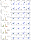
B-cell CLL/Lymphoma 10 promotes the proliferation of colorectal cancer cells in vitro and in vivo. A and B: Colony formation assays showing proliferation ability in SW480 and SW620 cell lines with BCL10 knockdown (SH-BCL10) or overexpression (OE-BCL10). NC1: Scrambled shRNA control; NC2: Empty vector control. cP < 0.001; C: Western blot analysis of MYC and Ki67 expression. bP < 0.01; cP < 0.001; D: Immunohistochemical staining of MYC and Ki67 in xenograft tumors. Scale bars: 50 μm (main panels, 20 × magnification); 20 μm (insets, 40 × magnification); E: EdU assay for cell proliferation detection. Scale bars: 50 μm.
Figure 2
Figure 2
B-cell CLL/Lymphoma 10 promotes invasion and migration without affecting baseline apoptosis. A-C: Wound healing assays showing migration ability in SW480 and SW620 cells. cP < 0.001; D-G: Transwell invasion assays with representative images (D and F) and quantification (E and G). cP < 0.001; H and I: Flow cytometry analysis of apoptosis using Annexin V/PI staining.
Figure 3
Figure 3
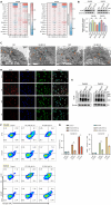
B-cell CLL/Lymphoma 10 regulates sensitivity to cuproptosis. A: Cell viability after elesclomol-Cu treatment (CCK-8 assay); B and C: Flow cytometry analysis of cell death using 7-AAD and Annexin V staining. TTM: Tetrathiomolybdate. bP < 0.01; cP < 0.001.
Figure 4
Figure 4
B-cell CLL/Lymphoma 10 affects cuproptosis sensitivity through DLAT regulation. A: Quantitative PCR analysis of cuproptosis-related genes; B: Western blot of B-cell CLL/Lymphoma 10 (BCL10) and DLAT expression. aP < 0.05; bP < 0.01; C: TEM images of mitochondrial morphology (orange arrows: Damaged mitochondria with cristae disruption); D: Immunofluorescence of indicated proteins after elesclomol-Cu treatment; E: Non-reducing Western blot of DLAT oligomers; F and G: Flow cytometry after DLAT knockdown. cP < 0.001.
Figure 5
Figure 5
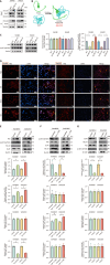
B-cell CLL/Lymphoma 10 regulates DLAT through NF-κB p65 phosphorylation. A: Co-IP of DLAT and B-cell CLL/Lymphoma 10 (BCL10); B: AlphaFold3 predicted binding model (ipTM < 0.6); C: Western blot of p65 and p-p65 (Ser536). cP < 0.001; D: Immunofluorescence of p65 (red) and DAPI (blue); orange arrows indicate nuclear translocation; E: Western blot after p65 transfection in BCL10-knockdown cells. cP < 0.001; F: Western blot after BAY 11-7082 treatment. aP < 0.05. bP < 0.01. cP < 0.001; G: Western blot after DLAT knockout. cP < 0.001.
Figure 6
Figure 6
In vivo effects of B-cell CLL/Lymphoma 10 on tumor growth and DLAT/p65 signaling. A: Experimental timeline; B-G: Tumor volume (B, D, E) and weight (C, F, G) measurements. aP < 0.05; bP < 0.01; cP < 0.001; H: Hematoxylin and eosin staining and IHC for BCL10, DLAT, p65, and p-p65 (Ser536). Scale bars: 50 μm (main panels, 20 × magnification); 20 μm (insets, 40 × magnification).
Figure 7
Figure 7
B-cell CLL/Lymphoma 10 modulates copper treatment sensitivity in vivo. A: Experimental timeline; B: Tumor volume and mouse weight changes; C-F: Quantification of tumor volume (C and D) and weight (E and F). cP < 0.001; G: Tumor growth inhibition rate. bP < 0.01.

References

    1. Bray F, Laversanne M, Sung H, Ferlay J, Siegel RL, Soerjomataram I, Jemal A. Global cancer statistics 2022: GLOBOCAN estimates of incidence and mortality worldwide for 36 cancers in 185 countries. CA Cancer J Clin. 2024;74:229–263. - PubMed
    1. Sun L, Xing J, Zhou X, Song X, Gao S. Wnt/β-catenin signalling, epithelial-mesenchymal transition and crosslink signalling in colorectal cancer cells. Biomed Pharmacother. 2024;175:116685. - PubMed
    1. Nguyen LH, Goel A, Chung DC. Pathways of Colorectal Carcinogenesis. Gastroenterology. 2020;158:291–302. - PMC - PubMed
    1. Li B, Ming H, Qin S, Zhou L, Huang Z, Jin P, Peng L, Luo M, Zhang T, Wang K, Liu R, Liou YC, Nice EC, Jiang J, Huang C. HSPA8 Activates Wnt/β-Catenin Signaling to Facilitate BRAF V600E Colorectal Cancer Progression by CMA-Mediated CAV1 Degradation. Adv Sci (Weinh) 2024;11:e2306535. - PMC - PubMed
    1. Li J, Li S, Xing X, Liu N, Lai S, Liao D, Li J. FTO-mediated ZNF687 accelerates tumor growth, metastasis, and angiogenesis in colorectal cancer through the Wnt/β-catenin pathway. Biotechnol Appl Biochem. 2024;71:245–255. - PubMed

MeSH terms