

A novel sialylation pathway mediated by extracellular vesicles in aggressive prostate cancer
- PMID: 40938891
- PMCID: PMC12431281
- DOI: 10.1371/journal.pone.0329014
A novel sialylation pathway mediated by extracellular vesicles in aggressive prostate cancer
Abstract
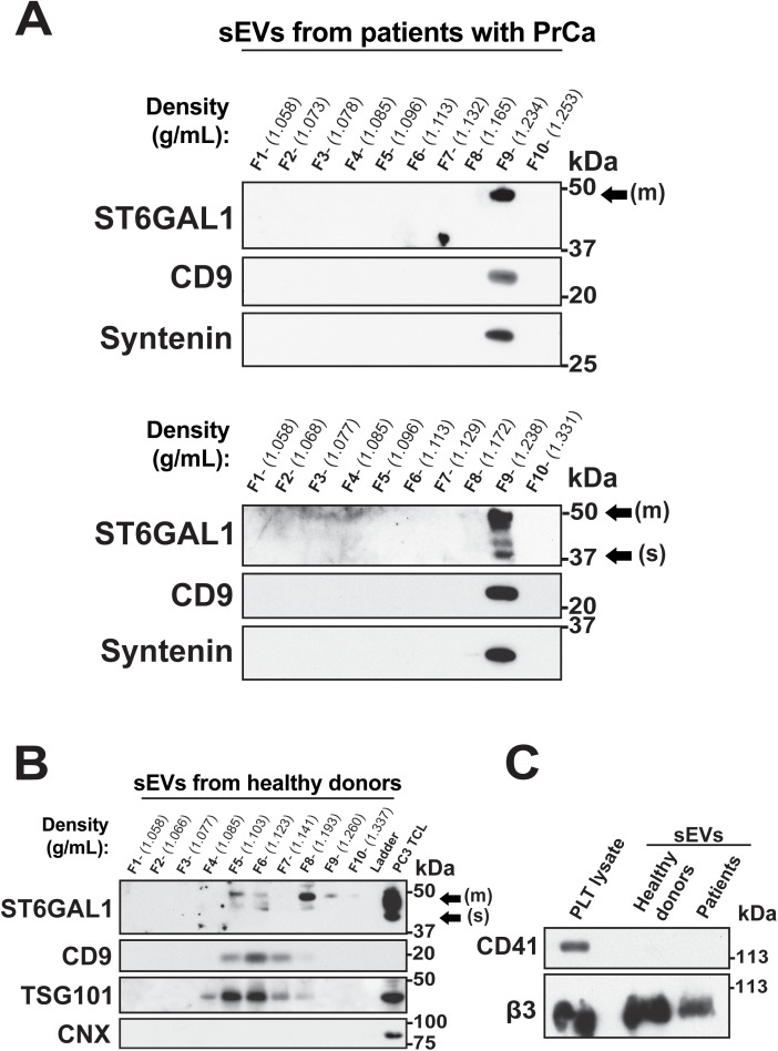
Altered cell surface glycosylation is a hallmark of cancer; among aberrant glycan structures, hypersialylated proteins contribute to disease progression. The enzyme ST6 β-galactoside α2,6-sialyltransferase 1 (ST6GAL1) mediates α2,6-linked sialylation of N-glycosylated proteins and is upregulated in many cancers, including prostate cancer (PrCa). We propose that ST6GAL1 may be released by cancer cells in small extracellular vesicles (sEVs) in the PrCa tumor microenvironment to potentially modulate cell surface sialylation in recipient cells. We isolated sEVs from PrCa cells by density gradient separation and characterized them by nanoparticle tracking analysis using ZetaView and immunoblotting analysis. We identified ST6GAL1 in both its membrane-bound and soluble forms, both active, in circulating sEVs from healthy donors and patients with PrCa. ST6GAL1 is also expressed in human PrCa cells (PC3, DU145, and C4-2B), and in murine cells (TRAMP-C2 and RM1) at different levels, which correlate with aggressive cell phenotypes. In addition to classic sEV markers, such as CD9, TSG101 and Syntenin, sEVs isolated from PrCa cell lines express PDL1, an immune checkpoint ligand. The soluble ST6GAL1 form is present in the sEVs released from DU145 and PC3 cells and can be transferred via sEVs to recipient PrCa cells. This transfer is prevented by expression of Nogo-66 receptor homolog 2 (NgR2) and β3 integrin, which are elevated in the aggressive neuroendocrine phenotype of the disease. The soluble form is absent in the sEVs released from the bone metastatic line C4-2B, which only contains the membrane-bound form. Our results suggest that ST6GAL1 in sEVs derived from PrCa cells may potentially play a role in promoting bone metastasis by facilitating the formation of the pre-metastatic niche.
Copyright: © 2025 Bach et al. This is an open access article distributed under the terms of the Creative Commons Attribution License, which permits unrestricted use, distribution, and reproduction in any medium, provided the original author and source are credited.
Conflict of interest statement
Competing interests N.M. Naranjo and C.E. Verrillo are currently employees of private companies, which has had no influence on this work. No disclosures were reported by other authors.
Figures


References
-
- Swindall AF, Londoño-Joshi AI, Schultz MJ, Fineberg N, Buchsbaum DJ, Bellis SL. ST6Gal-I protein expression is upregulated in human epithelial tumors and correlates with stem cell markers in normal tissues and colon cancer cell lines. Cancer Res. 2013;73(7):2368–78. doi: 10.1158/0008-5472.CAN-12-3424 - DOI - PMC - PubMed
MeSH terms
Substances
LinkOut - more resources
Full Text Sources
Medical
Research Materials
Miscellaneous
