Review
. 2025 Sep 6;14(17):1393.
doi: 10.3390/cells14171393.

Integrating Macrophages into Human-Engineered Cardiac Tissue

Affiliations
Review

Integrating Macrophages into Human-Engineered Cardiac Tissue

Yi Peng Zhao et al. Cells. .

Abstract

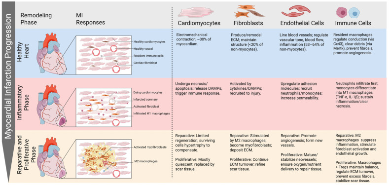
Heart disease remains a leading cause of morbidity and mortality worldwide, necessitating the development of in vivo models for therapeutic development. Advances in biomedical engineering in the past decade have led to the promising rise of human-based engineered cardiac tissues (hECTs) using novel scaffolds and pluripotent stem cell derivatives. This has led to a new frontier of human-based models for improved preclinical development. At the same time, there has been significant progress in elucidating the importance of the immune system and, in particular, macrophages, particularly during myocardial injury. This review summarizes new methods and findings for deriving macrophages from human pluripotent stem cells (hPSCs) and advances in integrating these cells into cardiac tissue. Key challenges include immune cell infiltration in 3D constructs, maintenance of tissue architecture, and modeling aged or diseased cardiac microenvironments. By integrating immune components, hECTs can serve as powerful tools to unravel the complexities of cardiac pathology and develop targeted therapeutic strategies.

Keywords: cardiac; engineered heart tissue; iPS; immune cell; macrophage.


Conflict of interest statement

The authors declare no conflicts of interest.

Figures

Figure 1
Figure 1
Phases of myocardial infarction and the role of each cell type. Cardiac remodeling after myocardial infarction progresses through inflammatory, reparative, and proliferative phases. Cardiomyocytes undergo necrosis and release DAMPs, activating immune responses. Neutrophils and M1 macrophages dominate the early inflammatory stage, while fibroblasts and endothelial cells contribute to scar formation and angiogenesis. During the reparative/proliferative phases, M2 macrophages and Tregs suppress inflammation, promote ECM deposition, and stabilize neovessels, ensuring balanced healing and scar maturation.
Figure 2
Figure 2
Function of macrophages during myocardial infarction. During myocardial infarction, resident macrophages and circulating monocytes first polarize into M1 macrophages, producing inflammatory cytokines in the early inflammatory phase. As repair progresses, macrophages transition to the M2 phenotype, which resolves inflammation, stimulates fibroblast activity, and promotes angiogenesis during the reparative and proliferative phases.
Figure 3
Figure 3
iPSC-derived macrophages: monolayer 2D cell culture. Schematic representation of the generation of macrophages from pluripotent stem cells using monolayer 2D cell culture method. iPSCs are cultured on Matrigel in feeder-free conditions to generate macrophages. The process includes mesoderm induction (BMP4, Activin A, CHIR99021), hematopoietic specification (VEGF, bFGF, SCF), progenitor expansion (VEGF, SCF, IL-3, IL-6), and macrophage differentiation and maturation (M-CSF, IL-34). Resulting cells express CD11b, IBA1, CD14, CD68, TMEM119, and P2RY12, resembling primary human macrophages.
Figure 4
Figure 4
hPSC-derived macrophages: co-culture with stromal cells. Schematic representation of the generation of macrophages from pluripotent stem cells using co-culture with stromal cells method. hPSCs are seeded onto a confluent OP9 stromal layer to induce mesoderm and hematopoietic specification (Day 0–9). Hematopoietic progenitors are harvested and cultured with M-CSF (±GM-CSF) to promote macrophage lineage commitment (after Day 12), followed by final maturation (after Day 21).
Figure 5
Figure 5
hPSC-derived macrophages: co-culture with stromal cells. Schematic representation of the generation of macrophages from pluripotent stem cells using embryoid body (EB) formation method. Stepwise differentiation of macrophages from human pluripotent stem cells (hPSCs) via embryoid body (EB) formation. The process mimics yolk sac hematopoiesis and includes mesoderm induction (BMP4, VEGF, SCF, FGF2), hematopoietic specification (VEGF, SCF, FLT3L), progenitor expansion (IL-3, M-CSF, SCF, FLT3L), macrophage differentiation (M-CSF, IL-34), and terminal maturation. Mature macrophages express canonical markers (CD11b, CD14, CD68, CD163), demonstrate phagocytic activity, and share MYB-independent ontogeny with yolk-sac-derived tissue-resident macrophages.

References

    1. Thygesen K., Alpert J.S., Jaffe A.S., Chaitman B.R., Bax J.J., Morrow D.A., White H.D., The Executive Group on behalf of the Joint European Society of Cardiology (ESC)/American College of Cardiology (ACC)/American Heart Association (AHA)/World Heart Federation (WHF) Task Force for the Universal Definition of Myocardial Infarction Fourth Universal Definition of Myocardial Infarction. Circulation. 2018;138:e618–e651. doi: 10.1161/CIR.0000000000000617. - DOI - PubMed
    1. Martin S.S., Aday A.W., Almarzooq Z.I., Anderson C.A.M., Arora P., Avery C.L., Baker-Smith C.M., Barone Gibbs B., Beaton A.Z., Boehme A.K., et al. 2024 Heart Disease and Stroke Statistics: A Report of US and Global Data From the American Heart Association. Circulation. 2024;149:e347–e913. doi: 10.1161/CIR.0000000000001209. - DOI - PMC - PubMed
    1. Salari N., Morddarvanjoghi F., Abdolmaleki A., Rasoulpoor S., Khaleghi A.A., Hezarkhani L.A., Shohaimi S., Mohammadi M. The global prevalence of myocardial infarction: A systematic review and meta-analysis. BMC Cardiovasc. Disord. 2023;23:206. doi: 10.1186/s12872-023-03231-w. - DOI - PMC - PubMed
    1. Pinto A.R., Ilinykh A., Ivey M.J., Kuwabara J.T., D’Antoni M.L., Debuque R., Chandran A., Wang L., Arora K., Rosenthal N.A., et al. Revisiting Cardiac Cellular Composition. Circ. Res. 2016;118:400–409. doi: 10.1161/CIRCRESAHA.115.307778. - DOI - PMC - PubMed
    1. Litviňuková M., Talavera-López C., Maatz H., Reichart D., Worth C.L., Lindberg E.L., Kanda M., Polanski K., Heinig M., Lee M., et al. Cells of the adult human heart. Nature. 2020;588:466–472. doi: 10.1038/s41586-020-2797-4. - DOI - PMC - PubMed

LinkOut - more resources