Panretinal Congenital Hypertrophy of the RPE in an 8-Year-Old Girl with an X-Linked STAG2 Mutation
- PMID: 40943870
- PMCID: PMC12429593
- DOI: 10.3390/jcm14176110
Panretinal Congenital Hypertrophy of the RPE in an 8-Year-Old Girl with an X-Linked STAG2 Mutation
Abstract
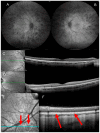
Introduction: Congenital hypertrophy of the retinal pigment epithelium (CHRPE) is a benign proliferation of the melanin-producing retinal pigment epithelium (RPE). Although often a benign and incidental finding, multifocal CHRPE may mimic lesions associated with familial adenomatous polyposis (FAP). Case Description: We describe an 8-year-old girl presenting with optic disc pallor and widespread multifocal bear track CHRPE observed bilaterally on dilated fundoscopy. Fundus autofluorescence (FAF) imaging showed uniform areas of hypoautofluorescence corresponding to the bear track lesions. Spectral domain optical coherence tomography (SD-OCT) demonstrated normal lamination without atrophy. The full-field electroretinogram (ffERG) was within normal limits. Whole-genome sequencing (WGS) revealed a likely pathogenic heterozygous variant in the STAG2 gene (c.3222dup, p.Ser1075IlefsTer12). Conclusions: We present a rare case of bilateral, panretinal bear track CHRPE in a child with a likely pathogenic variant in STAG2. Using multimodal imaging, we contrast bear track lesions of the retina with FAP-associated CHRPE. We also present possible ophthalmic manifestations in carriers of pathogenic STAG2 variants.
Keywords: autofluorescence; congenital hypertrophy of the retinal pigment epithelium (CHRPE); electroretinogram (ERG); inherited retinal disease (IRD); optical coherence tomography (OCT); stromal antigen 2 (STAG2).
Conflict of interest statement
S.H.T. receives financial support from Abeona Therapeutics, Inc., and Emendo is on the scientific and clinical advisory board for Nanoscope Therapeutics. The authors declare no conflicts of interest.
Figures


References
Publication types
Grants and funding
- Physician Scientist Award/Research to Prevent Blindness
- U01 EY030580/EY/NEI NIH HHS/United States
- 1R24EY027285-21/NH/NIH HHS/United States
- 1U01EY030580-20/NH/NIH HHS/United States
- 1U01EY034590-22/NH/NIH HHS/United States
- R01 EY033770/EY/NEI NIH HHS/United States
- NA/Gebroe Family Foundation
- R01 EY024698/EY/NEI NIH HHS/United States
- R24 EY027285/EY/NEI NIH HHS/United States
- 1R01EY033770-24/NH/NIH HHS/United States
- 1R01EY024698-22/NH/NIH HHS/United States
- NA/NYEE Foundation
- R24 EY028758/EY/NEI NIH HHS/United States
- NA/Lynette and Richard Jaffe Foundation
- U01 EY034590/EY/NEI NIH HHS/United States
- R01 EY018213/EY/NEI NIH HHS/United States
- 1R01EY018213-18/NH/NIH HHS/United States
- NA/Rosenbaum Family Foundation
- TA-GT-0321-0802-COLU-TRAP/FFB/Foundation Fighting Blindness/United States
- 1R24EY028758-22/NH/NIH HHS/United States
LinkOut - more resources
Full Text Sources
Miscellaneous

