Review
. 2025 Sep 4;14(17):6238.
doi: 10.3390/jcm14176238.

Diagnostic Accuracy of Exercise Stress Testing, Stress Echocardiography, Myocardial Scintigraphy, and Cardiac Magnetic Resonance for Obstructive Coronary Artery Disease: Systematic Reviews and Meta-Analyses of 104 Studies Published from 1990 to 2025

Affiliations
Review

Diagnostic Accuracy of Exercise Stress Testing, Stress Echocardiography, Myocardial Scintigraphy, and Cardiac Magnetic Resonance for Obstructive Coronary Artery Disease: Systematic Reviews and Meta-Analyses of 104 Studies Published from 1990 to 2025

Andrea Sonaglioni et al. J Clin Med. .

Abstract

Background: Since the 1990s, numerous investigations have assessed the diagnostic effectiveness-specifically sensitivity, specificity, and accuracy-of exercise stress testing (EST), stress echocardiography (SE), stress myocardial single-photon emission computed tomography (SPECT), and stress cardiac magnetic resonance imaging (CMR). However, the outcomes of these studies have often been inconsistent and inconclusive. To provide a clearer comparison, we conducted systematic reviews and meta-analyses aimed at quantitatively evaluating and comparing the aggregated diagnostic performance of these four commonly used techniques for detecting coronary artery disease (CAD). Methods: A comprehensive search of PubMed, Scopus, Embase, Cochrane Library, and Web of Science was conducted to identify cohort studies evaluating the diagnostic accuracy of EST, SE, stress myocardial SPECT, and stress CMR in symptomatic patients with suspected or confirmed CAD. The main goal was to compare their diagnostic value by pooling sensitivity and specificity results. Each study's data were extracted in terms of true positives, false positives, true negatives, and false negatives. Results: A total of 104 studies, comprising 16,824 symptomatic individuals with either suspected or known CAD, met the inclusion criteria. The pooled sensitivities for CAD detection were 0.66 (95% CI: 0.59-0.72, p < 0.001) for EST, 0.81 (95% CI: 0.79-0.83, p < 0.001) for SE, 0.82 (95% CI: 0.78-0.85, p < 0.001) for stress myocardial SPECT, and 0.83 (95% CI: 0.81-0.85, p < 0.001) for stress CMR. Corresponding specificities were 0.61 (95% CI: 0.55-0.67, p < 0.001), 0.85 (95% CI: 0.82-0.87, p < 0.001), 0.74 (95% CI: 0.70-0.78, p < 0.001), and 0.89 (95% CI: 0.86-0.92, p < 0.001), respectively. Considerable heterogeneity was observed across the studies, as reflected by I2 values ranging from 82.5% to 92.5%. Egger's generalized test revealed statistically significant publication bias (p < 0.05 for all methods), likely due to the influence of smaller studies reporting more favorable results. Despite this, sensitivity analyses supported the overall robustness and reliability of the pooled findings. Conclusions: Among the diagnostic tools assessed, EST demonstrated the lowest accuracy for detecting obstructive CAD, whereas stress CMR exhibited the highest. Although stress myocardial SPECT showed strong sensitivity, its specificity was comparatively limited. SE emerged as the most balanced option, offering good diagnostic accuracy combined with advantages such as broad availability, cost-effectiveness, and the absence of ionizing radiation.

Keywords: coronary artery disease; diagnostic accuracy; exercise stress testing; meta-analysis; myocardial SPECT; stress CMR; stress echocardiography.


Conflict of interest statement

Andrea Sonaglioni, Alessio Polymeropoulos, Massimo Baravelli, Gian Luigi Nicolosi, and Michele Lombardo declare no conflicts of interest. Giuseppe Biondi-Zoccai has consulted, lectured, and/or served as an advisory board member for Abiomed, Advanced Nanotherapies, Aleph, Amarin, AstraZeneca, Balmed, Cardionovum, Cepton, Crannmedical, Endocore Lab, Eukon, Guidotti, Innovheart, Meditrial, Menarini, Microport, Opsens Medical, Synthesa, Terumo, and Translumina, outside of the present work.

Figures

Figure 1
Figure 1
The PRISMA flow diagram used for identifying the included studies. CAD, coronary artery disease; CMR, cardiac magnetic resonance; EST, exercise stress testing; SE, stress echocardiography; and SPECT, single-photon emission computed tomography.
Figure 2
Figure 2
Forest plots illustrating the sensitivity and specificity of exercise stress testing are provided for the overall set of studies [26,27,28,29,30,31,32,33,34,35,36,37,38,39,40,41,42,43,44,45,46,47,48,49,50], as well as for the subgroups comprising female-only participants [31,34,39,40,45,47,48] and mixed-gender [26,27,28,29,30,32,33,35,36,37,38,41,42,43,44,45,46,49,50] cohorts.
Figure 3
Figure 3
(A) Crosshair plot illustrating the results of EST studies conducted in two distinct subgroups—female-only and mixed-gender populations. (B) The SROC curve represents the overall diagnostic performance of EST across all studies (AUC = 0.69; 95% CI: 0.62–0.76), as well as for studies including only women (AUC = 0.65; 95% CI: 0.52–0.80) and those involving both sexes (AUC = 0.71; 95% CI: 0.61–0.78). EST, exercise stress testing; SROC, summary receiver operating characteristic.
Figure 4
Figure 4
Begg’s funnel plot for the detection of publication bias in EST studies.
Figure 5
Figure 5
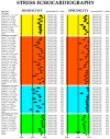
Forest plots illustrating the sensitivity and specificity of SE and its principal clinical applications, including ESE [51,52,53,54,55,56,57,58,59,60], DSE [26,28,29,30,33,36,37,44,53,54,55,61,62,63,64,65,66,67,68,69,70,71,72,73,74], dipSE [29,36,37,41,44,54,55,56,61,65,67,75,76], and dual-imaging SE [77,78,79,80,81,82,83,84]. DipSE, dipyridamole stress echocardiography; DSE, dobutamine stress echocardiography; ESE, exercise stress echocardiography; and SE, stress echocardiography.
Figure 6
Figure 6
(A) Crosshair plot displaying the sensitivity and specificity results from studies evaluating various forms of stress echocardiography, including ESE, DSE, dipSE, and dual-imaging SE. (B) SROC curve illustrating the overall diagnostic accuracy of SE (AUC = 0.85; 95% CI: 0.80–0.91) and the individual performance of each technique: ESE (AUC = 0.84; 95% CI: 0.69–0.91), DSE (AUC = 0.82; 95% CI: 0.66–0.89), dipSE (AUC = 0.71; 95% CI: 0.46–0.86), and dual-imaging SE (AUC = 0.86; 95% CI: 0.75–0.91). DipSE, dipyridamole stress echocardiography; DSE, dobutamine stress echocardiography; ESE, exercise stress echocardiography; SE, stress echocardiography; and SROC, summary receiver operating characteristic.
Figure 7
Figure 7
Begg’s funnel plot for the assessment of publication bias in studies evaluating sensitivity and specificity of SE for detecting obstructive CAD. CAD, coronary artery disease; SE, stress echocardiography.
Figure 8
Figure 8
Forest plots showing the sensitivity and specificity of stress myocardial SPECT and principal stress myocardial scintigraphic techniques adopted in clinical practice: exercise [38,43,46,49,51,52,67,68,69,73,85,86,87,88,89,90,91,92], dobutamine [30,36,37,62,66,72,74,89], dipyridamole [36,63,64,70,76,89,94,95,96], and adenosine [87,94,97,98,99,100,101] myocardial SPECT. SPECT, single-photon emission computed tomography.
Figure 9
Figure 9
(A) Crosshair plot presenting the sensitivity and specificity results from studies evaluating stress myocardial SPECT using exercise, dobutamine, dipyridamole, and adenosine protocols. (B) SROC curve illustrating the overall diagnostic accuracy of stress myocardial SPECT (AUC = 0.88; 95% CI: 0.79–0.92), along with the performance of individual techniques: exercise SPECT (AUC = 0.88; 95% CI: 0.79–0.93), dobutamine SPECT (AUC = 0.85; 95% CI: 0.66–0.94), dipyridamole SPECT (AUC = 0.89; 95% CI: 0.73–0.95), and adenosine SPECT (AUC = 0.75; 95% CI: 0.47–0.91). SPECT, single-photon emission computed tomography; SROC, summary receiver operating characteristic.
Figure 10
Figure 10
Begg’s funnel plot for the assessment of publication bias in studies evaluating sensitivity and specificity of stress myocardial SPECT for predicting obstructive CAD. CAD, coronary artery disease; SPECT, single-photon emission computed tomography.
Figure 11
Figure 11
Forest plots showing the sensitivity and specificity of stress CMR and principal stress CMR methods used in clinical practice: exercise [91,102,103], dobutamine [27,104,105,106,107,108,109,110,111,112], dipyridamole [113,114,115,116,117,118,119,120,121], and adenosine [92,100,101,106,122,123,124,125,126,127,128,129] CMR. CMR, cardiac magnetic resonance.
Figure 12
Figure 12
(A) Crosshair plot for stress CMR studies assessing sensitivity and specificity of exercise CMT, dobutamine CMR, dipyridamole CMR, and adenosine CMR. (B) SROC summarizing the overall performance of stress CMR (AUC = 0.85; 95%CI 0.60–0.94) and of each stress CMR technique: exercise (AUC = 0.74; 95%CI 0.30–0.95), dobutamine (AUC = 0.85; 95%CI 0.55–0.95), dipyridamole (AUC = 0.87; 95%CI 0.53–0.97), and adenosine (AUC = 0.85; 95%CI 0.57–0.95) CMR. CMR, cardiac magnetic resonance; SROC, summary receiver operating characteristic.
Figure 13
Figure 13
Begg’s funnel plot for the assessment of publication bias in studies evaluating sensitivity and specificity of stress CMR for identifying obstructive CAD. CAD, coronary artery disease; CMR, cardiac magnetic resonance.
Figure 14
Figure 14
Posterior estimates of sensitivity of the four methods analyzed (EST, SE, stress SPECT, and stress CMR) for detecting obstructive CAD. CAD, coronary artery disease; CMR, cardiac magnetic resonance; EST, exercise stress testing; SE, stress echocardiography; and SPECT, single-photon emission computed tomography.
Figure 15
Figure 15
Posterior estimates of specificity of the four methods analyzed (EST, SE, stress SPECT, and stress CMR) for detecting obstructive CAD. CAD, coronary artery disease; CMR, cardiac magnetic resonance; EST, exercise stress testing; SE, stress echocardiography; and SPECT, single-photon emission computed tomography.
Figure 16
Figure 16
Innovative screening strategy for symptomatic individuals with suspected CAD. A–P, antero–posterior; CAD, coronary artery disease; CCTA, coronary computed tomography angiography; MHI, modified Haller index; PTP, pre-test probability; and SE, stress echocardiography.

References

    1. GBD 2017 Causes of Death Collaborators Global, regional, and national age-sex-specific mortality for 282 causes of death in 195 countries and territories, 1980–2017: A systematic analysis for the Global Burden of Disease Study 2017. Lancet. 2018;392:1736–1788. doi: 10.1016/S0140-6736(18)32203-7. - DOI - PMC - PubMed
    1. Safiri S., Karamzad N., Singh K., Carson-Chahhoud K., Adams C., Nejadghaderi S.A., Almasi-Hashiani A., Sullman M.J.M., Mansournia M.A., Bragazzi N.L., et al. Burden of ischemic heart disease and its attributable risk factors in 204 countries and territories, 1990–2019. Eur. J. Prev. Cardiol. 2022;29:420–431. doi: 10.1093/eurjpc/zwab213. - DOI - PubMed
    1. Guan C., Wu S., Xu W., Zhang J. Global, regional, and national burden of ischaemic heart disease and its trends, 1990–2019. Public Health. 2023;223:57–66. doi: 10.1016/j.puhe.2023.07.010. - DOI - PubMed
    1. Khan M.A., Hashim M.J., Mustafa H., Baniyas M.Y., Al Suwaidi S.K.B.M., AlKatheeri R., Alblooshi F.M.K., Almatrooshi M.E.A.H., Alzaabi M.E.H., Al Darmaki R.S., et al. Global epidemiology of ischemic heart disease: Results from the Global Burden of Disease Study. Cureus. 2020;12:e9349. doi: 10.7759/cureus.9349. - DOI - PMC - PubMed
    1. Knuuti J., Wijns W., Saraste A., Capodanno D., Barbato E., Funck-Brentano C., Prescott E., Storey R.F., Deaton C., Cuisset T., et al. 2019 ESC Guidelines for the diagnosis and management of chronic coronary syndromes. Eur. Heart J. 2020;41:407–477. doi: 10.1093/eurheartj/ehz425. - DOI - PubMed

LinkOut - more resources