. 2025 Sep 13;74(10):305.
doi: 10.1007/s00262-025-04149-2.

Dual targeting of solid tumors using cytokine-induced killer cells modified with a CAR anti-tenascin C and a secretable EGFRxCD3 bispecific antibody

Affiliations

Dual targeting of solid tumors using cytokine-induced killer cells modified with a CAR anti-tenascin C and a secretable EGFRxCD3 bispecific antibody

Silvia Zaninelli et al. Cancer Immunol Immunother. .

Abstract

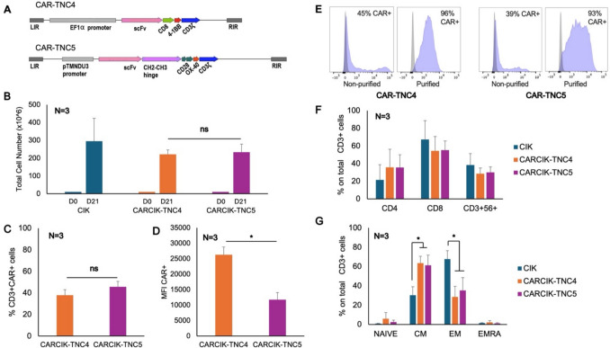
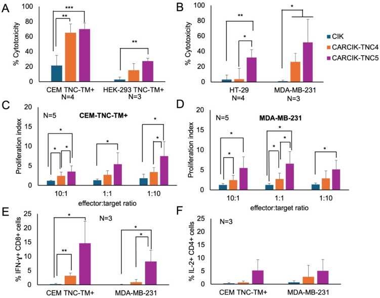
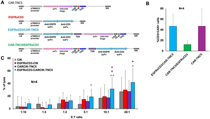
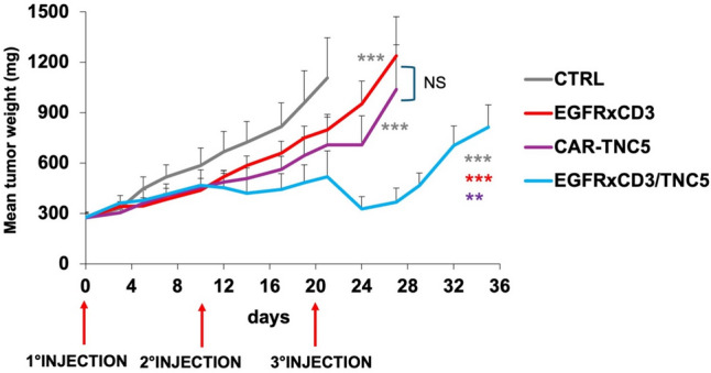
To generate CARCIK cells with enhanced targeting and penetration of solid tumors, we have designed new CAR molecules against tenascin C (TNC), an extracellular matrix and surface molecule, overexpressed in several tumor types. Two different anti-TNC CAR constructs were generated, sharing the anti-TNC scFv domain, fused to the CD3ζ fragment, but differing in co-signaling domains: CAR-TNC4 carrying the 4-1BB and CAR-TNC5 the CD28 and OX40 domains. Both CARs were introduced into cytokine-induced killer cells (CIK) by co-transfection with the sleeping beauty transposase plasmid. CARCIK-TNC cells were cytotoxic against TNC+ targets, proliferated and secreted the IFN-γ and IL-2 cytokines in response to target cell binding, with overall higher efficacy of CARCIK-TNC5 compared to CARCIK-TNC4. To enhance activity and specificity, we also generated a dual construct, carrying a secretable EGFRxCD3 bispecific T cell engager (sBiTE) antibody cDNA. CIK transfected with EGFRxCD3/CAR-TNC5 showed good expression of both molecules and synergistic killing of MDA-MB-231 cells in vitro compared to cells transfected with the single-targeting molecules. Also in vivo, in the subcutaneous TNC+ EGFR+ MDA-MB-231 xenograft model, significantly enhanced control of tumor growth was observed after injection of CIK cells transfected with dual, compared to single constructs. We conclude that CAR-TNC5 combined with EGFRxCD3 can endow CIK cells with different and potentially synergistic functions in vivo.

Keywords: Bispecific T cell engager; Chimeric antigen receptor; Cytokine induced killer; Tenascin C; solid tumor.


Conflict of interest statement

Declarations. Conflict of interests: AR has received honoraria for lectures or presentations from Novartis, Amgen, Pfizer, Astellas, Jazz, Janssen, Incyte, Kite-Gilead, Omeros, Sanofi, Italfarmaco; has received support for attending meetings and/or travel from Novartis, Amgen, Pfizer, Astellas, Jazz, Janssen, Incyte, Kite-Gilead, Omeros; participated on a Data Safety Monitoring Board or Advisory Board from Novartis, Amgen, Pfizer, Astellas, Jazz, Janssen, Incyte, Kite-Gilead, Roche, Omeros, Sanofi, Italfarmaco.

Figures

Fig. 1
Fig. 1
Generation of CARCIK-TNC cells and their characterization. A Schematic structure of the CAR-TNC constructs. B 10 × 106 PBMCs were co-transfected with the CAR-TNC4 and CAR-TNC5 + SB100X plasmids and expanded to CIK for 21 days. Data report total nucleated cells obtained. C-D CAR expression was analyzed at the end of culture by flow cytometry. Percentage CAR positive C and mean fluorescence intensity of CAR D are shown. E Examples of flow cytometry histograms of purified or non-purified CARCIK-TNC4 (MFI 29803 and 2086) or -5 (MFI 8928 and 4054). F-G Immunophenotype of the purified cellular products at the end of the culture, including CD4, CD8 and CD3+CD56+ populations F and effector-memory populations G, analyzed by flow cytometry. The results are the means and standard deviations of 3 experiments using different donors as starting material. The statistics refer to the comparison between CARCIK-TNC4 and CARCIK-TNC5 with unmodified CIK (*p < 0.05)
Fig. 2
Fig. 2
Functional activity of unmodified or CAR-modified CIK cells. A-B Cytotoxicity of unmodified or CAR-modified CIK in vitro was measured against two TNC stably transfected cell lines, at 5:1 E:T ratio for 4 h A or against TNC+ tumor cells lines at 1:1 E:T ratio for 24 h B. C, D Proliferation of CAR-modified, CFSE-stained CIK cells in vitro was measured by flow cytometry after stimulation with CEM TNC-TM+ C or MDA-MB-231 D. E, F IFN-γ E and IL-2 F production was determined by intracytoplasmic staining upon 6 h co-culture at 1:1 E:T ratio of unmodified or CAR-modified CIK cells. (*P < 0.05, **P < 0.01, ***P < 0.001)
Fig. 3
Fig. 3
Generation and characterization of armored CIK cells carrying anti-TNC CAR and secretable EGFRxCD3 bispecific antibody. A Schematic representation of the different constructs used. B TNC CAR expression on CIK cells genetically modified with the different constructs. C In vitro cytotoxic activity of unmodified or CAR/EGFRxCD3 modified CIK against the MDA-MB-231 cell line after 4 h of co-culture (N = 4). *: p ≤ 0.05 and **: p < 0.01. Statistical significance shown refers to armored CIK (i.e., EGFRxCD3/CARCIK-TNC5) against either unmodified CIK (gray *), EGFRxCD3-CIK (red *) or CARCIK-TNC5 (violet *)
Fig. 4
Fig. 4
In vivo therapeutic activity of armored CIK or CIK modified with a single-targeting molecule. NOD-SCID mice were inoculated subcutaneously with MDA-MB-231 tumor cells. When tumor reached ~ 250 mg, mice were randomized and inoculated iv with 10 × 106 gene-modified effector CARCIK cells, 3 times at 10 days intervals, as indicated in the graph. Tumor growth curves are shown. Statistical significance refers to armored CIK (i.e. EGFRxCD3-CARCIK-TNC5) against either EGFRxCD3-CIK (red *) or CARCIK-TNC5 (violet *). Significance was also measured against controls group (gray *). **: p < 0.01 and ***p < 0.001
Fig. 5
Fig. 5
Immunohistochemistry analysis of CIK cells infiltrating the MDA-MB-231 tumor in vivo 24 h after the last effector cells injection. A Analysis of human CD3+ CIK (upper panel) and HNA+ tumor cells (lower panel) on consecutive sections of tumor tissue (original magnification 10x). B Representative images of tumor tissue areas characterized by the presence of human CD3+ CIK cells. Top panels: mosaic reconstruction (original magnification 20x). Bottom panels: high magnification of representative tumor tissue portions (original magnification 40x). C High magnification of representative portions of tumor tissue reveals the presence of secretory granules containing human granzymes B. (original magnification 40x)

References

    1. Sterner RC, Sterner RM (2021) CAR-T cell therapy: current limitations and potential strategies. Blood Cancer J 11:1–11. 10.1038/s41408-021-00459-7 - DOI - PMC - PubMed
    1. June CH, O’Connor RS, Kawalekar OU, Ghassemi S, Milone MC (2018) CAR t cell immunotherapy for human cancer. Science 359:1361–1365. 10.1126/science.aar6711 - DOI - PubMed
    1. Nguyen DT, Ogando-Rivas E, Liu R, Wang T, Rubin J, Jin L, Tao H, Sawyer WW, Mendez-Gomez HR, Cascio M et al (2022) Car T cell locomotion in solid tumor microenvironment. Cells 11(12):1974. 10.3390/cells11121974 - DOI - PMC - PubMed
    1. Guha P, Heatherton KR, O’Connell KP, Alexander IS, Katz SC (2022) Assessing the future of solid tumor immunotherapy. Biomedicines 10:655. 10.3390/biomedicines10030655 - DOI - PMC - PubMed
    1. Xiao Y, Yu D (2021) Tumor microenvironment as a therapeutic target in cancer. Pharmacol Ther 221:107753. 10.1016/j.pharmthera.2020.107753 - DOI - PMC - PubMed

MeSH terms