. 2025 Aug 12;28(9):113343.
doi: 10.1016/j.isci.2025.113343. eCollection 2025 Sep 19.

KLK15 alters connective tissues in hypermobile Ehlers-Danlos syndrome

Affiliations

KLK15 alters connective tissues in hypermobile Ehlers-Danlos syndrome

Cortney Gensemer et al. iScience. .

Abstract

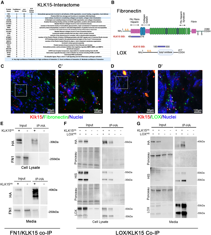
Hypermobile Ehlers-Danlos syndrome (hEDS) is a debilitating multisystem condition characterized by joint hypermobility, chronic pain, and diverse comorbidities, yet its genetic basis remains undefined. Whole-exome sequencing (WES) of 200 patients with hEDS revealed rare and low frequency variants in 14 of 15 kallikrein (KLK) genes, including a recurrent KLK15 missense variant (p.Gly226Asp) segregating in multiple families. KLK15, a secreted serine protease, is expressed in connective and immune tissues and interacts with extracellular matrix (ECM) components, including fibronectin and lysyl oxidase (LOX). A KLK15 knock-in mouse model recapitulated hEDS features in tendons and cardiac valves and exhibited dysregulated cytokine profiles. The variant altered KLK15 and LOX compartmentalization within the ECM, consistent with a dominant-negative effect. These findings identify KLK15 as a contributor to hEDS and reveal broader roles for KLK protease-ECM-immune crosstalk in connective tissue regulation. This study reframes hEDS as a condition involving matrix remodeling and immune signaling beyond collagen defects.

Keywords: Biological process; Body system; Molecular genetics; Non-infectious disease.


Conflict of interest statement

The authors declare no competing interests.

Figures

None
Graphical abstract
Figure 1
Figure 1
Kallikrein-15 enrichment and familial gene variant identification in hEDS (A) Pedigree of a multigenerational family with autosomal dominant hEDS. Black circles and squares represent those with a clinical hEDS diagnosis. Gray indicates those who are probable-hEDS. Unaffected individuals are black and unknown phenotypes and those whose DNA was not available are marked with an asterisk. Circles and squares represent females and males, respectively. WES was performed on the proband (IV-1) and IV-4 (arrow and arrowhead). (B) Chromatogram showing missense KLK15 variant (G/A). (C) Pedigree of family 2 showing genotype of KLK15G226D/+ allele (G/A) in affected I-1 and II-1. (D) KLK15 variants identified in WES analyses of 198 hEDS individuals. Consequence of the variants in changing splicing or coding is represented as are the MAF (gnomAD v2.1.1), SIFT, PolyPhen, and CADD (v1.7) scores.
Figure 2
Figure 2
Expression of KLK15 (A and B) RT-PCR showing KLK15 is expressed by human dermal fibroblasts, anterior cruciate ligament tissue (ACL), and in human mast cells. (C) Expression of Klk15 mRNA is observed in subset of murine tissues including thymus, skin, and Achilles tendon. (D) RNA scope showing positive signal throughout the murine adrenal gland and within the skin. RT (±) is presence or absence (negative control) of reverse transcriptase enzyme. Scale bars, 100 μm.
Figure 3
Figure 3
Identification of KLK15 interacting proteins (A) Results from two-hybrid screens identifying a total of 23 novel protein partners with FN1 and LOX being highlighted as highest priority hits relevant to the extracellular matrix. (B) Schematic of binding region where the KLK15 bait interacts with FN1 and LOX. s.s., signal sequence; SID, smallest interacting domain. (C and D) RNAscope showing punctate signals from all three probes appear in close proximity for KLK15, Fibronectin, and LOX in the skin, suggesting co-expression in the same cells. Scale bars are 30 and 120 μm. C′ and D′ are 4× zoom images of (C) and (D), respectively. (E) CoIP experiments demonstrating that KLK15 interacts with the secreted form of fibronectin. (F and G) CoIP experiments demonstrating that KLK15 interacts with transfected LOX in both cell extracts and media samples.
Figure 4
Figure 4
Pathogenicity of the familial KLK15 variant (A and B) Stress-strain curves from tensile testing of Klk15+/+ (n = 9) and Klk15G224D/+ (n = 8) tendons with a focus on the toe region (B). (C–E) Statistical analysis of transition point displacement (unpaired t test, p = 0.0253, Cohen’s d = −1.21), transition strain (Mann-Whitney U test, p = 0.036, rank-biserial r = −0.051) and toe modulus (unpaired t test, p = 0.0218, Cohen’s d = 1.24) from tensile testing curves. Error bars represent mean ± standard deviation. (F) Representative TEMs of Achilles tendon from Klk15+/+ and Klk15G224D/+ mice showing smaller collagen fibrils in the mutant tendons. (G) Quantification of fibril diameters from wildtype (n = 3,240) and mutant (n = 4,191) tendons showing a significant decrease in average fibril diameter in Klk15G224D/+ (84 vs. 101 nm). Statistical analysis was performed using a Mann-Whitney U test, ∗∗∗∗p < 0.0001, with an effect size (rank-biserial correlation, r) of −0.242. Bars represent data means. (H) Distribution of fibril diameters in 10 nm increments showing a leftward shift in diameter. (I) Echocardiography of 4-month-old wild-type (Klk15+/+;n = 5) and mutant mice (Klk15G224D/+; n = 6) showing mitral valve prolapse in 5/6 Klk15G224D/+ mice above the level of the annulus (yellow line). (J) Movats pentachrome stains revealed myxomatous mitral (arrow heads) and aortic leaflets (AV). Areas of chondrodysplasia and proteoglycan accumulation are evident in the hinge region of the aortic valve (arrows). Red, myocytes; blue, proteoglycans; black, elastin.
Figure 5
Figure 5
Dominant impact of KLK15 variant on LOX and KLK15 expression (A–C) Western analyses of tail biopsies from control (Klk15+/+; n = 3) and mutant (Klk15G224D/+; n = 5) mice probed for LOX expression in soluble or insoluble protein fractions. Significant shift in the ratio of LOX protein within the extracellular matrix is observed in the Klk15G224D/+ biopsies compared to controls (Unpaired t test, p = 0.0073, Cohen’s d = −0.252). (D–F) Western analyses of the same biopsies shown in (A–C) were probed for Klk15 showing an inverse relationship to LOX expression with significantly more Klk15 present in the insoluble fraction (unpaired t test, p = 0.008, Cohen’s d = −2.472) and a significant reduction in soluble to insoluble ratio (Welch’s t test, p = 0.0454, Cohen’s d = 3.112). Error bars represent means ± standard deviation. These data suggest a dominant impact of the Klk15 variant on molecular changes in the connective tissues.

References

    1. Gensemer C., Burks R., Kautz S., Judge D.P., Lavallee M., Norris R.A. Hypermobile Ehlers-Danlos syndromes: complex phenotypes, challenging diagnoses, and poorly understood causes. Dev. Dyn. 2021;250:318–344. doi: 10.1002/dvdy.220. - DOI - PMC - PubMed
    1. Petrucci T., Barclay S.J., Gensemer C., Morningstar J., Daylor V., Byerly K., Bistran E., Griggs M., Elliot J.M., Kelechi T., et al. Phenotypic clusters and multimorbidity in hypermobile Ehlers-Danlos syndrome. Mayo Clin. Proc. Innov. Qual. Outcomes. 2024;8:253–262. - PMC - PubMed
    1. Pavlopoulou A., Pampalakis G., Michalopoulos I., Sotiropoulou G. Evolutionary history of tissue kallikreins. PLoS One. 2010;5 doi: 10.1371/journal.pone.0013781. - DOI - PMC - PubMed
    1. Ahmed I.A., Jaffa M.A., Moussa M., Hatem D., El-Achkar G.A., Al Sayegh R., Karam M., Hamade E., Habib A., Jaffa A.A. Plasma Kallikrein as a modulator of liver injury/remodeling. Front. Pharmacol. 2021;12 doi: 10.3389/fphar.2021.715111. - DOI - PMC - PubMed
    1. Lenga Ma Bonda W., Lavergne M., Vasseur V., Brisson L., Roger S., Legras A., Guillon A., Guyétant S., Hiemstra P.S., Si-Tahar M., et al. Kallikrein-related peptidase 5 contributes to the remodeling and repair of bronchial epithelium. FASEB J. 2021;35 doi: 10.1096/fj.202002649R. - DOI - PubMed

LinkOut - more resources