Review
. 2025 Sep 12;6(9):e70385.
doi: 10.1002/mco2.70385. eCollection 2025 Sep.

Mitochondrial Diseases: Molecular Pathogenesis and Therapeutic Advances

Affiliations
Review

Mitochondrial Diseases: Molecular Pathogenesis and Therapeutic Advances

Jialun Mei et al. MedComm (2020). .

Abstract

Mitochondrial diseases are a heterogeneous group of inherited disorders caused by pathogenic variants in mitochondrial DNA (mtDNA) or nuclear genes encoding mitochondrial proteins, culminating in defective oxidative phosphorylation and multisystem involvement. Key pathogenic mechanisms include heteroplasmy driven threshold effects, excess reactive oxygen species, disrupted mitochondrial dynamics and mitophagy, abnormal calcium signaling, and compromised mtDNA repair, which together cause tissue-specific energy failure in high demand organs. Recent advances have expanded the therapeutic landscape. Precision mitochondrial genome editing-using mitochondrial zinc finger nucleases, mitochondrial transcription activator-like effector nucleases, DddA-derived cytosine base editor, and other base editing tools-enables targeted correction or rebalancing of mutant genomes, while highlighting challenges of delivery and off-target effects. In parallel, metabolic modulators (e.g., coenzyme Q10, idebenone, EPI-743) aim to restore bioenergetics, and mitochondrial replacement technologies and transplantation are being explored. Despite these promising strategies, major challenges remain, including off-target effects, precise delivery, and ethical considerations. Addressing these issues through multidisciplinary research and clinical translation holds promise for transforming mitochondrial disease management and improving patient outcomes. By bridging the understanding of mitochondrial dysfunction with advanced therapeutic interventions, this review aims to shed light on effective solutions for managing these complex disorders.

Keywords: base editing; gene therapy; genetic medicine; mitochondrial DNA (mtDNA); mitochondrial diseases; mitochondrial gene editing; therapeutic strategies.


Conflict of interest statement

The authors declare no conflicts of interest.

Figures

FIGURE 1
FIGURE 1
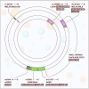
The mammalian mitochondrial genome: Mitochondria are small, double‐membraned organelles within cells, typically measuring 1–10 µm in size and often taking on elliptical or elongated cylindrical shapes. Inside mitochondria, the inner membrane is extensively folded into structures called cristae, which greatly increase its surface area. These cristae contain key components of the electron transport chain and the oxidative phosphorylation system, critical for generating cellular energy in the form of adenosine triphosphate (ATP). The mitochondrial matrix, located within the inner membrane, houses mitochondrial DNA, mitochondrial ribosomes, and machinery for protein synthesis, playing essential roles in various cellular processes. Mitochondrial DNA encodes essential components of the respiratory chain complexes I, III, and IV, as well as ATP synthase, all of which are vital for adenosine triphosphate (ATP) generation. Specifically, human mtDNA contains the genetic information for seven subunits of complex I (ND1–5, shown in purple), one subunit of complex III (CYB), three subunits of complex IV (COI–III, depicted in red), and two subunits of complex V (ATP8/6). Additionally, mtDNA encodes 22 transfer RNAs (tRNAs) and two ribosomal RNAs (rRNAs, represent in green), necessary for the translation of these mitochondrial proteins. This genetic information is densely packed and spans both DNA strands: a guanine‐rich heavy (H) strand and a cytosine‐rich light (L) strand. One major noncoding region (NCR) is present, housing the origin of heavy‐strand replication (OriH) and the promoters for heavy‐strand (HSP) and light‐strand (LSP) transcription. Another origin of replication dedicated to light‐strand synthesis (OriL) is situated approximately 11 kb away from OriH. The region between OriH and OriL is termed the major arc, while the smaller remaining region is referred to as the minor arc.
FIGURE 2
FIGURE 2
Mitochondrial dynamics and quality control in health and disease. Healthy mitochondria undergo continuous fusion (mediated by mitofusin 1/2 (MFN1/2) and optic atrophy protein 1 (OPA1) and fission (mediated by dynamin‐related protein 1 (DRP1) to maintain bioenergetic function, calcium (Ca2⁺) homeostasis, reactive oxygen species (ROS) balance, and regulation of apoptosis. Upon damage, PTEN‐induced putative kinase 1 (PINK1) accumulates on the outer mitochondrial membrane and recruits the E3 ubiquitin ligase Parkin, initiating selective mitochondrial autophagy (mitophagy). Damaged mitochondria are subsequently sequestered into autophagosomes and degraded in lysosomes, preserving mitochondrial quality and cellular health.
FIGURE 3
FIGURE 3
Overview of mitochondrial genome‐engineering strategies: Panel (A) illustrates the distinctive structure and functioning mechanism of mitochondrially targeted nucleases, which include restriction endonucleases (mitoRE), zinc finger nucleases (mtZFNs), and transcriptional activator‐like effectors nucleases (mitoTALENs). Within cells containing mutant mtDNA, these nucleases selectively identify mutated sequences (depicted in red). The induction of double‐strand breaks initiates the rapid degradation of the mutated mtDNA, leading to a reduction in the number of mutant copies. To restore the mtDNA copy count, the majority of the remaining mtDNA is of the wild‐type variety (depicted in blue). Consequently, this process replenishes the cells with normal mtDNA copies, ultimately enhancing the functionality of OXPHOS. While panel (B) illustrates the distinctive structure and functioning mechanism of base editors, which include DddA‐derived cytosine base editors (DdCBEs) and mtDNA base editors (mitoBEs). In cells containing a mixture of mutant and normal mt‐RNA, the base editor progressively rectifies the mutant mtDNA until the level of heteroplasmy falls below the threshold of pathogenicity.
FIGURE 4
FIGURE 4
The development history of mtDNA editing: The curve chart presented herein delineates a comprehensive overview of annual research publications spanning the past three decades in the domain of mitochondrial gene editing. Accompanying this visual representation are annotations highlighting key breakthroughs that have significantly influenced the advancement of this field. Furthermore, a specific emphasis has been placed on the meticulous development of animal models, enriching our understanding of mitochondrial gene editing's practical applications. mitoBEs, mitochondrial DNA base editors; DdCBE, DddA‐derived cytosine base editor; mitoRE, mitochondrially targeted restriction endonuclease; mitoTALEN; mitochondrially targeted transcription activator‐like effector nuclease; mtZFN, mitochondrially targeted zinc finger nuclease.
FIGURE 5
FIGURE 5
mtDNA mutations that have been or may be altered by gene editors: Summary of the mtDNA mutations corrected with available mitochondrial nucleases and in which tissues they have been successfully applied. ATP8/6, complex V subunits; CM, cardiomyopathy; CPEO, chronic progressive external ophthalmoplegia; DdCBE, DddA‐derived cytosine base editors; LHON, Leber hereditary optic neuropathy; MELAS, mitochondrial encephalomyopathy, lactic acidosis and stroke‐like episodes; MERRF, myoclonic epilepsy with ragged red fibers; MIDD, maternally inherited diabetes and deafness; MILS, maternally inherited Leigh syndrome; mitoBEs, mtDNA base editors; TALEN, targeted transcription activator‐like effector nuclease; ZFN, mitochondrially targeted zinc‐finger nuclease; NARP, neurogenic muscle weakness, ataxia, and retinitis pigmentosa; NCR, noncoding region; ND5, 6, complex I subunits; PMPS, Pearson marrow and pancreas syndrome.

References

    1. Muñoz‐Gómez S. A., Wideman J. G., Roger A. J., and Slamovits C. H., “The Origin of Mitochondrial Cristae From Alphaproteobacteria,” Molecular Biology and Evolution 34, no. 4 (2017): 943–956. - PubMed
    1. Gray M. W., Burger G., and Lang B. F., “Mitochondrial Evolution,” Science (New York, NY) 283, no. 5407 (1999): 1476–1481. - PubMed
    1. Gray M. W., “Mitochondrial Evolution,” Cold Spring Harbor Perspectives in Biology 4, no. 9 (2012): a011403. - PMC - PubMed
    1. Wu H., Dongchen W., Li Y., et al., “Mitogenomes Comparison of 3 Species of Asparagus L Shedding Light on Their Functions due to Domestication and Adaptative Evolution,” BMC Genomics [Electronic Resource] 25, no. 1 (2024): 1–16. - PMC - PubMed
    1. Takusagawa M., Misumi O., Nozaki H., et al., “Complete Mitochondrial and Chloroplast DNA Sequences of the Freshwater Green Microalga Medakamo hakoo,” Genes & Genetic Systems 98, no. 6 (2023): 353–360. - PubMed

LinkOut - more resources