. 2025 Sep 11;13(9):23259671251367060.
doi: 10.1177/23259671251367060. eCollection 2025 Sep.

Pediatric Meniscotibial Ligament Complex Anatomy and Biomechanics

Affiliations

Pediatric Meniscotibial Ligament Complex Anatomy and Biomechanics

Willemijn H van Deursen et al. Orthop J Sports Med. .

Abstract

Background: Meniscal repair is increasingly performed in pediatric patients, with capsular-based techniques remaining the gold standard despite limitations such as high failure rates and risk of meniscal extrusion. Recent studies highlight the potential role of accessory knee ligaments in improving meniscal stability and repair outcomes. The meniscotibial ligament complex (MTLC) has emerged as a potential area of interest to produce more normal anatomic and biomechanical meniscal function in meniscal repair.

Purpose: To evaluate the native anatomy and biomechanical strength of the MTLC of the medial and lateral meniscus of pediatric knees.

Study design: Descriptive laboratory study.

Methods: Fourteen fresh-frozen pediatric human knees (mean age, 7.5 years; range, 5-10 years; 6 male, 8 female) were used in this study. The depth of the recess between the MTLC and the meniscocapsular complex was measured. Subsequently, the medial and lateral menisci were divided into approximate thirds, creating anterior, central, and posterior testing zones for each meniscus. Each meniscus/MTLC complex underwent monotonic load-to-failure testing on an Instron 5944 test frame with a 2-kN load cell with load applied superiorly. Biomechanical properties were analyzed using linear mixed models with donor as a random factor and aspect (medial/lateral) and position (anterior/central/posterior) as fixed factors.

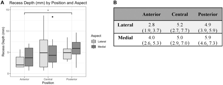
Results: The posterior recess depth was significantly larger (mean, 5.4 mm; 95% CI, 4.6-6.3 mm) than anterior (mean, 3.4 mm; 95% CI, 2.6-4.2 mm) (P = .049). Maximal load to failure in the posterior MTLC (mean, 93.5 N; 95% CI, 80.0-107.0 N) was significantly higher than anterior (mean, 69.2 N; 95% CI, 56.7-81.7 N) (P = .01).

Conclusion: This study defines a clear space in which the MTLC is distinct from the joint capsule, which is deepest in the posterior third of the medial and lateral meniscus. Our results demonstrate that the posterior region of the MTLC can withstand higher loads than the anterior region in pediatric knees.

Clinical relevance: These findings offer foundational insights into the native anatomy and biomechanics of the MTLC, guiding future studies involving the MTLC in meniscal repair. This knowledge may be particularly relevant to ramp lesions, other posterior meniscal tear patterns, and meniscal transplants.

Keywords: coronary ligament; meniscal instability; meniscal repair; meniscal tear; meniscotibial ligament complex; meniscus.


Conflict of interest statement

The authors declared that they have no conflicts of interest in the authorship and publication of this contribution. AOSSM checks author disclosures against the Open Payments Database (OPD). AOSSM has not conducted an independent investigation on the OPD and disclaims any liability or responsibility relating thereto. Ethical approval was deemed not necessary for the present study.

Figures

A visual compilation of images, including diagrams and surgical samples, showing the meniscotibial ligament complex (MTLC) and related anatomical structures for biomechanical testing
Figure 1.
Defining the meniscotibial ligament complex (MTLC) and isolating its anterior, central, and posterior aspects for biomechanical testing. (A) Schematic of a coronal cross section of half of a knee. The MTLC is depicted in red. The meniscocapsular recess space is indicated between the MTLC and the meniscocapsular complex (MCC, or joint capsule). The meniscocapsular recess space measurements were taken as indicated by the yellow line from the inferiormost point of the recess to the top of the meniscus, measured in the axial direction. (B) Gross examination of pediatric knee specimen indicating the distinct space that separates the MTLC and the MCC in the posterior aspect of the medial and lateral menisci. The distinct space also exists in the anterior and central regions but is only indicated in the posterior aspect in this image. The large white arrowhead indicates the popliteal hiatus. The small red lines indicate the MTLC, the small blue arrows indicate the MCC, and the small red circles show the meniscocapsular recess. MM, medial meniscus; LM, lateral meniscus; Po, popliteal tendon. (C) Axial schematic of radial meniscal transections, adapted from Dingel et al. Areas with red dashed line indicate where a scalpel was used to release the meniscus roots or transect the meniscus/MTLC complex into thirds. Numbered regions correspond to the segments of the meniscus/MTLC complex that subsequently underwent biomechanical testing: 1. Medial posterior; 2. Medial central; 3. Medial anterior; 4. Lateral anterior; 5. Lateral central; 6. Lateral posterior. ACL, anterior cruciate ligament; AL, anterolateral; AM, anteromedial. (D) Clamp on the anterior one-third of the lateral meniscus pulling vertically on the MTLC to failure. The tibia was potted in fiberglass resin and was vertically mounted on the Instron 5944 test frame.
Graph shows max load by position and meniscocapsular recess depth; box plot and table detail values for lateral and medial sections.
Figure 2.
A distinct separation exists between the meniscotibial ligament complex (MTLC) and the meniscal capsular complex. (A) Meniscocapsular recess depth of the anterior, central, and posterior regions of the lateral and medial menisci. Measurements were taken using a depth gauge and digital calipers at the deepest point of the recess between the meniscal capsular complex and the MTLC before the meniscus was transected into thirds to maintain the anatomic position of the capsule before biomechanical testing. Asterisk indicates significance at P < .05. Solid black dots indicate outliers beyond 1.5 times the interquartile range. (B) Summary table of mean recess depth for each unique aspect and position. Values are reported as mean (95% CI).
Structural biomechanical properties of the meniscus-tibial ligament complex (MTLC) by aspect and position. (A) Load to failure (N) of the anterior, central, and posterior regions of the lateral and medial MTLC. The posterior position was significantly higher than the anterior position (P = .01), with no difference between aspects. (B) Displacement (mm) at maximal load. After log transformation to correct for nonnormal residuals, the medial aspect was significantly lower than the lateral aspect (P < .001), and the anterior position was significantly lower than the posterior position (P = .04). (C) Stiffness of the MTLC by aspect and position as calculated from maximal slope of load-displacement curve showed no significant difference among aspect or position. (D) Work to maximal load (N·mm) of the anterior, central, and posterior segments of the lateral and medial MTLC, as calculated using the trapezoidal rule. The work to maximal load in the lateral aspect was significantly greater than the medial aspect (P = .03). Asterisk indicates significance at P < .05.
Figure 3.
Structural biomechanical properties of the meniscotibial ligament complex (MTLC) by aspect and position. (A) Load to failure (N) of the anterior, central, and posterior regions of the lateral and medial MTLC. The posterior position was significantly higher than the anterior position (P = .01), with no difference between aspects. (B) Displacement (mm) at maximal load. After log transformation to correct for nonnormal residuals, the medial aspect was significantly lower than the lateral aspect (P < .001), and the anterior position was significantly lower than the posterior position (P = .04). (C) Stiffness of the MTLC by aspect and position as calculated from maximal slope of load-displacement curve showed no significant difference among aspect or position. (D) Work to maximal load (N·mm, Newton-millimeters) of the anterior, central, and posterior segments of the lateral and medial MTLC, as calculated using the trapezoidal rule. The work to maximal load in the lateral aspect was significantly greater than the medial aspect (P = .03). Asterisk indicates significance at P < .05. Solid black dots indicate outliers beyond 1.5 times the interquartile range.
A: (A) Boxplot: Comparison of MTLC Failure Stress (MPa) by Position and Aspect. (B) Boxplot: Comparison of MTLC Strain at Max Load (mm/mm) by Position and Aspect. *Indicates p <0.05*.
Figure 4.
Material biomechanical properties of the meniscotibial ligament complex (MTLC) by aspect and position. (A) Failure stress (MPa) by aspect and position, as calculated by dividing the maximal by the maximal cross-sectional area. The central position was significantly greater than the anterior position (P = .004). (B) Strain of the MTLC at maximal load (mm/mm), as calculated by dividing the displacement at maximal load by the MTLC length at rest. The medial aspect was significantly higher as compared with the lateral aspect (P < .001). Asterisk indicates significance at P < .05. Solid black dots indicate outliers beyond 1.5 times the interquartile range.

References

    1. Abrams GD, Frank RM, Gupta AK, Harris JD, McCormick FM, Cole BJ. Trends in meniscus repair and meniscectomy in the United States, 2005-2011. Am J Sports Med. 2013;41(10):2333-2339. - PubMed
    1. Adams BG, Houston MN, Cameron KL. The epidemiology of meniscus injury. Sports Med Arthrosc Rev. 2021;29(3):e24. - PubMed
    1. Ahn JH, Bae TS, Kang KS, Kang SY, Lee SH. Longitudinal tear of the medial meniscus posterior horn in the anterior cruciate ligament–deficient knee significantly influences anterior stability. Am J Sports Med. 2011;39(10):2187-2193. - PubMed
    1. Bram JT, Mehta NN, Wright ML, Nguyen JC, Leska T, Ganley TJ. Pediatric ramp lesions. J Pediatr Orthop Soc N Am. 2020;2(1):48.
    1. Cavaignac E, Sylvie R, Teulières M, et al. What is the relationship between the distal semimembranosus tendon and the medial meniscus? A gross and microscopic analysis from the SANTI Study Group. Am J Sports Med. 2021;49(2):459-466. - PubMed

LinkOut - more resources