[Preprint]. 2025 Sep 4:2025.08.31.25334556.
doi: 10.1101/2025.08.31.25334556.

Autoantibodies neutralizing type I IFNs in 40% of patients with WNV encephalitis in seven new cohorts

Affiliations

Autoantibodies neutralizing type I IFNs in 40% of patients with WNV encephalitis in seven new cohorts

Adrian Gervais et al. medRxiv. .

Abstract

Mosquito-borne West Nile virus (WNV) infection is a growing global health problem. About 0.5% of infected individuals develop encephalitis. We previously showed that 40% of patients in six cohorts had WNV encephalitis because of circulating auto-antibodies (auto-Abs) neutralizing type I IFNs. In seven new cohorts, we found that the prevalence of auto-Abs was highest (40% [17-44%]) in patients with encephalitis, and very low in a small sample of individuals with asymptomatic or mild infection. In the 13 European, Middle-Eastern and American cohorts available, odds ratios for WNV encephalitis in individuals with these auto-Abs relative to those without them in a large sample of the general population untested for WNV infection range from ~20 (OR=17.7; 95% CI: 13.8-22.8, p<10-16) for auto-Abs neutralizing only 100 pg/mL IFN-α2 and/or IFN-ω to >2000 (OR=2218.4; 95% CI: 125.1-39337.7, p<10-16) for auto-Abs neutralizing high concentrations of IFN-α2 and high or low concentrations of IFN-ω. Pre-existing autoantibodies neutralizing type I IFNs are therefore causal for WNV encephalitis in about 40% of patients.


Conflict of interest statement

Declaration of interest J.L.-C. is an inventor on patent application PCT/US2021/042741, filed July 22, 2021, submitted by The Rockefeller University and covering the diagnosis of susceptibility to, and the treatment of, viral disease, and viral vaccines, including COVID-19 and vaccine-associated diseases.

Figures

Figure 1.
Figure 1.. Autoantibodies neutralzing type I IFNs in 318 West Nile virus (WNV)-infected individuals from seven new cohorts.
(A) Age and sex distribution of individuals in the WNVIC, WNVF, and WNVD groups. (B) Detection of autoantibodies against IFN-α2, IFN-β, and IFN-ω by ELISA. An OD value > 0.5 (dashed line) indicates a sample considered positive based on the signal typically observed for serum/plasma from healthy donors. (C) Luciferase-based neutralization assay for the detection of autoantibodies neutralizing 10 ng/mL IFN-α2, IFN-ω, or IFN-β. Neutralizing activity was defined as a luciferase signal below 15%. (D) Luciferase-based neutralization assay for detecting autoantibodies neutralizing 100 pg/mL IFN-α2 or IFN-ω, or 1 ng/mL IFN-β. Neutralizing activity was defined as a luciferase signal below 15%.
Figure 2.
Figure 2.. Proportions of individuals with auto-Abs neutralizing type I IFNs in seven new cohorts.
(A and B) Frequency of individuals carrying auto-Abs neutralizing type I IFNs at concentrations of 10 ng/mL (A) or 100 pg/mL (B), as determined in luciferase-based neutralization assays, in the three defined groups of WNV-infected individuals: WNVIC, WNVF, and WNVD. IFN-α, auto-Abs neutralizing IFN-α2 (regardless of their effects on other IFNs); IFN-ω, auto-Abs neutralizing IFN-ω (regardless of their effects on other IFNs); IFN-α ± ω ± β, auto-Abs neutralizing IFN-α2 and/or IFN-ω and/or IFN-β; IFN-α + ω, auto-Abs neutralizing both IFN-α2 and IFN-ω. (C) Number of type I IFNs neutralized in the three groups of WNV-infected individuals (WNVIC, WNVF, and WNVD), as determined with the luciferase-based neutralization assay. (D) Proportion of type I IFNs neutralized in the three groups of WNV-infected individuals (WNVIC, WNVF, and WNVD) according to the nature and combination of auto-Abs.
Figure 3.
Figure 3.
Proportions of patients with auto-Abs neutralizing type I IFNs by center and year of enrollment, and lineage in 13 cohorts from 5 countries. (A-B) Number and prevalence of individuals with auto-Abs neutralizing at least one type I IFN at a concentration of 100 pg/mL (IFN-α2, IFN-ω) or 1 ng/mL (IFN-β) in the three groups of individuals infected with WNV (WNVIC, WNVF, WNVD), by enrollment center. (C-D) Number and prevalence of individuals with auto-Abs neutralizing at least one type I IFN at a concentration of 100 pg/mL (IFN-α2, IFN-ω) or 1 ng/mL (IFN-β) in the three groups of individuals infected with WNV (WNVIC, WNVF, WNVD), by enrollment year. (E) Number of individuals with auto-Abs neutralizing at least one type I IFN at a concentration of 100 pg/mL (IFN-α2, IFN-ω) or 1 ng/mL (IFN-β) in subgroups of WNND patients: WNV encephalitis (WNE), WNV meningitis (WNM), acute flaccid paralysis (AFP) and unspecified neurological syndrome (UNS), by WNV lineage. (F) Distribution by year (x-axis), recruitment center (colors), and infecting WNV virus lineage (WNV-1, dashed boxes, WNV-2, dotted boxes) of the individuals with WNVD recruited.
Figure 4.
Figure 4.
Proportions of patients with auto-Abs neutralizing type I IFNs by sex and age in 13 cohorts from 5 countries. (A) Prevalence of individuals with auto-Abs neutralizing at least one type I IFN at a concentration of 100 pg/mL (IFN-α2, IFN-ω) or 1 ng/mL (IFN-β) in the three groups of individuals infected with WNV (WNVIC, WNVF, WNVD), by sex. (B) Prevalence of individuals with auto-Abs neutralizing at least one type I IFN at a concentration of 100 pg/mL (IFN-α2, IFN-ω) or 1 ng/mL (IFN-β) in the three groups of WNV-infected individuals (WNVIC, WNVF, WNVD), by age class.
Figure 5.
Figure 5.
Frequency of auto-Abs against type I IFNs in the WNVIC, WNVF, and WNVD groups and the two WNVD subgroups (WNVD without evidence of neuroinvasive disease, and WNND) relative to the general population in the seven new cohorts and the overall study population consisting of all 13 cohorts. Horizontal bars represent the 95% CI limits. IFN-α: autoantibodies neutralizing IFN-α2 (regardless of effects on other IFNs); IFN-ω: autoantibodies neutralizing IFN-ω; IFN-α ± ω ± β: autoantibodies neutralizing IFN-α2 and/or IFN-ω and/or IFN-β; IFN-α + ω: autoantibodies neutralizing both IFN-α2 and IFN-ω. ns: non-significant; ****p < 10−4.
Figure 6.
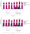
Figure 6.. Odds ratios for the presence of auto-Abs in the WNV groups relative to the general population.
Odds ratios (ORs) for the presence of auto-Abs in the WNVIC group relative to the general population, adjusted for age and sex with Firth’s bias-corrected logistic regression, in the seven new cohorts (A) and the overall study population consisting of 13 cohorts (B). ORs for the presence of auto-Abs in individuals with WNVF and WNVD in the seven new cohorts (C) and the overall study population consisting of 13 cohorts (D) relative to the general population, also adjusted for age and sex via logistic regression. Horizontal bars represent the 95% CI limits. IFN-α: autoantibodies neutralizing IFN-α2 (regardless of effects on other IFNs); IFN-ω: autoantibodies neutralizing IFN-ω; IFN-α ± ω ± β: autoantibodies neutralizing IFN-α2 and/or IFN-ω and/or IFN-β; IFN-α + ω: autoantibodies neutralizing both IFN-α2 and IFN-ω. Ns: non-significant; ****p < 10−4.
Figure 7.
Figure 7.. Odds ratios for the presence of auto-Abs in the WNVD group relative to the general population.
Odds ratios (ORs) for the presence of auto-Abs in the WNVD group and the two WNVD subgroups (WNVD without evidence of neuroinvasive disease, and WNV neuroinvasive disease; WNND) relative to the general population, adjusted for age and sex with Firth’s bias-corrected logistic regression, in the seven new cohorts (A) and the overall study population consisting of 13 cohorts (B). Odds ratios (ORs) for the presence of auto-Abs in the WNND group and the four WNND subgroups (encephalitis; WNE, meningitis; WNM, acute flaccid paralysis; AFP, and unspecified neurological syndrome; UNS) in the seven new cohorts (C) and in overall study population consisting of 13 cohorts (D) relative to the general population, also adjusted for age and sex via logistic regression. Horizontal bars represent the 95% CI limits. IFN-α: autoantibodies neutralizing IFN-α2 (regardless of effects on other IFNs); IFN-ω: autoantibodies neutralizing IFN-ω; IFN-α ± ω ± β: autoantibodies neutralizing IFN-α2 and/or IFN-ω and/or IFN-β; IFN-α + ω: autoantibodies neutralizing both IFN-α2 and IFN-ω. ns: non-significant; ****p < 10−4.
Figure 8.
Figure 8.. Odds ratios for the presence of auto-Abs in the WNV groups relative to the general population, by age group.
ORs for the presence of auto-Abs in patients with WNVD relative to the general population, as determined by logistic regression, stratified for age group, for auto-Abs neutralizing different combinations of high concentrations (A) and low concentrations (B) of type I IFNs in the seven new cohorts, and for auto-Abs neutralizing different combinations of high concentrations (C) and low concentrations (D) of type I IFNs in the overall study population consisting of 13 cohorts. ORs were calculated separately for patients aged ≤65 and >65 years. Horizontal bars represent the 95% CI limits. IFN-α: autoantibodies neutralizing IFN-α2 (regardless of effects on other IFNs); IFN-ω: autoantibodies neutralizing IFN-ω; IFN-α ± ω ± β: autoantibodies neutralizing IFN-α2 and/or IFN-ω and/or IFN-β; IFN-α + ω: autoantibodies neutralizing both IFN-α2 and IFN-ω. ns: non-significant; ****p < 10−4.

References

    1. Young J.J., Haussig J.M., Aberle S.W., Pervanidou D., Riccardo F., Sekulić N., Bakonyi T., and Gossner C.M.. Epidemiology of human West Nile virus infections in the European Union and European Union enlargement countries, 2010 to 2018. Euro Surveill. 26:2001095. 2021. doi: 10.2807/1560-7917.ES.2021.26.19.2001095 - DOI - PMC - PubMed
    1. Postler T.S., Beer M., Blitvich B.J., Bukh J., de Lamballerie X., Drexler J.F., Imrie A., Kapoor A., Karganova G.G., Lemey P., Lohmann V., Simmonds P., Smith D.B., Stapleton J.T., and Kuhn J.H.. Renaming of the genus Flavivirus to Orthoflavivirus and extension of binomial species names within the family Flaviviridae. Arch Virol. 168:224. 2023. doi: 10.1007/s00705-023-05835-1 - DOI - PubMed
    1. Petersen L.R., Carson P.J., Biggerstaff B.J., Custer B., Borchardt S.M., and Busch M.P.. Estimated cumulative incidence of West Nile virus infection in US adults, 1999–2010. Epidemiol Infect. 141:591–5. 2013. doi: 10.1017/S0950268812001070 - DOI - PMC - PubMed
    1. Chancey C., Grinev A., Volkova E., and Rios M.. The global ecology and epidemiology of West Nile virus. Biomed Res Int. 2015:376230. 2015. doi: 10.1155/2015/376230 - DOI - PMC - PubMed
    1. Chowdhury P., and Khan S.A.. Global emergence of West Nile virus: Threat & preparedness in special perspective to India. Indian J Med Res. 154:36–50. 2021. doi: 10.4103/ijmr.IJMR_642_19 - DOI - PMC - PubMed

Publication types

LinkOut - more resources