

Managing malignant middle cerebral artery infarction with open scalp incision and partial hemispherectomy: illustrative case
- PMID: 40953523
- PMCID: PMC12435381
- DOI: 10.3171/CASE2523
Managing malignant middle cerebral artery infarction with open scalp incision and partial hemispherectomy: illustrative case
Abstract
Background: Malignant middle cerebral artery (MCA) infarctions result in cerebral edema that can lead to brain herniation and death. Standard management includes decompressive hemicraniectomy (DHC) and comprehensive neurocritical care. Some patients may continue to decline despite these measures. Reopening of the scalp incision may allow for additional decompression and provide mortality benefit.
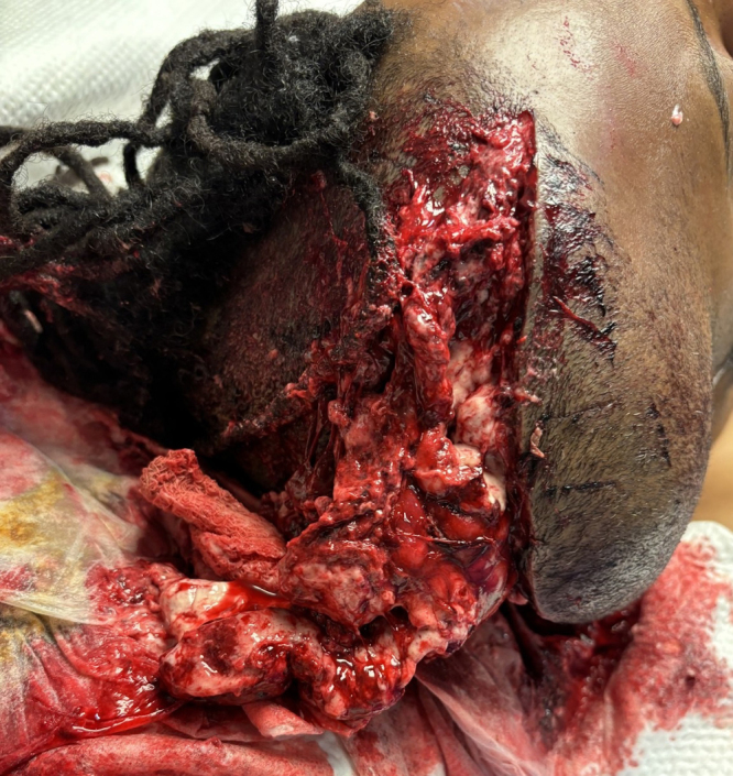
Observations: A 47-year-old man developed malignant right MCA territory infarction following intravenous thrombolysis and unsuccessful mechanical thrombectomy. Despite aggressive hyperosmolar therapy and a large DHC, the patient continued to exhibit clinical decline and radiological progression of cerebral edema. In response, the scalp incision was reopened to facilitate maximal external cerebral herniation, a strategy the authors believe was critical in managing the brain swelling. The patient was maintained on prophylactic antibiotics during this period, given the increased infection risk associated with exposed brain surface and potential CSF leakage. Following the resolution of cerebral swelling, a right partial hemispherectomy was performed to excise the infarcted hemisphere. Notably, the patient achieved significant functional recovery following the intervention and an extended period of rehabilitation.
Lessons: This case highlights the complexities encountered in the surgical management of malignant cerebral infarction, particularly when standard decompressive measures fail. https://thejns.org/doi/10.3171/CASE2523.
Keywords: decompressive hemicraniectomy; hemispherectomy; ischemic brain edema; malignant MCA infarction; open scalp incision.
Figures


References
-
- Liebeskind DS Jüttler E Shapovalov Y Yegin A Landen J Jauch EC.. Cerebral edema associated with large hemispheric infarction. Stroke. 2019;50(9):2619-2625. - PubMed
-
- Hacke W Schwab S Horn M Spranger M De Georgia M von Kummer R.. “Malignant” middle cerebral artery territory infarction: clinical course and prognostic signs. Arch Neurol. 1996;53(4):309-315. - PubMed
-
- Jüttler E, Schwab S, Schmiedek P.Decompressive Surgery for the Treatment of Malignant Infarction of the Middle Cerebral Artery (DESTINY): a randomized, controlled trial. Stroke. 2007;38(9):2518-2525. - PubMed
-
- Schwab S, Steiner T, Aschoff A.Early hemicraniectomy in patients with complete middle cerebral artery infarction. Stroke. 1998;29(9):1888-1893. - PubMed
LinkOut - more resources
Full Text Sources
