

Choroid plexus volume in brain disorders: a systematic review
- PMID: 40954490
- PMCID: PMC12439388
- DOI: 10.1186/s12987-025-00702-4
Choroid plexus volume in brain disorders: a systematic review
Abstract
Background: The choroid plexus is a highly vascularized structure located in the lateral, third, and fourth ventricles of the brain. Recent studies suggest that volumetric changes in choroid plexus volume are associated with progression in various brain diseases. Segmentation algorithms have significantly improved our ability to study choroid plexus volumetrics in relation to various pathologies. Thus, the specific purpose of this review was to describe to what extent choroid plexus volume estimation provides clinically relevant information in brain diseases.
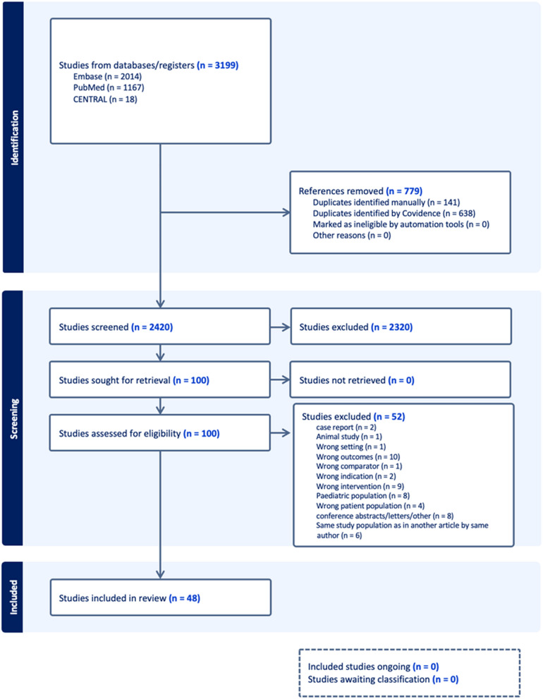
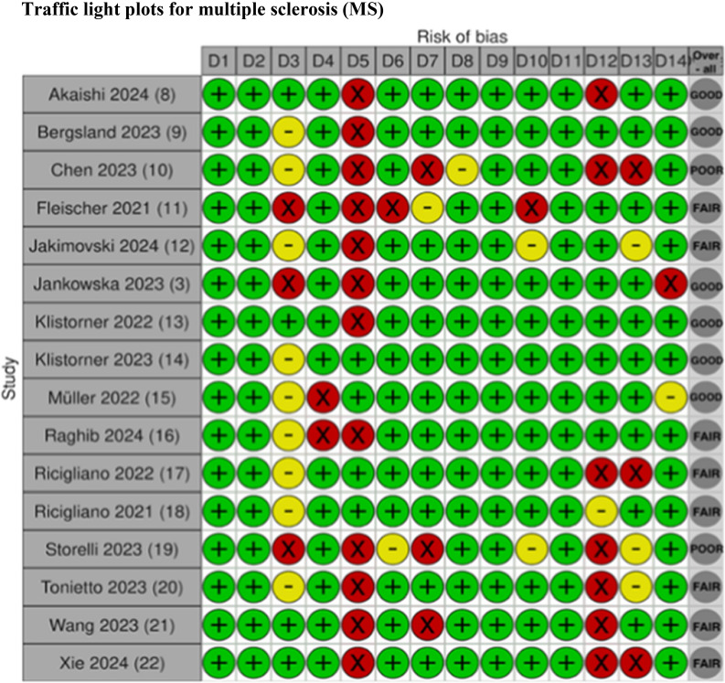
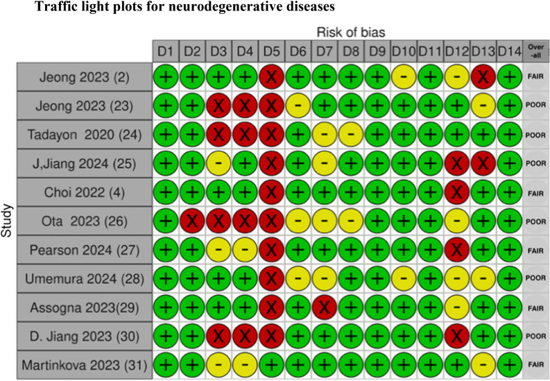
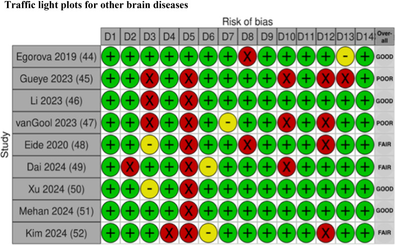
Methods: An extensive literature search was conducted across Pubmed, Embase and Cochrane databases. A comprehensive, detailed qualitative descriptive analysis, and a thorough risk-of-bias assessment were performed for the included studies.
Results: Forty-eight studies were included in this systematic review in the categories of multiple sclerosis, neurodegenerative diseases, psychiatric disorders, healthy populations and a group categorized as "other" for all other brain diseases that did not fit into the other categories.
Conclusion: For many of the studies included, the patients had a larger choroid plexus volume compared to healthy controls. Evidence is currently insufficient to determine whether CPV enlargement correlates with clinical severity or functional scores. The most common segmentation technique was the automatic segmentation method, followed by manual correction of the segmented choroid plexus. Thus, this review highlights the growing interest choroid plexus volume, its segmentation, and its potential as a biomarker for numerous brain diseases.
Keywords: Brain disorders; Choroid plexus volume; Magnetic resonance imaging; Segmentation techniques; Systematic review.
© 2025. The Author(s).
Conflict of interest statement
Declarations: Ethics approval and consent to participate. Not applicable. Competing interest: The authors declare no competing interests.
Figures


References
Publication types
MeSH terms
Grants and funding
LinkOut - more resources
Full Text Sources
Medical
