. 2025 Sep 16;135(22):e192925.
doi: 10.1172/JCI192925. eCollection 2025 Nov 17.

Maintenance DNA methylation is required for induced Treg reparative function following viral pneumonia in mice

Affiliations

Maintenance DNA methylation is required for induced Treg reparative function following viral pneumonia in mice

Anthony M Joudi et al. J Clin Invest. .

Abstract

FOXP3+ natural regulatory T cells (nTregs) promote resolution of inflammation and repair of epithelial damage following viral pneumonia-induced lung injury, thus representing a cellular therapy for patients with severe viral pneumonia and the acute respiratory distress syndrome. Whether in vitro-induced Tregs (iTregs), which can be rapidly generated in substantial numbers from conventional T cells, also promote lung recovery is unknown. nTregs require specific DNA methylation patterns maintained by the epigenetic regulator ubiquitin-like with PHD and RING finger domains 1 (UHRF1). Here, we tested whether iTregs promote recovery following viral pneumonia and whether iTregs require UHRF1 for their pro-recovery function. We found that adoptive transfer of iTregs to mice with influenza virus pneumonia promotes lung recovery and that loss of UHRF1-mediated maintenance DNA methylation in iTregs leads to reduced engraftment and a delayed repair response. Transcriptional and DNA methylation profiling of adoptively transferred UHRF1-deficient iTregs that had trafficked to influenza-injured lungs demonstrated transcriptional instability with gain of transcription factors that define effector T cell lineage. Strategies to promote the stability of iTregs could be leveraged to further augment their pro-recovery function during viral pneumonia and other causes of severe lung injury.

Keywords: Epigenetics; Immunology; Inflammation; Influenza; Pulmonology; T cells.


Conflict of interest statement

Conflict of interest: NM is currently an employee of and owns stock in Vertex Pharmaceuticals. BDS holds United States Patent US 10,905,706 B2, Compositions and Methods to Accelerate Resolution of Acute Lung Inflammation, and serves on the Scientific Advisory Board of Zoe Biosciences.

Figures

Figure 1
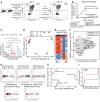
Figure 1. Adoptive transfer of iTregs promotes survival following viral pneumonia.
Foxp3GFP-DTR mice were treated with DTx every 48 hours beginning 2 days before inoculation with 6.5 PFU of influenza A/WSN/33 H1N1 virus and at 5 DPI received retro-orbital adoptive transfer of 1 × 10⁶ nTregs, iTregs, or Tconv cells or PBS. Additional controls received DTx but no influenza inoculation (DTx no flu) or influenza inoculation but no diphtheria toxin (no DTx). (A) Schematic of experimental design. (B) Mice were followed over time for arterial oxyhemoglobin saturation (SpO2) measured via dorsal collar clip. (C) Survival of mice that received indicated treatments. (DG) Mice were euthanized at 24 DPI, and lungs were analyzed by flow cytometry for frequency of epithelial, CD326+CD31CD45 cells (D); frequency of ATII, CD326+MHCII+T1A cells (E); frequency of KRT5+CD326+ epithelial cells (F); and frequency of Ki-67+CD326+MHCII+T1A cells (G). Data from recipients of iTregs are derived from post-caval lobe; data from all other groups are derived from whole-lung suspensions. (B, DTx no flu n = 4, no DTx n = 6, nTreg n = 11, PBS n = 9, Tconv n = 12, iTreg n = 18; C, DTx no flu n = 4, no DTx n = 9, nTreg n = 27, PBS n = 21, Tconv n = 25, iTreg n = 18; D and E, no DTx n = 3, nTreg n = 15, PBS n = 11, Tconv n = 10, iTreg n = 9; F, no DTx n = 3, nTreg n = 15, PBS n = 11, Tconv n = 8, iTreg n = 7; G, no DTx n = 3, nTreg n = 14, PBS n = 9, Tconv n = 10, iTreg n = 9). Data in B generated from 4 independent experiments. Data in C generated from 5 independent experiments. Data in DG generated from 4 independent experiments. Survival curve (C) P value was determined using log-rank (Mantel-Cox) test, **P < 0.005. Data presented as mean and SD with *q < 0.05 according to multiple Mann-Whitney tests and correcting for multiple comparisons using the 2-stage linear step-up procedure of Benjamini, Krieger, and Yekutieli with Q = 5% (DG).
Figure 2
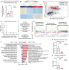
Figure 2. UHRF1 is dispensable for iTreg FOXP3 expression and suppressive capacity but is required for transcriptional and epigenetic stability in vitro.
(AD) Natural and induced Tregs derived from Uhrf1+/+ and Uhrf1fl/fl mice were exposed to tamoxifen to delete UHRF1 from day 0 to day 5 (“early”) or day 6 to day 12 (“delayed”) of culture, then harvested on days 5 and 12 for flow cytometry and RNA-Seq analysis. (A) Schematic. (B) Frequency of Foxp3-GFP+tdTomato+ iTregs and Foxp3-GFPtdTomato+ (ex-FOXP3) cells. (C) Percent suppression of CD4+CTV+Foxp3-GFP splenic responder T cells cocultured for 72 hours at indicated ratios of experimental Tregs. (D) PCA of 6,978 DEGs, identified from ANOVA-like testing with FDR q < 0.05. (EI) Induced Tregs derived from Uhrf1+/+ and Uhrf1fl/fl mice were exposed to tamoxifen to delete UHRF1 from day 6 to day 12 (“delayed”) of culture, then harvested on day 12 for RNA-Seq and DNA methylation analysis. (E) K-means clustering of 127 genes with an FDR q < 0.05 with k = 2. (F) MA plot comparing gene expression between groups. Genes of interest are annotated. (G) Enrichment plots of gene sets (P < 0.05, FDR q < 0.25) generated through GSEA pre-ranked testing of the expressed genes. (H) PCA of 81,179 differentially methylated cytosines identified from ANOVA-like testing with FDR q < 0.05. Ellipses represent normal contour lines with 1 SD probability. (I) Cumulative distribution function plot of differentially methylated cytosines expressed as β scores, with 0 representing unmethylated and 1 representing fully methylated; a shift in the cumulative distribution function up and to the left represents relative hypomethylation. (B, n = 3 per group; C, n = 3 per group; DI, n = 2 for Uhrf1fl/fl, n = 3 for Uhrf1+/+). B and C representative of 3 independent biological replicates. *q < 0.05 according to 2-way ANOVA with 2-stage linear step-up procedure of Benjamini, Krieger, and Yekutieli with Q = 5 (C).
Figure 3
Figure 3. Loss of UHRF1 is sufficient to impair repair capabilities of iTregs during viral pneumonia.
Foxp3GFP-DTR mice were treated with DTx every 48 hours beginning 2 days before inoculation with 6.5 PFU of influenza A/WSN/33 H1N1 virus, and then received retro-orbital adoptive transfer of 1 × 10⁶ Uhrf1fl/fl or Uhrf1+/+ iTregs at 5 DPI as in Figure 1. iTregs were treated with tamoxifen from culture day 0 to day 3 and harvested for adoptive transfer on culture day 5. Mice were euthanized 24 DPI, and lungs were analyzed by flow cytometry. Epithelial cell data are derived from post-caval lobes. (A) Survival of Foxp3GFP-DTR mice that received Uhrf1+/+ or Uhrf1fl/fl iTregs. (B) SpO2 over time in mice from A. (C and D) CD326+CD31 cell frequency (C) and total number (D). (E and F) CD326+MHCII+T1A cell frequency (E) and total number (F). (G and H) KRT5+CD326+ cell frequency (G) and total number (H). (I and J) Ki-67+CD326+MHCII+ cell frequency (I) and total number (J). (A and B, Uhrf1+/+ iTreg recipients n = 18, Uhrf1fl/fl iTreg recipients n = 15; CF, Uhrf1+/+ iTreg recipients n = 9, Uhrf1fl/fl iTreg recipients n = 6; G and H, Uhrf1+/+ iTreg recipients n = 7, Uhrf1fl/fl iTreg recipients n = 6; I and J, Uhrf1+/+ iTreg recipients n = 9, Uhrf1fl/fl iTreg recipients n = 6.) Survival curve (A) P value was determined using log-rank (Mantel-Cox) test, *P < 0.05. *P < 0.05 or *q < 0.05 according to mixed-effects model (restricted maximum likelihood) with 2-stage linear step-up procedure of Benjamini, Krieger, and Yekutieli with Q = 5% (B). Data presented as mean and SD with *P < 0.05, **P < 0.005 according to Mann-Whitney U test (CJ). Data from recipients of Uhrf1+/+ iTregs are re-presented from results shown in Figure 1 and were generated in contemporaneous experiments as the data generated from mice that received Uhrf1fl/fl iTregs. Data in A and B generated from 4 independent experiments. Data in CJ generated from 2 independent experiments.
Figure 4
Figure 4. UHRF1-deficient iTregs promote an insufficient tissue-protective response during peak lung injury.
Foxp3GFP-DTR mice were treated with DTx every 48 hours beginning 2 days before inoculation with 6.5 PFU of influenza A/WSN/33 H1N1 virus and received retro-orbital adoptive transfer of 1 × 10⁶ Uhrf1fl/fl or Uhrf1+/+ iTregs at 5 DPI. iTregs were treated with tamoxifen from day 0 to day 3 of culture and harvested for adoptive transfer on culture day 5. Recipient mice were euthanized 11 DPI, and lungs and spleen were analyzed by flow cytometry. Epithelial cell data are derived from the post-caval lobes. Transferred iTreg quantification is derived from the remaining lung lobes. (A) Total number of ATII cells (n = 15 per group). (B) Total number of Ki-67+ ATII cells (Uhrf1+/+ iTregs n = 13, Uhrf1fl/fl iTregs n = 14). (C) Total number of CD326CD31+ (endothelial) cells (Uhrf1+/+ iTregs n = 14, Uhrf1fl/fl iTregs n = 15). (D) Frequency of tdTomato+ cells in lung (n = 16 per group). (E) Total number of tdTomato+ cells in lung (n = 16 per group). (F) Frequency of Foxp3-GFPtdTomato+ (ex-FOXP3) cells in lung (Uhrf1+/+ n = 15, Uhrf1fl/fl n = 16). (G) Frequency of tdTomato+ cells in spleen (n = 8 per group). (H) Total number of tdTomato+ cells in spleen (n = 4 per group). (I) Frequency of Foxp3-GFPtdTomato+ cells in spleen (n = 8 per group). Data presented as mean and SD. *P < 0.05, **P < 0.005, according to Mann-Whitney U test. Data in AF generated from 2 independent experiments. Data in GI generated from 1 independent experiment.
Figure 5
Figure 5. UHRF1 is required for iTreg phenotypic stability and lung tissue engraftment following viral pneumonia.
DTx-treated, influenza A–infected Foxp3GFP-DTR recipient mice received retro-orbital adoptive transfer of 1 × 10⁶ Foxp3-GFP+tdTomato+ Uhrf1fl/fl or Uhrf1+/+ iTregs at 5 DPI. iTregs were cultured with tamoxifen from day 0 to day 3, then harvested for adoptive transfer, as in Figure 1. Transferred Foxp3-GFP+tdTomato+ Uhrf1fl/fl or Uhrf1+/+ iTregs were sorted from the lungs of recipient Foxp3GFP-DTR mice 24 DPI for quantification and profiling via bulk RNA-Seq. (A) Frequency of Foxp3-GFPtdTomato+ (ex-FOXP3) cells. (B) K-means clustering of 1,187 genes with FDR q < 0.05 comparing recovered Uhrf1+/+ and Uhrf1fl/fl iTregs with k = 2. (C) MA plot comparing gene expression of recovered Uhrf1+/+ and Uhrf1fl/fl iTregs. Genes of interest are annotated. (D) GSEA dot plot highlighting key statistics (FDR q value and normalized enrichment score [NES]) and enriched gene sets. Red dots denote gene sets with a positive enrichment score or enrichment at the top of the ranked list. Blue dots denote gene sets with a negative enrichment score or enrichment at the bottom of the ranked list. (E) Enrichment plots of hallmark gene sets generated through GSEA pre-ranked testing of the expressed genes in Uhrf1+/+ and Uhrf1fl/fl iTregs. All gene sets displayed significant enrichment with FDR q value < 0.25. (F) Selected Gene Ontology (GO) processes from 945 and 105 total enriched gene sets with FDR q < 0.25 in Uhrf1fl/fl and Uhrf1+/+ iTregs, respectively. Gene sets are annotated and ranked by –log10-transformed FDR q value. (G) Frequency of tdTomato+ iTregs recovered. (H) Total number of tdTomato+ iTregs recovered. ***P < 0.0005 according to Mann-Whitney U test (AF, Uhrf1+/+ n = 9, Uhrf1fl/fl n = 7; G, Uhrf1+/+ n = 8, Uhrf1fl/fl n = 7; H, Uhrf1+/+ n = 8, Uhrf1fl/fl n = 6). Data generated from 2 independent experiments.
Figure 6
Figure 6. Loss of UHRF1 results in delayed transcriptional changes over the course of viral pneumonia.
Foxp3GFP-DTR mice were treated with DTx every 48 hours beginning 2 days before inoculation with 6.5 PFU of influenza A/WSN/33 H1N1 virus and then received retro-orbital adoptive transfer of 1 × 10⁶ Foxp3-GFP+tdTomato+ Uhrf1fl/fl or Uhrf1+/+ iTregs at 5 DPI as in Figure 1. iTregs were treated with tamoxifen from day 0 to day 3 of culture, then harvested for adoptive transfer on culture day 5. Adoptively transferred Foxp3-GFP+tdTomato+ Uhrf1fl/fl or Uhrf1+/+ iTregs were sorted from the lungs of recipient Foxp3GFP-DTR mice 11 or 24 DPI and were compared via RNA transcriptomic and DNA methylation analysis. (A) PCA of 2,117 DEGs identified from ANOVA-like testing with FDR q < 0.05. Independent biological replicates are shown. (B) GSEA dot plot highlighting key statistics (FDR q value and NES) and enriched gene sets for Uhrf1+/+ iTregs at 11 DPI. Blue dots denote gene sets with a negative enrichment score or enrichment at the bottom of the ranked list. (C) Selection of representative enriched GO processes from 40 total enriched gene sets with an FDR q < 0.25 in Uhrf1+/+ iTregs at 11 DPI. Gene sets are annotated and ranked by –log10-transformed FDR q value. (D) K-means clustering of 34 differentially methylated regions with a difference of ≥10% between Uhrf1+/+ and Uhrf1fl/fl iTregs. (AD, 24 DPI Uhrf1+/+ iTreg recipients n = 9, Uhrf1fl/fl iTreg recipients n = 7, 11 DPI Uhrf1+/+ and Uhrf1fl/fl iTreg recipients n = 8).

Update of

References

    1. Sakaguchi S, et al. Immunologic self-tolerance maintained by activated T cells expressing IL-2 receptor alpha-chains (CD25). Breakdown of a single mechanism of self-tolerance causes various autoimmune diseases. J Immunol. 1995;155(3):1151–1164. doi: 10.4049/jimmunol.155.3.1151. - DOI - PubMed
    1. Sakaguchi S, et al. Regulatory T cells and immune tolerance. Cell. 2008;133(5):775–787. doi: 10.1016/j.cell.2008.05.009. - DOI - PubMed
    1. D’Alessio FR, et al. CD4+CD25+Foxp3+ Tregs resolve experimental lung injury in mice and are present in humans with acute lung injury. J Clin Invest. 2009;119(10):2898–2913. doi: 10.1172/JCI36498. - DOI - PMC - PubMed
    1. Garibaldi BT, et al. Regulatory T cells reduce acute lung injury fibroproliferation by decreasing fibrocyte recruitment. Am J Respir Cell Mol Biol. 2013;48(1):35–43. doi: 10.1165/rcmb.2012-0198OC. - DOI - PMC - PubMed
    1. Mock JR, et al. Foxp3+ regulatory T cells promote lung epithelial proliferation. Mucosal Immunol. 2014;7(6):1440–1451. doi: 10.1038/mi.2014.33. - DOI - PMC - PubMed

MeSH terms

Substances

LinkOut - more resources