. 2025 Sep 16:14:RP103107.
doi: 10.7554/eLife.103107.

Prophage regulation of Shewanella fidelis 3313 motility and biofilm formation with implications for gut colonization dynamics in Ciona robusta

Affiliations

Prophage regulation of Shewanella fidelis 3313 motility and biofilm formation with implications for gut colonization dynamics in Ciona robusta

Ojas Natarajan et al. Elife. .

Abstract

Lysogens, bacteria with one or more viruses (prophages) integrated into their genomes, are abundant in the gut of animals. Prophages often influence bacterial traits; however, the influence of prophages on the gut microbiota-host immune axis in animals remains poorly understood. Here, we investigate the influence of the prophage SfPat on Shewanella fidelis 3313, a persistent member of the gut microbiome of the model marine tunicate, Ciona robusta. Establishment of a SfPat deletion mutant (ΔSfPat) reveals the influence of this prophage on bacterial physiology in vitro and during colonization of the Ciona gut. In vitro, deletion of SfPat reduces S. fidelis 3313 motility and swimming while increasing biofilm formation. To understand the in vivo impact of these prophage-induced changes in bacterial traits, we exposed metamorphic stage 4 Ciona juveniles to wildtype (WT) and ΔSfPat strains. During colonization, ΔSfPat localizes to overlapping and distinct areas of the gut compared to the WT strain. We examined the differential expression of various regulators of cyclic-di-GMP, a secondary signaling molecule that mediates biofilm formation and motility. The pdeB gene, which encodes a bacterial phosphodiesterase known to influence biofilm formation and motility by degrading cyclic-di-GMP, is upregulated in the WT strain but not in ΔSfPat when examined in vivo. Expression of the Ciona gut immune effector, VCBP-C, is enhanced during colonization by ΔSfPat compared to the WT strain; however, VCBP-C binding to the WT strain does not promote the excision of SfPat in an SOS-dependent pathway. Instead, VCBP-C binding significantly reduces the expression of a phage major capsid protein. Our findings suggest that SfPat influences host perception of this important colonizing commensal and highlights the significance of investigating tripartite dynamics between prophages, bacteria, and their animal hosts to better understand the gut microbiota-host immune axis.

Keywords: Ciona robusta; Shewanella fidelis 3313; VCBP-C; ciona robusta (c. intestinalis type a); gut microbiome; infectious disease; innate immunity; microbiology; prophage; tunicate.


Conflict of interest statement

ON, SG, MY, SL, FN, NP, CA, AL, EK, BL, MB, JG, LD No competing interests declared

Figures

Figure 1.
Figure 1.. General prophage deletion scheme.
(a) Location of upstream, downstream, and flanking primers used in the deletion of SfPat, primer orientation shown with respect to the prophage. (b) Deletion of SfPat from S. fidelis 3313 identified after assembling Illumina (short-read) sequenced genomes and mapping onto the improved (short and long-read, PacBio, sequencing) WT genome. The figure illustrates SfPat deletion as revealed by subsequent Illumina sequencing. The solid gray areas on the SfPat genome indicate regions that share identity with the WT.
Figure 2.
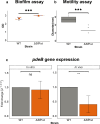
Figure 2.. Effects of prophages on biofilm and swimming in S. fidelis 3313.
(a) Influence of prophages on in vitro biofilm formation over 24 hours quantified with crystal violet assay (n=3), (b) role of prophages in swimming quantified as the diameter of spread on soft agar after 24 hours (n=6), and (c) fold-change of pdeB (with Rho as internal control) from 24 hour biofilm (in vitro) (n=4) and 24 hour in vivo (n=4). *p-value<0.05, **p-value<0.01; ns = not significant.
Figure 2—figure supplement 1.
Figure 2—figure supplement 1.. qPCR showing relative fold change gene expression with Rho as an endogenous control.
(a) Cyclic-di-GMP regulators expression in WT and ΔSfPat in vitro after 24 hours of exposure (n=4), (b) Cyclic-di-GMP regulators expression in WT and ΔSfPat in vivo of Ciona MS4 after 24 hours of exposure (n=4). ns = not significant.
Figure 3.
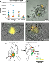
Figure 3.. The influence of SfPat prophage on gut colonization in Ciona.
(a) Results of six biological replicates (n=6, each replicate being a pool of ten juvenile tunicates) of the experimental exposure of Ciona MS4 juveniles to either WT or ΔSfPat strains for 1 hour and 24 hours; retention quantification displayed as a Beeswarm plot of colony-forming units (CFUs). There is significant retention observed in WT after 24 hours. The MS4 juveniles reveal differential colonization of WT and ΔSfPat after 1 hour of exposure (b–e), where WT strain is stained with BacLight Red and ΔSfpat is stained with BacLight Green reveal (b) WT is seen localized in the lower esophagus to anterior stomach, while the ΔSfPat deletion strain localized to the hindgut, while (c) the WT is seen localized mostly as a fecal pellet in the center of the stomach, while ΔSfPat prefers to localize to the stomach wall. (d) The WT strain is retained in the pyloric cecum. (e) Summary schematic of asymmetric bilateral views of MS4 animals; top of image is anterior and stomach is posterior. The ventral side is the ‘En’ side, and the dorsal side is the opposite side. The findings can be summarized as such: WT is retained in E and S, in PC, and also in the HG, while the ΔSfPat is retained in the stomach folds, MG, and portions of the HG. Some overlap in signal is noted with yellow coloring. En = endostyle, E = esophagus, S = stomach, MG = mid gut, HG = hind gut, PC = pyloric cecum.
Figure 3—figure supplement 1.
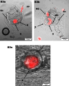
Figure 3—figure supplement 1.. BacLight-stained WT localization in Ciona MS4 after 1-hour exposure.
WT stained with BacLight Red exposed to Ciona MS4 for 1 hour. (a) WT found in esophagus (E), stomach (S), and fecal pellet (FP). (b) WT also found to occupy the hind gut (HG). (c) WT is retained in the center of the stomach but not the stomach walls.
Figure 3—figure supplement 2.
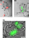
Figure 3—figure supplement 2.. BacLight-stained ΔSfPat localization in Ciona MS4 after 1-hour exposure.
ΔSfPat localization in Ciona MS4 after 1-hour exposure. (a) Stained with BacLight Red, ΔSfPat is found adhered to the stomach (S) and mid gut (MG). (b) Stained with BacLight Green, ΔSfPat is found in the mid gut and hind gut (HG). (c) ΔSfPat is more localized in the stomach fold than the center of the stomach, with presence in the mid gut.
Figure 4.
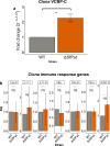
Figure 4.. The influence of prophages on host gene expression.
(a) VCBP-C gene expression in MS4 juveniles after 1 hour of exposure to S. fidelis 3313 strains (n=4). (b) Survey of additional innate immune gene expression in MS4 juveniles after 24-hour exposure to WT or ΔSfPat mutant strains (n=3). Actin is the internal control. *p-value<0.05, **p-value<0.01, ns = not significant.
Figure 5.
Figure 5.. Lysogen gene expression in response to host immune effector binding.
Gene expression of SfPat structural protein P5, recA and lexA of WT strain grown as a 24-hour biofilm while exposed to 50 μg/ml VCBP-C. Rho is the internal control (n=4). *p-value<0.05, ns = not significant.
Figure 5—figure supplement 1.
Figure 5—figure supplement 1.. RefFinder identifying Rho as the stable endogenous control.
Rho is the most stable gene across strains when tested in vitro (n=3).
Author response image 1.
Author response image 1.. WT S.
fidelis 3313 was exposed in vitro to 50 µg/ml VCBP-C in stationary cultures. Biofilms were observed for 24hrs. At 12 hrs, the presence of VCBP-C increased the amount of biofilms, whereas reduced biofilms were observed at 4 and 24hrs. Our findings (manuscript Fig 2a) reveal that SfPat contributes to biofilm formation, exposure to SfPat deletion mutants increases host VCBP-C expression (manuscript Fig. 4a), and VCBP-C binding to WT S. fidelis 3313 reduces the expression of SfPat P5 capsid protein (manuscript Fig. 5). These findings suggest that in vivo exposure/ colonization assays benefit from detailed time-course observations to be further explored in follow-up, future experiments.

Update of

  • doi: 10.1101/2022.11.23.517592
  • doi: 10.7554/eLife.103107.1
  • doi: 10.7554/eLife.103107.2

References

    1. Allen HK, Looft T, Bayles DO, Humphrey S, Levine UY, Alt D, Stanton TB. Antibiotics in feed induce prophages in swine fecal microbiomes. mBio. 2011;2:e00260-11. doi: 10.1128/mBio.00260-11. - DOI - PMC - PubMed
    1. Andersen CL, Jensen JL, Ørntoft TF. Normalization of real-time quantitative reverse transcription-PCR data: a model-based variance estimation approach to identify genes suited for normalization, applied to bladder and colon cancer data sets. Cancer Research. 2004;64:5245–5250. doi: 10.1158/0008-5472.CAN-04-0496. - DOI - PubMed
    1. Aron EJ, Trimble The bee swarm plot, an alternative to stripchart. d641db5Github. 2021 https://github.com/aroneklund/beeswarm
    1. Aziz RK, Edwards RA, Taylor WW, Low DE, McGeer A, Kotb M. Mosaic prophages with horizontally acquired genes account for the emergence and diversification of the globally disseminated M1T1 clone of Streptococcus pyogenes. Journal of Bacteriology. 2005;187:3311–3318. doi: 10.1128/JB.187.10.3311-3318.2005. - DOI - PMC - PubMed
    1. Banks DJ, Lei B, Musser JM. Prophage induction and expression of prophage-encoded virulence factors in group A Streptococcus serotype M3 strain MGAS315. Infection and Immunity. 2003;71:7079–7086. doi: 10.1128/IAI.71.12.7079-7086.2003. - DOI - PMC - PubMed

LinkOut - more resources