

doi: 10.1016/j.yaoo.2025.02.010.
Epub 2025 Jun 27.
Ocular Manifestations of Giant Cell Arteritis
Affiliations
- PMID: 40959132
- PMCID: PMC12435531
- DOI: 10.1016/j.yaoo.2025.02.010
Item in Clipboard
Ocular Manifestations of Giant Cell Arteritis
Adv Ophthalmol Optom.
2025 Aug.
No abstract available
Keywords: Giant cell arteritis; irreversible blindness; ischemic vasculitis; ocular imaging modalities; ocular manifestation.
Conflict of interest statement
Disclosure Statement: The Authors have nothing to disclose
Figures
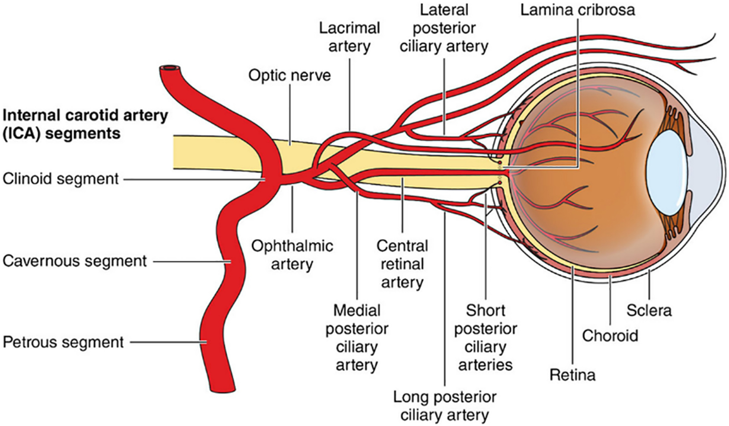
Blood supply to the retina. From Stroke.2021;52:e282-e294 ©2021 American Heart Association, Inc., with permission.

[A] is a normal OPTOS widefield image of the right eye which is the unaffected eye. [B] is an OPTOS widefield image of the left eye, which was affected with a cilioretinal artery occlusion. The cotton wool spot (yellow arrow) can be appreciated along the inferior aspect of the macula. [C] is an optical coherence tomography of the left eye affected with the cilioretinal artery occlusion and a paracentral acute middle maculopathy (red arrow) can be appreciated by the hyperreflective (whitening) band-like lesion in the inner nuclear layer of the retina along the inferior aspect of the macula. [D] is a fluorescein angiogram of the left eye affected with the cilioretinal artery occlusion demonstrating delayed filling (green arrow) in the inferior aspect of the macula.

CDUS of a TAB specimen demonstrating classical hypoechoic halo sign of GCA (arrows). (Left) Cross sectional view. (Right) Longitudinal view. From “The impact of temporal artery biopsy on surgical practice” by A. Cristaudo 2016 with permission.
References
Grants and funding
LinkOut - more resources
Full Text Sources