. 2025 Aug 30;21(12):5586-5608.
doi: 10.7150/ijbs.116513. eCollection 2025.

O-GlcNAcylated Hsp47 as a predictive biomarker in colorectal cancer: Kaempferol targets OGT-collagen axis for therapeutic intervention

Affiliations

O-GlcNAcylated Hsp47 as a predictive biomarker in colorectal cancer: Kaempferol targets OGT-collagen axis for therapeutic intervention

Chishun Zhou et al. Int J Biol Sci. .

Abstract

Colorectal cancer (CRC) is a highly lethal gastrointestinal malignancy, and its progression is closely related to abnormal protein O-GlcNAcylation modifications, especially during extracellular matrix (ECM) remodeling. Kaempferol is a natural flavonoid with medicinal value that can inhibit CRC progression through various pathways. However, it is unclear whether its mechanism of action involves O-GlcNAc-driven metabolic reprogramming. This study confirmed that kaempferol can significantly inhibit CRC growth both in vitro and in vivo and effectively reduce the overall protein O-GlcNAcylation levels. Mechanistic studies indicate that kaempferol reduces the levels of substrate uridine diphosphate N-acetylglucosamine (UDP-GlcNAc) and downregulates the expression of O-GlcNAc transferase (OGT), thereby decreasing the O-GlcNAcylation levels of proteins. This leads to a reduction in the O-GlcNAc modification of downstream heat shock protein 47 (Hsp47), which in turn affects the expression and intracellular localization of Hsp47, ultimately inhibiting the maturation and secretion of type I collagen, thereby blocking CRC progression. This study reveals a new mechanism by which kaempferol inhibits CRC by targeting the O-GlcNAcylation pathway. The study results suggest that O-GlcNAc-modified Hsp47 could serve as a potential therapeutic target for CRC and propose a treatment strategy guided by flavonoid biomarkers based on the inhibition of the OGT-collagen axis.

Keywords: Colorectal Cancer; Hsp47; Kaempferol; O-GlcNAc; OGT.


Conflict of interest statement

Competing Interests: Linlin Lu reported receiving funding from the National Natural Science Foundation of China during the conduct of the study. Zhongqiu Liu also reported receiving funding from the National Natural Science Foundation of China during the study. No funding was reported by the other authors.

Figures

Figure 1
Figure 1
Upregulation of O-GlcNAcylation in CRC tissues and cells. A, Analysis of a digestive system tissue microarray. B, Immunohistochemical detection of O-GlcNAcylation levels of proteins in cancer and adjacent tissues from 20 clinical CRC tissue samples. C, Fluorescence analysis of O-GlcNAcylation of proteins from four CRC patients at different TNM stages. DAPI (blue) marks the nucleus, phalloidin (red) marks the cytoskeleton, and O-GlcNAcylation is shown in green. D, Western blotting analysis of O-GlcNAcylation of proteins from four CRC patients at different TNM stages, where N represents adjacent normal tissue and T represents tumor tissue. E, O-GlcNAcylation in two colonic epithelial cell lines (HCoEpiC and NCM460) and six CRC cell lines (DLD-1, HCT116, RKO, HT-29, SW480, and Caco2). F, Analysis of O-GlcNAcylation in APCMin/+ mouse tissues. G, Kaplan‒Meier survival curve depicting the overall survival of 1,336 CRC patients stratified by O-GlcNAcylation level, where overall survival was defined as the time from the date of surgery to the date of death or last follow-up. The data are presented as the means ± SDs, * P < 0.05, ** P < 0.01, *** P < 0.001, ns, nonsignificant.
Figure 2
Figure 2
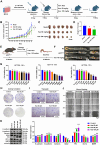
Kaempferol effectively inhibits CRC both in vitro and in vivo. A, MC38 cells were subcutaneously implanted into the right axillary region of C57BL/6 mice to establish a xenograft tumor model, followed by kaempferol treatment; each group in the animal experiment consisted of 6 mice (n=6). B, Effect of kaempferol on tumor volume in mice during treatment. C, Images of excised tumors from mice. D, Tumor weights of the mice. E, In the APCMin/+ mouse model, kaempferol was administered orally for 10 weeks in the treatment group. F, Tumor formation in APCMin/+ mice. G, The effect of kaempferol on the proliferation of HCT116 cells at 24 h, 48 h, and 72 h was assessed via the sulforhodamine B (SRB) assay. H, A colony formation assay was used to evaluate the effect of kaempferol on the stemness of HCT116 cells. I, The impact of kaempferol on HCT116 cell migration was examined via a Transwell assay. J, Wound healing assay was used to assess the effect of kaempferol on HCT116 cell migration. K, Western blotting analysis was performed to detect the influence of kaempferol on proteins related to metastasis, stemness, and proliferation in HCT116 cells. The data are presented as the means ± SDs, * P < 0.05, ** P < 0.01, *** P < 0.001, ns indicates no statistically significant difference.
Figure 3
Figure 3
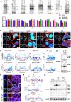
Kaempferol Inhibits protein O-GlcNAcylation in CRC. A, Detection and quantification of O-GlcNAcylation levels after treatment with kaempferol (25, 50, 100 µM) in two normal colon epithelial cell lines and six CRC cell lines. B, LSCM (laser scanning confocal microscopy) was used to detect the expression and localization of protein O-GlcNAcylation in HCT116, SW480, Caco2, and RKO cells. The nuclear marker DAPI is shown in blue, the cytoskeletal marker phalloidin is shown in red, and O-GlcNAcylation is shown in cyan. C, Analysis of the effect of kaempferol on protein O-GlcNAcylation in the four CRC cell lines. D, Western blotting analysis of the effect of kaempferol on protein O-GlcNAcylation in the APCMin/+ mouse model. E, LSCM detection of the effect of kaempferol on the expression and localization of protein O-GlcNAcylation in APCMin/+ mice. F, Analysis of the effects of kaempferol on protein O-GlcNAcylation in APCMin/+ mice via LSCM. G, Western blotting analysis of the effects of kaempferol on protein O-GlcNAcylation in the xenograft tumor tissues of mice. The data are presented as the means ± SDs, * P < 0.05, ** P < 0.01, *** P < 0.001, ns indicates no statistically significant difference.
Figure 4
Figure 4
The impact of OGA/OGT on the inhibition of protein O-GlcNAcylation by kaempferol in CRC. A, The process of protein O-GlcNAcylation catalyzed by OGT/OGA. B, Effects of various concentrations of kaempferol on glucose uptake in HCT116 cells. C, Effect of kaempferol on the mRNA expression of HBP-related catalytic enzymes. D, LC‒MS analysis of the effect of kaempferol on UDP-GlcNAc levels. E, Effects of kaempferol at different concentrations and time points on OGT/OGA expression. F, LSCM detection of the effects of kaempferol on OGT/OGA expression and cellular localization. G, Effects of various concentrations of kaempferol combined with OSMI-1 on protein O-GlcNAcylation. H, Effects of kaempferol combined with OSMI-1 and TMG on CRC cell proliferation. I, Colony formation assay to evaluate the effect of kaempferol combined with OSMI-1 and TMG on the colony-forming ability of CRC cells. J, Transwell assay to examine the effect of kaempferol combined with OSMI-1 and TMG on CRC cell migration. K, The subcutaneous tumor implantation experiment in mice was conducted to examine the effects of kaempferol treatment alone and its combination with the OGT inhibitor OSMI-4 on mouse tumors. The data are presented as the means ± SDs, * P < 0.05, ** P < 0.01, *** P < 0.001, ns indicates no statistically significant difference.
Figure 5
Figure 5
Kaempferol Targets OGT to Exert its Anti-CRC Effects. A, Effects of OGT knockdown and overexpression on kaempferol-mediated inhibition of HCT116 cell proliferation. B, Effects of OGT knockdown and overexpression on kaempferol-mediated inhibition of HCT116 cell stemness. C, Effects of OGT knockdown and overexpression on kaempferol-mediated inhibition of HCT116 cell migration. D, Fluorescence colocalization of kaempferol and OGT; DAPI labels the nucleus in blue, kaempferol shows intrinsic green fluorescence, and OGT is marked in red. E, SIP assay to detect the binding between kaempferol and OGT. F, CETSA confirming the interaction between kaempferol and OGT. G, Root mean square deviation (RMSD) of kaempferol and OGT during the 300 ns simulation. The RMSD measures the average displacement of selected atoms in a given frame relative to a reference frame, providing insights into protein structural dynamics and equilibrium during the simulation. For globular proteins, deviations within 1-3 Å are acceptable. H, Molecular docking between kaempferol and OGT. I, Conservation analysis of OGT amino acids. J, OGT protein expression after amino acid site mutation. K, The effects of the mutations on OGT stability were computed via PoPMusic 2.1 and I-mutant 3.0. L, Effects of mutations at the detection site on OGT enzyme activity as analyzed via the UDP-Glo™ glycosyltransferase assay kit. M, Effects of OGT amino acid site mutation on the ability of kaempferol to inhibit OGT expression. The data are presented as the means ± SDs, * P < 0.05, ** P < 0.01, *** P < 0.001, ns indicates no statistically significant difference.
Figure 6
Figure 6
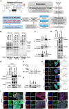
Kaempferol Reduces O-GlcNAcylation of Hsp47. A, Bioinformatics analysis and screening workflow for O-GlcNAcylated proteins in CRC. B, Effect of kaempferol on protein O-GlcNAcylation in the cytoplasm and nucleus of HCT116 cells. C, Immunoprecipitation validation of glycosylations in NNMT, Hsp47, and ARL8A. D, Immunoprecipitation analysis of the effect of kaempferol on Hsp47 glycosylation. E, Verification of the effect of kaempferol on the glycosylation of Hsp47 at the animal level. F, Click-iT enzyme labeling enrichment assay to evaluate the effect of kaempferol on Hsp47 glycosylation. G, Effect of kaempferol on Hsp47 protein expression. H, LSCM analysis of the effect of kaempferol on Hsp47 expression. The nuclear marker DAPI is shown in blue, Hsp47 in green, the cytoskeletal marker phalloidin in red, and O-GlcNAcylation in cyan. I, LSCM analyzes the expression of Hsp47 in cancer and matched adjacent non-cancerous tissues.
Figure 7
Figure 7
The role of Hsp47 in kaempferol-mediated inhibition of CRC. A, SRB assay assessing the effect of Col003 on the proliferative activity of CRC cells. B, Transwell assay evaluating the impact of Col003 on the migration of CRC cells. C, In the APCMin/+ mouse model, the treatment groups included oral administration of 50 mg/kg and 100 mg/kg of kaempferol, intraperitoneal injection of 0.5 mg/kg OSMI-1 and 1.0 mg/kg CoI003, as well as the combined use of kaempferol with OSMI-1 and CoI003, administered for 10 weeks. D, Tumor formation in APCMin/+ mice. E, HE staining was used to evaluate the pathological features of the colonic tissues of the animals. F, LSCM detection of Hsp47 and collagen I expression in mouse tissues; DAPI was used to label the nuclei in blue, collagen I in red, and Hsp47 in green. G, LSCM detection of the effect of kaempferol on the localization of Hsp47 within the cytoplasm, endoplasmic reticulum, and Golgi apparatus; DAPI is used to label the nuclei in blue, and the cytoplasm, endoplasmic reticulum, and Golgi apparatus are marked in red (phalloidin, calnexin, GM130), with Hsp47 in green. H, LSCM detection of the effect of Hsp47 knockdown on the localization of collagen I in the Golgi apparatus. The data are presented as the means ± SDs, * P < 0.05, ** P < 0.01, *** P < 0.001, ns indicates no statistically significant difference.
Figure 8
Figure 8
The O-GlcNAc modification site Ser76 on Hsp47 is crucial for the CRC inhibitory effect of kaempferol. A, Prediction of O-GlcNAcylation sites on Hsp47 via the O-GlcNAc Database v2.0, YinOYang-1.2, GPP, O-GlcNAcAtlas, and O-GlcNAcPRED-DL. B, Prediction of protein stability after mutation at potential glycosylation sites via PoPMuSiC 2.1 and I-Mutant 3.0. C, Immunoprecipitation to verify the effects of Hsp47 mutants S53A and S76A on the O-GlcNAcylation of Hsp47 protein. D, Immunofluorescence detection of the effects of Hsp47 mutants S53A and S76A on the O-GlcNAcylation of the Hsp47 protein. E, The Transwell experiment verifies the effect of Hsp47 mutants S53A and S76A on cell migration. F, Edu experiment verifies the effect of Hsp47 mutants S53A and S76A on cell proliferation. G, Immunofluorescence validation of the effects of Hsp47 mutants S53A and S76A on collagen I secretion function in cells. H, Western blotting verifies the functional impact of the mutant Ser76 on the interaction between Hsp47 and pro-collagen. I, CETSA verifies the effect of the S76A mutant on the thermal stability of Hsp47. J, Immunofluorescence analysis was performed to investigate the distribution of collagen I and procollagen I within the endoplasmic reticulum and Golgi apparatus in cells treated with kaempferol or OGT knockdown. K, Immunofluorescence analysis demonstrated that the blockade of O-GlcNAcylation at the Ser-76 site on Hsp47 inhibits kaempferol-mediated secretion of collagen type I. The data are presented as the means ± SDs, * P < 0.05, ** P < 0.01, *** P < 0.001, ns indicates no statistically significant difference.

References

    1. Siegel RL, Miller KD, Wagle NS, Jemal A. Cancer statistics, 2023. CA Cancer J Clin. 2023 Jan;73(1):17–48. - PubMed
    1. Fernández-Ponce C, Geribaldi-Doldán N, Sánchez-Gomar I, Quiroz RN, Ibarra LA, Escorcia LG, Fernández-Cisnal R, Martinez GA, García-Cózar F, Quiroz EN. The Role of Glycosyltransferases in Colorectal Cancer. Int J Mol Sci. 2021 May 30;22(11):5822. - PMC - PubMed
    1. Ma J, Wu C, Hart GW. Analytical and Biochemical Perspectives of Protein O-GlcNAcylation. Chem Rev. 2021;121(3):1513–1581. - PubMed
    1. Pinho SS, Reis CA. Glycosylation in cancer: mechanisms and clinical implications. Nat Rev Cancer. 2015;15(9):540–555. - PubMed
    1. Fromme JE, Zigrino P. The Role of Extracellular Matrix Remodeling in Skin Tumor Progression and Therapeutic Resistance. Front Mol Biosci. 2022 Apr 26;9:864302. - PMC - PubMed