. 2025 Sep 1.
doi: 10.1039/d5sc04734j. Online ahead of print.

Engineering ultrapotent trivalent anticoagulants through hybridisation of salivary peptides from multiple haematophagous organisms

Affiliations

Engineering ultrapotent trivalent anticoagulants through hybridisation of salivary peptides from multiple haematophagous organisms

Joshua W C Maxwell et al. Chem Sci. .

Abstract

Haematophagous organisms are a rich source of salivary anticoagulant polypeptides that exert their activity by blocking the catalytic site and one of two positively charged exosites on the host protease thrombin. Here, we describe a molecular engineering approach to hybridise post-translationally sulfated polypeptides from different blood-feeding organisms to enhance anticoagulant activity. This led to the discovery of a triply sulfated hybrid anticoagulant, XChimera, possessing fragments from flea, leech, and fly salivary polypeptides that exhibits femtomolar inhibitory activity against thrombin. The crystallographic structure of a complex of XChimera with thrombin shows that it displays a trivalent binding mode in which it simultaneously blocks three functional sites of the protease, the active site and exosites I and II. This trivalent chimera exhibited ultrapotent anticoagulant activity in a suite of in vitro clotting assays and was also shown to possess potent in vivo antithrombotic activity in a murine model of thrombosis.


Conflict of interest statement

During the performance of all research studies I. A. and S. M. S. were employed by the Heart Research Institute, and affiliated with the University of Sydney, receiving salaries funded by academic grants. These authors are currently employed by ThromBio Pty Ltd, with the latter employment commencing after initial submission of the manuscript. S. P. J. is the founder and director of ThromBio Pty Ltd.

Figures
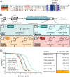
Fig. 1
Fig. 1. Engineering of the C-terminal region of flea thrombin inhibiting anticoagulant XC43. (A) Alignment of amino acid sequences of XC43 (UniProt entry A2IAB2), hirudin(40–65) (UniProt entry P09945), and variegin (UniProt entry P85800). (B) Schematic depicting the synthesis of a library of XC43 derivatives (1–7) using Fmoc-SPPS as outlined in the methods and SI. (C) Inhibition profiles of XC43-derived peptides (1–7) against α-thrombin using a chromogenic substrate assay. Data are expressed as the mean of two technical replicates and error bars plotted as SD. The inhibition constants (Ki) for the peptides (± standard errors) were determined by fitting the inhibited steady-state velocity data to the Morrison model.
Fig. 2
Fig. 2. Rational design, synthesis and profiling of a trivalent thrombin inhibitor. (A) Overlay of mature peptide and protein sequences of hirudin(55–65) (UniProt entry P09945), XC43 (UniProt entry A2IAB2) and TTI (UniProt entry O97373) and design of a chimeric inhibitor. (B) Synthesis of TTI-XC43-Hiru (8) and XChimera (9) using Fmoc-SPPS as outlined in the methods and SI (final isolated yields are provided in parentheses over all solid-phase synthesis steps). (C) Inhibition profiles of XC43-derived peptides 1, 7, 8 and 9 against γ-thrombin using a chromogenic substrate assay. Data are expressed as the mean of two independent experiments and error bars plotted as SD. The inhibited steady-state velocity data were fitted to the Morrison model. (D) Perturbations to thermal shift of thrombin in the presence of XC43-derived inhibitors. Melting temperatures (Tm) were determined as the point of inflection in the fluorescence emission ratio of 350/330 and the difference in melting temperature (ΔTm) was determined as the difference with thrombin alone. Data are representative of 4–6 technical replicates.
Fig. 3
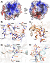
Fig. 3. XChimera (9) is a trivalent binder of human α-thrombin. (A) The acidic N-terminal segment (residues 1–12) of XChimera (9) (stick model with nitrogen atoms blue, oxygen red, sulfur yellow, and carbon tan; PDB entry 8RTN) binds to the exosite II (left) of thrombin (solid surface representation with mapped surface electrostatic potential [blue, positive; red, negative]), whereas the C-terminal segment (residues 22–48) occupies the active-site cleft and recognises the exosite I of the proteinase (right). The 2Fo–Fc electron density map (contoured at 0.9σ) for XChimera (9) is displayed as a black mesh. Thrombin orientations in the left and right panels are related through a 120° rotation around y. (B) Main interactions between the sulfotyrosine-containing motif (residues 7–12) of XChimera (9) (coloured as in (A)) and the exosite II of thrombin (transparent tan cartoon with selected residues as sticks, colour-coded as for XChimera (9)). The structural superposition of human α-thrombin in complex with TTI (mauve; PDB entry 6TKG) is represented as for the XChimera (9) complex. (C) Close-up view of the interactions between the sulfated Y46 of XChimera (9) and the exosite I of the proteinase (selected residues are represented and coloured as in (B)). Superposed human α-thrombin in complex with hirudin (orange; PDB entry 2PW8) is shown with selected residues of the proteinase represented as for the XChimera (9) complex. For clarity, only Y63 of hirudin is shown. Water molecules involved in the recognition of sulfate groups of Y46 (XChimera (9)) or Y63 (hirudin) are represented as red and orange spheres, respectively. (D) Close-up on the active site of the superposed α-thrombin molecules complexed to either XChimera (9) or XC43 (pale cyan; PDB entry 7MJ5). Residues at positions P2, P1, and P1′ from both inhibitors (in XChimera P27, K28, and L29, respectively) are represented as sticks and colour-coded as in (B). The catalytic triad (H57, D102 and S195) and D189 at the bottom of the S1 specificity pocket of the proteinase are represented as sticks and colour-coded as for the inhibitors. (E) Structural differences between XC43 and XChimera (9) (represented as in (D)) at the exosite I due to the substitution of the XC43 C-terminal region (residues V29-A36) by the C-terminal and tyrosine-sulfated region of hirudin (Y63-Q65). Dotted black lines represent hydrogen bonds and superscripts T, H, X, and (9) denote residues corresponding to α-thrombin, hirudin, XC43, and XChimera (9) molecules, respectively.
Fig. 4
Fig. 4. In vitro assessment of the anticoagulant potential of thrombin inhibitors 1, 7 and 9. (A) Effect of the thrombin inhibitors on the aPTT of human plasma in vitro. See (B) for key. (B) Calibrated automated thrombogram of human plasma in the presence of 120 nM 1, 7 and 9. (C) ESI mass spectrum of XChimera (9) following incubation in pooled human plasma for 24 hours at 200 μM. Data are presented as the average mass spectrum over the total ion current. (D) Analytical HPLC chromatograms (λ = 214 nm) of XChimera (9) prior to (t = 0) and following incubation with human α-thrombin for 4 h at 37 °C. (E) Examination of the activity of XChimera (9) on α- and γ-thrombin-induced platelet aggregation. Washed human platelets isolated as described in the methods section were preincubated with vehicle, or the indicated concentration of XChimera (9) or sulfo-XC43-Hiru (7), prior to addition of α- or γ-thrombin (0.5 or 20 nM, respectively). Platelet aggregation was analysed as described in the methods section. Data are represented as the mean ± SEM of % maximal platelet aggregation in response to α- (left) and γ-thrombin (right) resulting from experiments performed with at least four independent blood donors (n = 4–6).
Fig. 5
Fig. 5. In vivo evaluation of XChimera (9) in a murine model of thrombosis. (A) Ex vivo aPTT measurements following bolus intravenous administration of XChimera (9) at varying doses. (B) Average fibrin intensity over duration of measurement. (C) Average thrombus volume over duration of measurement. (D) Time course of fibrin intensity following injury with and without treatment. (E) Time course of thrombus volume following injury with and without treatment. (F) Exemplar images of thrombus following needle injury after treatment with 9 or hirudin. Images were acquired 15 min post injury. Fibrin is represented in green and platelets in red.

References

    1. Townsend N. et al. . Nat. Rev. Cardiol. 2022;19:133–143. - PubMed
    1. Magnussen C. et al. . N. Engl. J. Med. 2023;389:1273–1285. - PMC - PubMed
    1. Vogel B. et al. . Lancet. 2021;397:2385–2438. - PubMed
    1. Feigin V. L. et al. . Lancet Neurol. 2021;20:795–820. - PMC - PubMed
    1. Campbell B. C. V. et al. . Nat. Rev. Dis. Primers. 2019;5:70. - PubMed

LinkOut - more resources