. 2025 Sep;14(18):e71227.
doi: 10.1002/cam4.71227.

Focal Adhesion Kinase Intersects With the BRD4-MYC Axis and YAP1 to Drive Tumor Cell Growth, Phenotypic Plasticity, Stemness, and Metastatic Potential in Colorectal Cancer

Affiliations

Focal Adhesion Kinase Intersects With the BRD4-MYC Axis and YAP1 to Drive Tumor Cell Growth, Phenotypic Plasticity, Stemness, and Metastatic Potential in Colorectal Cancer

Rongbo Han et al. Cancer Med. 2025 Sep.

Abstract

Objective: Colorectal cancer (CRC) remains one of the leading causes of cancer-related death worldwide due to the lack of effective therapies. Here we explored the clinical basis and therapeutic promise of the integrin-focal adhesion kinase (FAK)-dependent pathway for CRC.

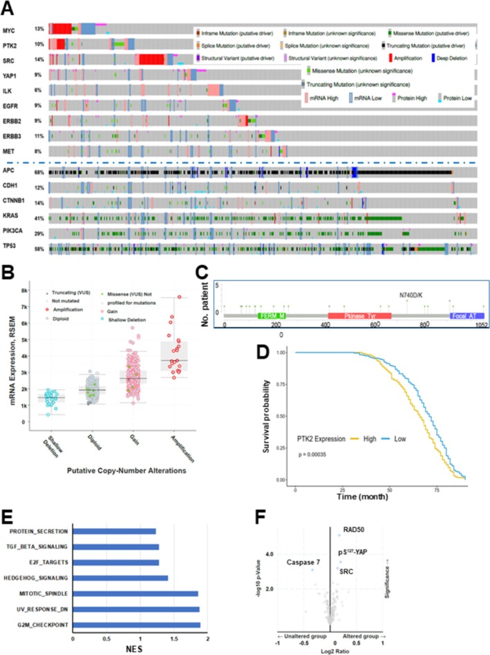
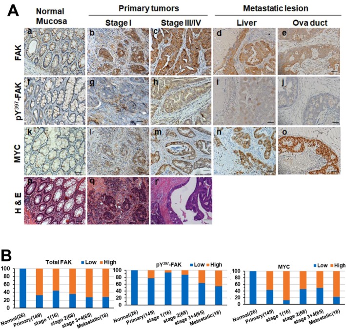
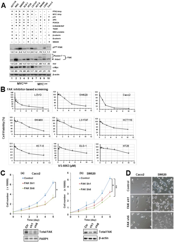
Methods and results: Our bioinformatic and histological analyses showed that FAK was markedly upregulated at both mRNA and protein and signaling levels in the two CRC patient cohorts. Particularly, the portion of carcinomas carrying active FAK (Y397phosphorylation) increased by threefold from stage I to III/IV tumors or metastatic lesions. Consistent with this clinic landscape, FAK inhibition via knockdown or chemical inhibitors suppressed tumor cell growth largely in the subset of CRC cell lines with low MYC expression. In contrast, the FAK inhibition was less effective in the cell line pool with high MYC expression. The resistance to FAK targeting diminished upon a co-inhibition of BRD4 via BET inhibitors. It coincided with an induction of cell cycle arrest at G1-S and G2-M phases, elevated apoptosis and chemosensitivity (paclitaxel and oxaliplatin), and impaired stemness. Mechanistically, the BET inhibitor induced an EMT-like phenotype, tilting tumor cell dependence toward the integrin-FAK axis. Moreover, inhibiting FAK alone or in combination with SRC or BRD4 markedly suppressed cell motility and the YAP or MYC activation, and restored the expression of the long isoform BRD4. Also, co-genomic/genetic dysregulations of FAK and YAP1 or SRC strongly correlated with poor disease-free patient survival.

Conclusion: Overall, our study highlights the potent pro-malignant role of the integrin-FAK axis in CRC, fueling its targeting as a single agent or synthetic lethal-based therapy.

Keywords: BRD4; FAK; MYC; colorectal cancer; metastasis; tumor growth.


Conflict of interest statement

The authors declare no conflicts of interest.

Figures

FIGURE 1
FIGURE 1
Probing genomic and transcriptional dysregulation of integrin‐dependent pathways and associated oncogenic pathways in a colorectal cancer patient cohort from the TCGA database. (A) Tumor samples with mutations and CNA data (N = 594 samples/patients) from a colorectal adenocarcinoma cohort (TCGA, PanCancer Atlas), which spanned all stages of CRC primary tumors (N = 594), were probed for altered level of mRNA, gene mutations, structural variants of FAK (PTK2) and other integrin‐dependent signaling effectors as well as oncogenic drivers via use of c‐Bioportal software. The threshold of z‐score relative to normal samples (log RNA Seq V2 RSEM) was set at ±2. (B) Association between the gene copy alterations of PTK2 (FAK) and mRNA expression in the cohort. (C) Occurrence of point mutations in FAK gene in the cohort. (D–F) The overall patient survival was plotted for the populations with high or low mRNA expression of FAK in the TCGA cohort. Y axis: Probability of patient survival. X axis: Duration of patient survival (months). Patient survival curves were compared using log‐rank test to assess differences in disease free survival (DFS) between the high vs. low expression groups. Hazard ratios (HR) with 95% CI were calculated to quantify the risk associated with high vs. low expression of relevant genes. The data was normalized by transcripts per million (TPM). A list of pathways and genes from the gene enrichment analysis via the Hallmark‐based logarithm (E, F). (F) X axis: Values of NES (FDR value < 0.05).
FIGURE 2
FIGURE 2
IHC analysis of total and active FAK and MYC expression in adenocarcinomas and distant metastatic lesions from a local CRC patient cohort. (A) Representative images of H&E and IHC staining for FAK, active FAK (pY397), and MYC in normal colonic mucosa or tumor‐adjacent tissues (a, f, k and p), stage I (b, g, I and q) and stage III/IV (c, h, m and r) colorectal adenocarcinomas, as well as liver (d, I and n) and oviduct (e, j and o) metastatic lesions. Panels show H&E (p, q, r), total FAK (a–e), pY397‐FAK (f–j), and MYC (k–o) staining. (B) Proportion of tumors with low (blue) or high (red) protein expression of total FAK, pY397‐FAK, and MYC across tumor stages in the CRC patient cohort (%). The x‐axis indicates the source of tissues and number of tumors examined. Antibody staining values are shown separately for cytoplasmic and nuclear expression of FAK, pY397‐FAK, and MYC. Scale bar: 100 μm.
FIGURE 3
FIGURE 3
FAK dependence in colorectal adenocarcinoma cells. (A) Aberrant expression of total FAK, active FAK (pY397), and MYC across a panel of CRC cell lines analyzed by immunoblotting. Tumor cells were cultured in 6‐well plates, lysed, and probed with the indicated antibodies. Protein expression differences between cell lines were quantified by densitometry (OD values) relative to HCT116 (lane 1), normalized to β‐actin loading. Status of oncogenic drivers is indicated: Amp, gene amplification; additional oncogenes harboring point mutations or deletions are listed. Putative PTK2–TRAPPC9 fusion gene products are also noted. (B) CRC cell line responses to escalating doses of the FAK inhibitor VS‐6063. Cells seeded in 48‐ or 96‐well plates were pre‐cultured overnight in 10% FBS and treated for 72 h with the indicated doses of VS‐6063. Viability was determined by MTT assay. Y‐axis: cell viability (% of control, 0.1% DMSO); mean ± SEM, n = 3. (C) Effect of FAK knockdown on proliferation. Caco2 and SW620 cells with (w) or without (w/o) stable FAK knockdown were seeded in 96‐well plates with 10% FBS and monitored for changes in cell number over time. Y‐axis: cell number; mean ± SEM, n = 3. Validation of FAK knockdown was confirmed by immunoblotting; β‐actin served as a loading control. Control: parental cells or cells treated with scramble shRNA. (D) Representative images of Caco2 (a–c) and SW620 (d–f) cells with or without stable FAK knockdown. Cells were seeded in 12‐well plates, cultured overnight, and imaged microscopically. p < 0.05; p < 0.01.
FIGURE 4
FIGURE 4
Crosstalk between the integrin–FAK and BRD4–MYC axes in CRC cell growth. Human and mouse CRC cell lines were probed pharmacologically to assess interactions between the integrin–FAK axis and MYC or other oncogenic pathways. (A, D) Cell viability analyses. HCT116 and MC38 cells were treated for 72 h with the indicated doses of various inhibitors, either alone or in combination, followed by MTT assay. Y‐axis: cell viability (mean ± SEM, n = 3). (B, C) Cell cycle and apoptosis analyses. CRC cells cultured under 5% FBS were treated with the indicated inhibitors and examined for effects on (B) cell cycle distribution by flow cytometry or (C) apoptosis/biochemical changes by immunoblotting. For cell cycle analysis, triplicate cultures were treated with control (0.1% DMSO) or the indicated inhibitors for 48 h before analysis. Data are shown as mean % (± SEM, n = 3) of cells in each phase. Groups with the same letters differ significantly in % G2/M phase or apoptosis (p < 0.05, multiple comparisons). For immunoblotting, tumor cells were seeded in 12‐well plates, treated with inhibitors for 24 h, lysed in RIPA buffer, and probed with the indicated antibodies. β‐actin served as loading control. Top panel: LS174T cells treated with VS‐6063 (lanes 2–3, 6–9) and JQ1 (lanes 4–9). Bottom panel: HCT116 cells treated with VS‐6063 (lanes 2–4, 7–12) and JQ1 (lanes 5–12). Lane 1: 0.1% DMSO control.
FIGURE 5
FIGURE 5
Links between FAK and E‐cadherin, β‐catenin, MYC, BRD4, and YAP1 in pro‐metastatic functions and distant metastasis in vivo. (A) Representative wound‐healing–like migration assays (n = 3) showing the effects of FAK inhibition (VS‐6063, “VS”) or BET inhibition (JQ1) on cell motility compared to DMSO control in HT‐29 (a) and HCT116 (b) cells. (B) Representative images of IHC staining of E‐cadherin (a–e), β‐catenin (f–j), and BRD4 (k–o) in normal intestinal mucosa (a, f and k), stage I primary tumors (b, g and l), stage III primary tumors (c, h and m),and distant metastatic lesions in the liver (d, j and n) or oviduct (e, j and o). (C) IHC staining of FAK (a), pY397‐FAK (b), E‐cadherin (c), β‐catenin (d), and MYC (e) in paraffin‐embedded, cell‐enriched pellets derived from patient pleural fluids or ascites. (D) IHC staining of fibronectin in CRC tissues from stage I (a), stage II (b), and stage III (d–e) tumors. Scale bar : 100 μm (all panels).
FIGURE 6
FIGURE 6
Role of the integrin–FAK signaling axis in CRC stemness and drug sensitivity. (A, B) Effect of FAK inhibition on tumor cell chemosensitivity. (A) Cell viability analysis. CRC cell lines (SW620, HCT116, HT‐29) were seeded in 48‐ or 96‐well plates (triplicates), cultured in 5% FBS overnight, and treated with chemotherapeutic agents (paclitaxel, oxaliplatin, irinotecan) in the presence or absence of FAK inhibitor (VS‐6063, “VS”) for 48–72 h prior to MTT assay. For comparison, the effects of Wnt pathway inhibition (ICG001) or BET inhibition (JQ1) were also tested with paclitaxel and oxaliplatin. X‐axis: drug dose; Y‐axis: cell viability (% of DMSO control, < 0.1% DMSO), mean ± SEM, n = 3. (B) Representative images of tumor cells treated with VS‐6063 with or without irinotecan for 24 h. (C) Effect of FAK and BET inhibition on tumorsphere formation (stemness). HCT116 cells were seeded into ultra‐low–attachment 24‐well plates (n = 2–3), incubated overnight, and treated with DMSO or the indicated doses of VS‐6063 and JQ1. Tumorspheres were imaged microscopically after 48–60 h. Representative microscopic fields (2–3 per condition) are shown for DMSO and inhibitor treatments (a–i).
FIGURE 7
FIGURE 7
Clinical association of FAK, SRC, and YAP1 with therapeutic relevance in CRC. (A–D) Disease‐free survival (DFS) analysis in the TCGA CRC cohort (PanCancer Atlas, N = 594). (a) Kaplan–Meier DFS curves comparing patients with or without genomic alterations, mutations, or dysregulated mRNA expression (z‐score ±2 relative to normal; log RNA Seq V2 RSEM) of FAK, SRC, or YAP1. Y‐axis: patient survival (%); X‐axis: DFS time (months). p values are indicated. (b) Volcano plots of aberrant protein expression in altered vs unaltered groups. Genes with marked differences are labeled. (c) Profiles of key oncogenic driver gene alterations across groups. (E) Correlation analysis of FAK, c‐Src, and YAP1 expression in the TCGA cohort. (F) Representative IHC staining of YAP1 in normal mucosa (a), stage I tumors (b), stage III/IV tumors (c), and metastatic lesions from liver (d) and oviduct (e). Scale bar: 100 μm. (G) Effect of FAK knockdown ± chemotherapeutic agents on YAP1 and SFK (SRC family kinase) activation. SW620 cells with or without stable FAK knockdown were cultured overnight, treated with paclitaxel (PTX) or 5‐fluorouracil (5‐FU) for 24 h, lysed in RIPA buffer, and immunoblotted. β‐actin served as a loading control. Fold changes are indicated. (H) Proposed working model of functional interactions between the integrin–dependent pathways, YAP1, SRC and the BRD4–MYC axis in CRC progression.

References

    1. Bray F., Laversanne M., Sung H., et al., “Global Cancer Statistics 2022: GLOBOCAN Estimates of Incidence and Mortality Worldwide for 36 Cancers in 185 Countries,” CA: A Cancer Journal for Clinicians 74, no. 3 (2024): 229–263. - PubMed
    1. Cornish A. J., Gruber A. J., Kinnersley B., et al., “The Genomic Landscape of 2,023 Colorectal Cancers,” Nature 633, no. 8028 (2024): 127–136. - PMC - PubMed
    1. Cancer Genome Atlas Network , “Comprehensive Molecular Characterization of Human Colon and Rectal Cancer,” Nature 487, no. 7407 (2012): 330–337. - PMC - PubMed
    1. Ma Y. S., Huang T., Zhong X. M., et al., “Proteogenomic Characterization and Comprehensive Integrative Genomic Analysis of Human Colorectal Cancer Liver Metastasis,” Molecular Cancer 17, no. 1 (2018): 139. - PMC - PubMed
    1. Dienstmann R., Vermeulen L., Guinney J., Kopetz S., Tejpar S., and Tabernero J., “Consensus Molecular Subtypes and the Evolution of Precision Medicine in Colorectal Cancer,” Nature Reviews. Cancer 17, no. 4 (2017): 268. - PubMed

MeSH terms