. 2025 Sep 15;152(18):dev204814.
doi: 10.1242/dev.204814. Epub 2025 Sep 22.

Automated, high-throughput in situ hybridization of sea urchin (Lytechinus pictus) embryos

Affiliations

Automated, high-throughput in situ hybridization of sea urchin (Lytechinus pictus) embryos

Yoon Lee et al. Development. .

Abstract

Despite the reach of in situ hybridization (ISH) in developmental biology, it is rarely used at scale. The major bottleneck is the throughput of the assay, which relies upon labor-intensive manual steps. The goal of this study was to develop a high-throughput, automated hybridization chain reaction (HCR) pipeline for the sea urchin (Lytechinus pictus). Our method, which we term high-throughput (HT)-HCR, can process 192 gene probe sets on whole-mount embryos within 32 h. The physical properties of sea urchin embryos enabled us to utilize a 96-well plate format, miniaturized reaction volumes, a general-purpose robotic liquid handler and automated confocal microscopy. Using this approach, we produced high quality localization data for 101 target genes across three developmental stages. The results reveal the localization of previously undescribed physiological genes, as well as canonical developmental transcription factors. HT-HCR represents an order of magnitude increase in the throughput of spatial expression profiling studies utilizing the sea urchin. This will enable more-sophisticated perturbation analyses and drug-screening efforts in this emerging animal model.

Keywords: In situ hybridization (ISH); Lytechinus pictus; High-throughput; Hybridization chain reaction (HCR); Sea urchin; Spatial transcriptomics; Transcription factor; Transporter.


Conflict of interest statement

Competing interests The authors declare no competing or financial interests.

Figures
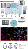
Fig. 1.
Fig. 1.
Diagram of a fully automated HCR workflow. (A) Automated sample processing using the Opentrons Flex robot. (A1) The consumable probe plate as the starting vessel. A 96-well plate, where each well contains 5 μl of a pool of probes for four mRNA targets is used as the starting vessel for sample processing. For all liquid handling steps throughout the protocol, dispense speeds are set at 200 μl/s and aspiration speeds at 10 μl/s. All aspirations are set to use a tip to well bottom distance of 3 mm. (A2) The probe hybridization step. 10 μl of fixed embryos in hybridization buffer are transferred to each well to start the probe hybridization step. The inset illustrates transferability of sea urchin embryos using standard automation pipette tips. (A3) The probe wash step. Probes are washed from the sample using a formamide-based probe wash buffer. The insets illustrate the liquid handling speeds and tip to bottom distances allow for resuspension and mixing of embryos throughout washes. (A4) The amplification step. 10 μl of embryos are transferred to a plate of pre-plated amplifier hairpins in dextran sulfate. (A5) The amplifier wash step. Amplifier hairpins are washed using 5×SSCT, and 150 μl of samples in 5×SSCT are subsequently transferred to a glass bottom high-content imaging plate. Samples are centered using a plate shaker module set at 450 rpm for 30 min. (B) Automated confocal image acquisition using the ImageXpress HT.ai. The imaging plates containing centered samples are placed into the confocal microscope. Depending on data storage availability, the center 4 or 9 are imaged for each well. An example of an output of the demultiplexed and multiplexed versions of a single site of a positive control sample well is shown. (C) A schematic of developmental stages imaged in this study to be used as a developmental staging key. PMCs, primary mesenchyme cells.
Fig. 2.
Fig. 2.
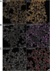
Comparison of results from manual and automated assays. (A,B) Images show the localization of several positive control targets in 24 hpf embryos. Manually processed samples (A) and robotically processed samples (B) appear identical. Nuclear staining using Hoechst 33342 appears in gray. Scale bars: 100 μm.
Fig. 3.
Fig. 3.
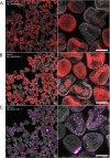
Validation of HT-HCR by localization of previously described transcription factor genes. All images contain a mix of 12, 24 and 36 hpf embryos, and are automatically demultiplexed and stitched outputs resulting from an automated image processing workflow. Each image contains between 50 and 100 embryos, which are annotated with the NCBI RefSeq ID for the mRNA sequence targeted by probe oligonucleotides. (A) onecut2: expression is sparse in 12 hpf embryos. In 24 hpf and 36 hpf embryos, expression is concentrated in the ciliary band. (B) scratch: expression is either sparse or absent in 12 hpf embryos. At 24 hpf, expression is concentrated in the archenteron. At 36 hpf, expression is restricted to the coelomic pouches. (C) homeobrain: at 12 hpf, expression is present in the apical ectoderm. At 24 and 36 hpf, expression is in the apical ectoderm and stomodeum. In more developed 36 hpf embryos, expression can be seen in the upper region of the developing oral hood. (D) XHOX-3 and eve2: expression is most enriched in 12 hpf embryos in a ring of cells in the aboral ectoderm. Sparse expression is visible in some 24 hpf embryos. (E) dri: expression is most enriched in 24 hpf embryos and is restricted to the oral ectoderm. Sparse expression is visible along the ciliary band in 36 hpf embryos. (F) six1: expression is restricted to the coelomic pouches in 36 hpf embryos. Scale bars: 100 μm.
Fig. 4.
Fig. 4.
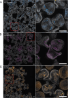
HT-HCR to localize previously undescribed cellular structure, enzyme and transcription factor genes. All images contain a mix of 12, 24 and 36 hpf embryos, and are automatically demultiplexed and stitched outputs resulting from an automated image processing workflow. Each image contains between 50 and 100 embryos, and are annotated with the NCBI RefSeq ID for the mRNA sequence targeted by probe oligonucleotides. (A) Genes involved in cellular structure: gelsolin-like protein 2 and mucin-2. gelsolin-like protein 2: expression does not appear to be present in 12 hpf embryos. In 24 hpf embryos, expression is visible in PMCs and SMCs. mucin-2: expression is faint or absent in 12 hpf embryos. Expression is visible in the ectoderm in 24 and 36 hpf embryos. (B) Enzymes: D-dopachrome decarboxylase and hematopoietic prostaglandin D synthase. D-dopachrome decarboxylase: expression is visible in the NSM in 12 hpf embryos. Expression is visible in mesodermal cells in 24 hpf embryos. Expression is faint in 36 hpf embryos – some samples show signal in mesodermal cells and the midgut. hematopoietic prostaglandin D-synthase: expression is not present in 12 hpf embryos. In 24 hpf embryos, expression is visible in the gut tube. By 36 hpf, expression is localized in the midgut and foregut. (C) Transcription factors: thyroid transcription factor 1 and odd-skipped related 1 protein. thyroid transcription factor 1: expression is either faint or absent in 12 hpf embryos. In 24 hpf embryos, expression is visible in the apical ectoderm. In 36 hpf embryos, expression is visible in the oral hood. odd-skipped related 1 protein: expression is faint or absent in 12 hpf embryos. In 24 hpf embryos, expression appears to concentrate in the gut tissue. By 36 hpf, expression is restricted to the cardiac sphincter. Scale bars: 100 μm.
Fig. 5.
Fig. 5.
Previously unreported ABC transporter expression patterns revealed by HT-HCR. Images were collected and annotated as in Fig. 4. (A) ABCB7: expression is low in 12 hpf embryos. Expression is somewhat ubiquitous but concentrated in mesodermal cells in 24 hpf embryos. Expression is restricted to pigment cells by 36 hpf. (B) ABCA3: expression is ubiquitous in 12 hpf and 24 hpf embryos. Expression is concentrated in the midgut in 36 hpf embryos. (C) ABCG12: expression is concentrated in the NSM in 12 hpf embryos. At 24 hpf and 36 hpf, expression is concentrated in mesodermal cells. Scale bars: 100 μm.
Fig. 6.
Fig. 6.
Previously unreported SLC transporter expression patterns revealed by HT-HCR. Images were collected and annotated as in Fig. 4. (A) SLC5A7: expression is either sparse or absent in 12 hpf embryos. At 24 hpf, expression is present in what appears to be neuronal precursor cells. At 36 hpf, expression is either sparse or absent. (B) SLC18A3: expression is either sparse or absent in 12 hpf embryos. At 24 hpf, expression is concentrated in what appears to be neuronal precursor cells. Expression is concentrated in cells distributed throughout the ciliary band in 36 hpf embryos. (C) SLC6A14: expression is either sparse or absent in 12 hpf and 24 hpf embryos. At 36 hpf, expression is concentrated in one of the coelomic pouches. Scale bars: 100 μm.
Fig. 7.
Fig. 7.
Aquaporin-8 and Strp-4 expression patterns revealed by HT-HCR. Images were collected and annotated as in Fig. 4. (A) aquaporin-8: expression is either sparse or absent in 12 hpf embryos. At 24 hpf and 36 hpf, expression concentrated in the gut. (B) short transient receptor potential channel 4: at 12 hpf, expression is concentrated in the NSM. At 24 hpf and 36 hpf, expression is concentrated in mesodermal cells. Scale bars: 100 μm.

Update of

References

    1. Agre, P. and Kozono, D. (2003). Aquaporin water channels: molecular mechanisms for human diseases. FEBS Lett. 555, 72-78. 10.1016/S0014-5793(03)01083-4 - DOI - PubMed
    1. Allikmets, R., Raskind, W. H., Hutchinson, A., Schueck, N. D., Dean, M. and Koeller, D. M. (1999). Mutation of a putative mitochondrial iron transporter gene (ABC7) in X-linked sideroblastic anemia and ataxia (XLSA/A). Hum. Mol. Genet. 8, 743-749. 10.1093/hmg/8.5.743 - DOI - PubMed
    1. Amore, G., Yavrouian, R. G., Peterson, K. J., Ransick, A., McClay, D. R. and Davidson, E. H. (2003). Spdeadringer, a sea urchin embryo gene required separately in skeletogenic and oral ectoderm gene regulatory networks. Dev. Biol. 261, 55-81. 10.1016/S0012-1606(03)00278-1 - DOI - PubMed
    1. Anderson, S. L., Hose, J. E. and Knezovich, J. P. (1994). Genotoxic and developmental effects in sea urchins are sensitive indicators of effects of genotoxic chemicals. Environ. Toxicol. Chem. 13, 1033-1041. 10.1002/etc.5620130704 - DOI
    1. Anderson, C. M. H., Ganapathy, V. and Thwaites, D. T. (2008). Human solute carrier SLC6A14 is the beta-alanine carrier: SLC6A14 is the β-alanine carrier. J. Physiol. 586, 4061-4067. 10.1113/jphysiol.2008.154500 - DOI - PMC - PubMed

MeSH terms

LinkOut - more resources