. 2025 Sep 26;137(8):1117-1132.
doi: 10.1161/CIRCRESAHA.125.326758. Epub 2025 Sep 3.

Septin4 Regulates Cardiac Fibrosis After Pressure Overload

Affiliations

Septin4 Regulates Cardiac Fibrosis After Pressure Overload

Doğacan Yücel et al. Circ Res. .

Abstract

Background: In response to cardiac injury the mammalian heart undergoes ventricular remodeling to maintain cardiac function. These changes are initially considered compensatory, but eventually lead to increased cardiomyocyte apoptosis, reduced cardiac function and fibrosis which are important contributors to the development of heart failure. The small GTPase Sept4 (Septin4) has previously been implicated in the regulation of regeneration and apoptosis in several organs. However, the role of Sept4 in regulating the response of the heart to stress is unknown.

Methods: Ten-week-old wild-type (WT) and Sept4 knockout mice were subjected to transverse aortic constriction to induce cardiac injury. Genotype-dependent differences were investigated at baseline and at 1- and 4-week postinjury time points. To definitively establish the fibroblast-specific cardioprotective effects of Sept4, we generated a fibroblast-specific Sept4 conditional knockout model.

Results: Under homeostatic conditions Sept4 knockout mice showed normal cardiac function comparable with WT controls. In response to transverse aortic constriction, WT mice developed reduced cardiac function and heart failure, accompanied by an increase in cardiomyocyte apoptosis. In contrast, knockout mice were protected against injury with maintenance of normal cardiac function and reduced levels of cardiomyocyte apoptosis. Both at baseline and after transverse aortic constriction, knockout hearts exhibited decreased levels of cardiac extracellular matrix deposition and fibrosis compared with WT controls. In support of these data, the level of myofibroblast activation was lower after injury in knockout mice. Furthermore, the knockout group showed higher levels of cardiac compliance and improved diastolic function compared with WT controls. Mechanistically, we identified reduced fibrosis development due to alterations in calcineurin-dependent signaling in fibroblasts. These results were further verified in fibroblast-specific conditional Sept4 knockout mice subjected to cardiac pressure overload.

Conclusions: We identified Sept4 as an important regulator of extracellular matrix remodeling in the heart. Sept4 controls the conversion of fibroblast to myofibroblast through calcineurin-dependent mechanisms.

Keywords: cardiovascular disease; constriction; fibrosis; heart failure; myofibroblast.


Conflict of interest statement

None.

Figures

Figure 1.
Figure 1.
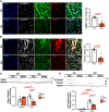
Septin 4 expression in the heart. A, mRNA expression of Sept4 in wild-type (WT) and knockout (KO) mice hearts (N=3 per group, 2-tailed Student t test). B, Western blot image shows a lack of Sept4 protein in KO cardiac lysates. C, Relative Sept4 mRNA expression in different cell types from WT hearts (N=3 per cell population, 2-tailed Student t test). D, Immunohistochemistry for Septin4 (red) and markers of cardiomyocytes (CMs; Troponin T), cardiac fibroblast (CFB; vimentin), and endothelial cells (CD31) shows Sept4 expression mainly in CFBs and endothelial cells. E, UMAP plot of single-cell sequencing shows different clusters. F, Assessment of Sept4 mRNA in all major cell types in the heart shows Sept4 expression is limited to fibroblasts, smooth muscle cells, pericytes, and endothelial cells. CD31+ indicates endothelial cells; and CD45+ indicates immune cells. UMAP indicates uniform manifold approximation and projection.
Figure 2.
Figure 2.
Septin 4 deletion results in changes in extracellular matrix (ECM) gene and protein expression. A, Heatmap of mRNA sequencing showing ECM genes that are downregulated in knockout (KO) mice under homeostatic conditions (N=3 per group). B, Enriched GO-term biological program analysis reveals alterations in ECM- and TGFβ (transforming growth factor β)-related pathways. C, Heatmap of mass spectrometry-based relative quantification of proteins detected in decellularized hearts from wild-type (WT) and KO hearts under homeostatic conditions (N=4 and 7, respectively). D, Quantification of protein abundance for COL1A1 (collagen type 1 alpha 1, left), COL1A2 (collagen type 1 alpha 2, middle), and COL6A2 (collagen type 6 alpha 2, right) based on relative molar mass of proteins detected within a given sample (N=4 and 7 for WT and KO, respectively, 2 tailed Student t test). GO indicates gene ontology.
Figure 3.
Figure 3.
Septin 4 deletion enhances cardiac compliance. Representative images of electron micrographs of decellularized cardiac tissue from wild-type (WT) and knockout (KO) hearts (A, top) with masks generated by the TWOMBLI algorithm (A, bottom) used to quantify extracellular matrix (ECM) structure and organization. B, Quantification of high-density matrix, a measure of overall abundance of ECM bundles (N=10 for each group, 2-tailed Student t test). C, Quantification of end points, a measure of collagen cross-linking (N=10 for each group, 2-tailed Student t test). D, Representative Stress/Strain curve of WT and KO hearts based on deformity of the cardiac tissue on probe compression and quantification of compressive modulus based on stress-strain curves, N=7 for WT and N=10 for KO groups, 2-tailed Student t test. E, Representative pressure volume (PV) loop tracings of WT and KO hearts under homeostatic conditions. F, Invasive hemodynamics-based assessment of left ventricular end diastolic pressure (LVEDP) and the time constant of isovolumic relaxation (Tau) under uninjured conditions (N=6 and 7 for WT and KO, respectively; 2-tailed Student t test).
Figure 4.
Figure 4.
Preserved cardiac function and reduced fibrosis in Septin 4 knockout (KO) mice after pressure overload. A, Schematic depiction of the study. B, left, Ejection fraction (right) fractional shortening; (C, left) left ventricular internal diastolic diameter (LVIDd); and (C, right) left ventricular internal systolic diameter (LVIDs) from wild-type (WT) and KO mice measured 4-weeks after Sham or TAC surgery (N=4, 7, 17, 22 for WT, KO Sham, WT, KO TAC, respectively, 2 tailed Student t test). D, Lung weight to body weight (LW/BW) and (E) heart weight to body weight (HW/BW) ratios of WT and KO mice 4 weeks after Sham or TAC surgery (N=4, 6, 17, 20 for WT, KO Sham, WT, KO TAC, respectively, Mann-Whitney U test). F, left, Representative image of cardiomyocyte (CM) cross-sectional area staining 4 weeks after sham/TAC by WGA (red) staining, and (F, right) quantification of the average cross-sectional area per CM (N=4, 5, 8, 10 for WT, KO Sham, WT, KO TAC, respectively, 2-tailed Student t test). G, left, Representative images of Sirius Red-Fast Green (SRFG) staining on WT and KO hearts, and (G, right) quantification of the fibrotic scar (red) in interstitial areas 4 weeks after TAC injury (N=14 per group, 1-tailed Mann-Whitney U test). H, left, Representative images of SRFG staining on perivascular areas of WT and KO hearts 4 weeks after TAC injury, and (right) quantification of perivascular fibrosis (red) after 4 weeks after TAC injury (N=14 per group, 1-tailed Mann-Whitney U test). Ex vivo invasive hemodynamics-based measurement of (I) invasive hemodynamics-based measurement of left ventricular end diastolic pressure and (J) isovolumetric relaxation constant (Tau) in WT and KO hearts 4 weeks after TAC injury (N=7, 6 for WT, KO, respectively, 2-tailed Student t test. K, Invasive hemodynamics-based measurement of end diastolic pressure volume relation slope in response to dobutamine in WT and KO hearts 4 weeks after TAC injury (N=7 and 6 for WT, KO, respectively, 2-tailed Student t test). CSA indicates cell surface area on cross-sections; EDPVR, end-diastolic pressure volume relationship; and WGA, wheat germ agglutinin.
Figure 5.
Figure 5.
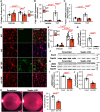
Septin 4 deletion blunts cardiac TGF-β and fibroblast activation after pressure overload. A, left, Representative images of αSMA positive cardiac fibroblast (CFBs) in wild-type (WT) and knockout (KO) hearts 1 week after TAC (blue=DAPI, gray=vimentin, green=WGA, red=αSMA) and their relative abundance (A, right) in each group N=4 for each group, 2 tailed Student t test). B, left, Representative staining of Postn (periostin) in WT and KO hearts 1-week after TAC (blue=DAPI, gray=vimentin, green=WGA, red=Postn; right) quantification of Postn positive areas (N=4 for each group, 2-tailed Student t test). C, top, Western blotting image for TGFBR1 (transforming growth factor beta receptor 1) and (bottom) quantification of the band intensities for each group 1 week after TAC (N=3, 4 for each sham, TAC group, respectively, 2-tailed Student t test. D, top, Western blotting images and (bottom) quantification for phosphorylated SMAD2 (p-SMAD2) and total SMAD2 demonstrate higher levels of SMAD2 phosphorylation in the WT group 1 week after TAC (N=3 and 4 for each sham, TAC group, respectively, 2-tailed Student t test). TGF-β indicates transforming growth factor-beta; and WGA, wheat germ agglutinin.
Figure 6.
Figure 6.
Septin 4 regulates cardiac fibroblast TGF-β stimulation and activation. A, Amount of collagen produced by cultured wild-type (WT) and knockout (KO) cardiac fibroblast (CFBs) 24 hours after TGF-β (10 ng/mL) or vehicle treatment (N=8 for each group, 2-tailed t-test). Relative (B) Ctgf (connective tissue growth factor) and (C) Postn (periostin) mRNA expressions in WT and KO cultured CFBs 24 hours after TGF-β (10 ng/mL) or vehicle treatment (N=5 for baseline groups and N=4 for KO and N=6 for WT TGF groups, 2-tailed Student t test). D, Representative images of αSMA positive myofibroblasts 24 hours after TGF-β (1 ng/mL) or vehicle treatment, and (E) quantification of abundance of myofibroblasts (αSMA+Vim [vimentin]+ cells, N=17 for KO, N=20 for WT baseline groups, N=21 for KO and N=23 for WT TGF groups, Mann-Whitney U test). F, Western blot image for SEPT4 and GAPDH (glyceraldehyde-3-phosphate dehydrogenase) in lysates of CFBs treated with Scrambled or Sept4 siRNA (Sept4 KD). G, Relative quantification of SEPT4 protein expression normalized to GAPDH in CFBs treated with Scrambled or Sept4 siRNA (N=6 for each group, 2-tailed Student t test). H, Western blot images for αSMA, POSTN, and GAPDH 48 hours after scrambled or Sept4 siRNA treatment. I–J, Quantification of the relative amounts of αSMA (I) and Postn (J) protein normalized to GAPDH (N=6 for each group, 2-tailed Student t test). K, left, Representative images of collagen scaffolds containing control and S4 KD CFBs 4 hours after release. K, right, Percent Gel contraction of collagen gels containing Control or S4 KD CFBs (N=5 for WT and N=6 for KO groups, 2-tailed Student t test). TGF-β indicates transforming growth factor-beta.
Figure 7.
Figure 7.
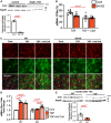
Septin 4 dependent defect in myofibroblast differentiation involves Calcineurin. A, top, Western blot image for CnA (calcineurin A) and GAPDH in lysates of cardiac fibroblast (CFBs) treated with scrambled (control [Cont]) or Sept4 siRNA (S4 KD). A, bottom, Relative quantification of CnA protein expression normalized to GAPDH (glyceraldehyde-3-phosphate dehydrogenase) in CFBs treated with Scrambled or Sept4 siRNA (N=3 for each group, 2-tailed Student t test). B, Quantification of the abundance of αSMA (α smooth muscle actin) positive myofibroblasts 24 hours after TGF (transforming growth factor)-β (1 ng/mL) ±cyclosporine A (1 ng/mL) for Scrambled or Sept4 siRNA (S4 KD) treated CFBs (N=5 for each group, 2-way ANOVA followed by Tukey post hoc test). C, Representative images of CFBs transduced with adenoviral particles expressing activated CnA (calcineurin A) stained for αSMA and Vim (vimentin), 24 hours after treatment with TGF-β (transforming growth factor-beta) (10 ng/mL) or vehicle. D, Quantification of αSMA positive activated CFBs (N=4 for Cont, N=6 for TGF and TGF+ Ad CnA, 2-way ANOVA followed by Tukey post hoc test). E, top, Western blot images and (bottom) quantification of CnA band intensity normalized to GAPDH from wild-type (WT) and knockout (KO) hearts, 4 weeks after TAC injury (N=6 for each group, 2-tailed Student t test).
Figure 8.
Figure 8.
Fibroblast-specific deletion of Septin 4 inhibits fibrosis after pressure overload. A, Schematic depiction of the study. B, left, Western blot images and (right) quantification of Sept4 expression from Septin4fl/fl cultured cardiac fibroblast (CFBs) without (Cre−) or with (Cre+) Cre-mediated deletion (n=3 for each group, 2-tailed Student t test). C, left, Representative images of Picrosirius red staining and (right) quantification of total collagen deposition from Septin4fl/fl (Cre−) and Septin4fl/fl Tcf21MCM/+ (Cre+) hearts 1 week after sham or transverse aortic constriction (TAC) injury (n=6 for sham Cre−, n=7 for TAC Cre−, N=5 for sham Cre+, N=6 for TAC Cre+, 2-way ANOVA followed by Tukey post hoc test). D, top, Representative images and (bottom) quantification of relative abundance of αSMA positive myofibroblasts from Septin4fl/fl (Cre−) and Septin4fl/fl Tcf21MCM/+ (Cre+) hearts 1 week after sham or TAC surgery (N=6 for sham Cre−, N=7 for TAC Cre−, N=5 for sham Cre+, N=6 for TAC Cre+, 2-way ANOVA followed by Tukey post hoc test). E, top, Western blot images and (bottom) quantification of αSMA expression from Septin4fl/fl (Cre−) and Septin4fl/fl Tcf21MCM/+ (Cre+) hearts 1 week after sham or TAC injury (N=4 for sham Cre− or Cre+, N=5 for TAC Cre− or Cre+, 2-way ANOVA followed by Tukey post hoc test). Gapdh indicates glyceraldehyde-3-phosphate dehydrogenase.

References

    1. Tsao CW, Aday AW, Almarzooq ZI, Anderson CAM, Arora P, Avery CL, Baker-Smith CM, Beaton AZ, Boehme AK, Buxton AE, et al. ; American Heart Association Council on Epidemiology and Prevention Statistics Committee and Stroke Statistics Subcommittee. Heart disease and stroke statistics-2023 update: a report from the American Heart Association. Circulation. 2023;147:e93–e621. doi: 10.1161/CIR.0000000000001123 - PMC - PubMed
    1. Xie M, Burchfield JS, Hill JA. Pathological ventricular remodeling: therapies: part 2 of 2. Circulation. 2013;128:1021–1030. doi: 10.1161/CIRCULATIONAHA.113.001879 - PMC - PubMed
    1. Burchfield JS, Xie M, Hill JA. Pathological ventricular remodeling: mechanisms: part 1 of 2. Circulation. 2013;128:388–400. doi: 10.1161/CIRCULATIONAHA.113.001878 - PMC - PubMed
    1. Gogiraju R, Xu X, Bochenek ML, Steinbrecher JH, Lehnart SE, Wenzel P, Kessel M, Zeisberg EM, Dobbelstein M, Schafer K. Endothelial p53 deletion improves angiogenesis and prevents cardiac fibrosis and heart failure induced by pressure overload in mice. J Am Heart Assoc. 2015;4:e001770. doi: 10.1161/JAHA.115.001770 - PMC - PubMed
    1. Revelo XS, Parthiban P, Chen C, Barrow F, Fredrickson G, Wang H, Yucel D, Herman A, van Berlo JH. Cardiac resident macrophages prevent fibrosis and stimulate angiogenesis. Circ Res. 2021;129:1086–1101. doi: 10.1161/CIRCRESAHA.121.319737 - PMC - PubMed

MeSH terms