. 2025 Sep 17;16(1):8264.
doi: 10.1038/s41467-025-63805-4.

GALNTL5 binds GalNAc and is required for migration through the uterotubal junction and sperm-zona pellucida binding

Affiliations

GALNTL5 binds GalNAc and is required for migration through the uterotubal junction and sperm-zona pellucida binding

Taichi Noda et al. Nat Commun. .

Abstract

More than 20 genes expressed in the male reproductive tract have been identified as essential factors for sperm migration to and through the utero-tubal junction (UTJ), and they are divided into ADAM3-dependent and ADAM3-independent pathways. In parallel, sperm having UTJ migration defects also show impaired binding to the zona pellucida (ZP). Herein, we demonstrate that knockout of Galntl5, encoding a sperm surface protein, causes impaired sperm binding with the UTJ and ZP, and null males have severe infertility. GALNTL5 appreciably disappears in sperm lacking Adam3 or Lypd4, required for ADAM3-dependent and ADAM3-independent pathways, and GALNTL5 binds to N-acetylgalactosamine (GalNAc) distributed on the UTJ and ZP. Blockage of GalNAc decreases the number of sperm binding to the UTJ and ZP. Thus, we unveil that GALNTL5 is a responsible factor for UTJ migration and sperm-ZP binding, and that sperm bind to the UTJ and ZP through interaction of GALNTL5 and GalNAc.


Conflict of interest statement

Competing interests: The authors declare no competing interests.

Figures
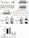
Fig. 1
Fig. 1. Male mice disrupted for both variants of Galntl5 are nearly sterile.
a gRNA design. There are two variants for mouse Galntl5. Blue-colored and yellow-colored “ATG” show the 1st Methionine for variants 1 and 2, respectively. The underlined sequence was used as the gRNA. Upper-case and lower-case letters show exon and intron regions, respectively. b Detection of Galntl5 mRNA using mouse multi-tissues. Hypoxanthine guanine phosphoribosyl transferase (Hprt) is used as the control. The data reproducibility was checked by two technical replicates. Br: brain, He: heart, In: intestine, Ki: kidney, Li: liver, Lu: lung, Ov: ovary, Sp: spleen, St: stomach, Te: testis, and Ut: uterus. c Detection of Galntl5 mRNA using mouse testes at 5–42 days of birth. The data reproducibility was checked by two technical replicates. d Detection of GALNTL5 mRNA using human multi-tissues. Glyceraldehyde-3-phosphate dehydrogenase (GAPDH) is used as the control. The data reproducibility was checked by two technical replicates. Cap: caput, Cau: cauda, Cor: corpus, Epi: epididymis. e Direct sequencing. The 17 and 25 nucleotides (nts) were deleted in the em1 and em2 alleles of Galntl5, leading to the appearance of a premature stop codon by the frameshift mutation (see Supplementary Fig. 1c). wt: wild-type, em: enzyme mutation. f Detection of GALNTL5 proteins in testicular germ cells (TGC) and sperm. We used antibodies to recognize N- or C-termini of mouse GALNTL5 (Supplementary Fig. 1d and Supplementary Table 4). In the control (ctrl) TGC, two bands were detected at the molecular sizes of variants 1 and 2 (~50 kDa and ~46 kDa, respectively) (see blue arrows). Using the C-terminus antibody, we also found non-specific bands of ~50 and ~37 kDa (red arrows) in both control and KO TGCs. In the ctrl sperm, GALNTL5 was detected at ~37 kDa only when we used an antibody to recognize the C-terminus (a yellow arrow). These specific bands disappeared in Galntl5 KO TGC and sperm. IZUMO1 was used as the loading control. The data reproducibility was checked by four biological replicates. g Male fertility. The females mated with Galntl5 KO males lacking variants 1 and 2 (v1 + v2) rarely delivered pups.
Fig. 2
Fig. 2. Spermatogenesis and sperm morphology, and motility in Het and KO males of Galntl5 variants 1 and 2 are normal.
a Testis morphology. b Testis weight. There was no difference in the testis weight between Galntl5 Het [3.15 ± 0.56 (em2/wt)] and KO mice [3.2 ± 1.1 (em2/em2)] (Mann–Whitney test, p = 0.94). ns: not significant. c Histological analysis of testes with Periodic Acid Schiff (PAS)-Hematoxylin staining. The area dotted by the yellow color was magnified (right panels). The data reproducibility was checked by two biological replicates. d Sperm morphology. The microscopic observation of sperm from Galntl5 Het and KO mice was comparable to WT sperm. The data reproducibility was checked by two biological replicates. e Sperm motility analysis with CASA. After 10- and 120-min incubation, each parameter of the sperm motility was measured. There were no differences among the groups [Kruskal–Wallis test, sperm motility: p = 0.99 (10 min), and p = 0.55 (2 h), sperm progressive motility: p = 0.83 (10 min), and p = 0.50 (2 h)].
Fig. 3
Fig. 3. Galntl5 KO sperm lacking variants 1 and 2 show defects in ZP binding and migration into the oviduct.
a Fertilization rates with cumulus-intact eggs. Sperm from Galntl5 KO mice could fertilize eggs at a comparable level as sperm from Galntl5 Het mice [96.9 ± 2.7% (em2/wt), 100.0% (em2/em2)] (Mann–Whitney test, p = 0.40). ns: not significant. b Observation of sperm binding to the ZP. c Number of sperm bound to the ZP. Sperm from Galntl5 KO mice hardly bind to the ZP [sperm number/egg: 14.3 ± 4.3 (em2/wt), 1.3 ± 1.0 (em2/em2)] (Mann–Whitney test, p = 0.02). *p < 0.05. d Observation of sperm behavior in the female reproductive tract. Though the fluorescence-labeled sperm from Galntl5 KO mice abundantly exist in the uterus, these KO sperm hardly pass through the UTJ. The area dotted by the yellow color was magnified (lower panels). The data reproducibility was checked by five biological replicates. e Observation of sperm binding at the UTJ. The fluorescence-labeled WT sperm bind to the UTJ, but Galntl5 KO sperm hardly bind to the UTJ. Scale bars were 200 µm. Yellow-dotted lines show the border between the uterus and the oviduct. The data reproducibility was checked by two biological replicates (WT) and three biological replicates (KO).
Fig. 4
Fig. 4. GALNTL5 almost disappears in KO sperm of UTJ migration-related genes.
a Detection of testicular proteins required for sperm migration through the UTJ. IZUMO1 was used as the loading control. The data reproducibility was checked by two biological replicates. b Detection of sperm ADAM3. ADAM3 is slightly decreased in Galntl5 KO sperm, but ADAM3 is absent in Adam3 KO (also see Supplementary Fig. 4a). The data reproducibility was checked by five biological replicates. c Detection of sperm proteins required for sperm migration through the UTJ. The data reproducibility was checked by two biological replicates. d Observation of ADAM3 localization in sperm. IZUMO1 and ZPBP1 were used as the loading controls of the detergent-enriched and detergent-depleted phases. The data reproducibility was checked by two biological replicates. e Detection of testicular GALNTL5. Variants 1 and 2 (blue arrows) are detected in Adam3 KO and Lypd4 KO TGC. IZUMO1 was used as the loading control. The data reproducibility was checked by five biological replicates. f Detection of sperm GALNTL5. GALNTL5 almost disappears in Adam3 KO and Lypd4 KO sperm (yellow arrow) (also see Supplementary Fig. 4b). The data reproducibility was checked by five biological replicates.
Fig. 5
Fig. 5. GALNTL5 variant 1 is more important for male fertility.
a gRNA design to generate mice lacking Galntl5 variant 1. Blue-colored and yellow-colored letters show the first methionine for variants 1 and 2, respectively. To delete the 1st Met for Galntl5 variant 1, we designed a gRNA (underlined sequence). b Direct sequencing. The indel mutations were present in the em3 (8 nt deletion and 3 nt insertion) and em4 (2 nt insertion) alleles of Galntl5, leading to the disruption of the 1st Met for Galntl5 variant 1 (blue-colored letters) (also see Supplementary Fig. 1c). The lowercase letters show the inserted nucleotides. c Detection of GALNTL5. Only the upper band, a protein from GALNTL5 variant 1, disappeared in the TGC of Galntl5em3 and Galntl5em4 KO mice. Furthermore, GALNTL5 at ~37 kDa disappeared in these KO sperm, confirming that only variant 1 of GALNTL5 was disrupted in Galntl5em3 and Galntl5em4 KO mice, and the processed protein from variant 1 existed in sperm. Arrows show GALNTL5. IZUMO1 was used as the loading control. The data reproducibility was checked by two biological replicates. d Male fertility. Galntl5em3 and Galntl5em4 KO males are near sterile [pups/plug: 7.2 ± 0.6 (Ctrl), 0.2 ± 0.4 (em3/em3), 0.1 ± 0.2 (em4/em4)], and comparable to the male infertility of Galntl5em1 and Galntl5em2 KO mice. e Localization of GALNTL5 protein in sperm. If targeted proteins on the sperm surface are biotinylated, the molecular weight shifts up slightly. GALNTL5 was slightly shifted up, indicating that GALNTL5 is localized on the sperm surface. ADAM3 (sperm surface protein), and acetylated tubulin (cytoskeletal protein) were used for positive control (P.C.) and negative control (N.C.), respectively. The data reproducibility to detect GALNTL5 was checked by three biological replicates.
Fig. 6
Fig. 6. GALNTL5 binds N-acetylgalactosamine (GalNAc).
a Lectin blot analysis. The pattern of glycan modifications in Galntl5 KO sperm was comparable to Het sperm. BASIGIN was used as the loading control. The data reproducibility was checked by two technical replicates. b Binding assay of GALNTL5 and sugars using sugar-immobilized gold nanoparticles. By cross-linking the molecules of interest and sugar-immobilized gold nanoparticles, the complexes are precipitated, leading to a decrease in the absorbance at 530 nm. When the synthesized GALNTL5 protein was mixed with GalNAc-binding particles, a decrease in absorbance was observed (1-way ANOVA). *p < 0.05, ***p < 0.01. c Binding assay of GALNTL5 and sugars using carbohydrate gel. After adding the molecules of interest in the column equipped with sugar-immobilized beads, only the molecules to specifically bind with the sugar are trapped in the beads. The trapped molecules are eluted by adding the inhibiting sugars. To examine the GalNAc-binding ability of GALNTL5, the proteins of ~50 kDa (immature form) and ~37 kDa (expected mature form) of mouse GALNTL5 (see Supplementary Fig. 5a) were incubated in a column equipped with GalNAc-binding beads. Both immature and mature GALNTL5 proteins were detected in the elution buffer. Con A and GS-I were used for N.C. and P.C., respectively. The data reproducibility to detect GALNTL5 was checked by two biological replicates.
Fig. 7
Fig. 7. Blockage of GalNAc on the UTJ and ZP surface decreased the sperm number bound to the UTJ and ZP.
a Detection of GalNAc in the UTJ. By the lectin histochemistry with Rhodamine-labeled Dolichos biflorus agglutinin (DBA) which recognizes the terminal α-GalNAc, DBA bound to the epithelium in UTJ. The Rhodamine-labeled DBA preabsorbed with GalNAc was used as the N.C. The area dotted by the white color was magnified (right panels). The data reproducibility was checked by three biological replicates. b Detection of GalNAc in the ZP. By live cell staining with Rhodamine-labeled DBA, DBA bound to the surface of ZP. The Rhodamine-labeled DBA preabsorbed with GalNAc was used as the N.C. The data reproducibility was checked by three biological replicates. c Sperm-UTJ binding assay. To evaluate the possibility of sperm-UTJ binding through GalNAc residues on the UTJ, the UTJ was incubated with DBA or DBA preabsorbed with GalNAc. After washing with TYH medium, the UTJ was inseminated with sperm pre-stained with Hoechst 33342. After fixation and washing, the sperm bound to the UTJ were observed. The Hoechst signal (the display changed to grayscale) in the area dotted by the black color was magnified (right panels). The data reproducibility was checked by four biological replicates. d Sperm-ZP binding assay. To evaluate the possibility of sperm-ZP binding through GalNAc on the ZP, eggs were incubated with DBA or DBA preabsorbed with GalNAc (see Supplementary Fig. 6). The eggs were washed with TYH medium and then inseminated with sperm for 30 min. After fixation and washing, the eggs were observed (left panel), and the sperm number bound to ZP was counted (right panel) (DBA: 6.8 ± 5.3, DBA + GalNAc: 13.4 ± 7.5) (Mann–Whitney test, p < 0.0001). Center line within the box: median, whiskers in box-and-whiskers plots: minimum to maximum, ****p < 0.001.

References

    1. Lillie, F. R. The mechanism of fertilization. Science38, 524–528 (1913). - PubMed
    1. Wassarman, P. M. Zona pellucida glycoproteins. Annu. Rev. Biochem.57, 415–442 (1988). - PubMed
    1. Wassarman, P. M. Towards molecular mechanisms for gamete adhesion and fusion during mammalian fertilization. Curr. Opin. Cell Biol.7, 658–664 (1995). - PubMed
    1. Bleil, J. D. & Wassarman, P. M. Mammalian sperm-egg interaction: identification of a glycoprotein in mouse egg zonae pellucidae possessing receptor activity for sperm. Cell20, 873–882 (1980). - PubMed
    1. Rankin, T. et al. Mice homozygous for an insertional mutation in the Zp3 gene lack a zona pellucida and are infertile. Development122, 2903–2910 (1996). - PubMed

MeSH terms

LinkOut - more resources