. 2025 Oct;646(8087):1167-1177.
doi: 10.1038/s41586-025-09487-w. Epub 2025 Sep 17.

A neuronal architecture underlying autonomic dysreflexia

Affiliations

A neuronal architecture underlying autonomic dysreflexia

Jan Elaine Soriano et al. Nature. 2025 Oct.

Abstract

Autonomic dysreflexia is a life-threatening medical condition characterized by episodes of uncontrolled hypertension that occur in response to sensory stimuli after spinal cord injury (SCI)1. The fragmented understanding of the mechanisms underlying autonomic dysreflexia hampers the development of therapeutic strategies to manage this condition, leaving people with SCI at daily risk of heart attack and stroke2-5. Here we expose the neuronal architecture that develops after SCI and causes autonomic dysreflexia. In parallel, we uncover a competing, yet overlapping neuronal architecture activated by epidural electrical stimulation of the spinal cord that safely regulates blood pressure after SCI. The discovery that these adversarial neuronal architectures converge onto a single neuronal subpopulation provided a blueprint for the design of a mechanism-based intervention that reversed autonomic dysreflexia in mice, rats and humans with SCI. These results establish a path towards essential pivotal device clinical trials that will establish the safety and efficacy of epidural electrical stimulation for the effective treatment of autonomic dysreflexia in people with SCI.


Conflict of interest statement

Competing interests: G.C., A.A.P., J.W.S., J.B., R.D. and S.L. hold various patents in relation to the present work. G.C., A.A.P. and R.D. are consultants of ONWARD Medical. G.C., A.A.P., J.W.S., J.B. and S.L. are minority shareholders of ONWARD, a company with direct relationships with the presented work. The other authors declare no competing interests.

Figures

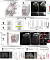
Fig. 1
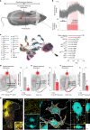
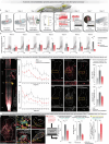
Fig. 1. Autonomic dysreflexia triggers transcriptional activity in the lumbosacral and lower thoracic spinal cord.
a, Experimental model to quantify the severity of autonomic dysreflexia in mice with complete SCI. b, Changes in blood pressure during an episode of autonomic dysreflexia elicited by a controlled colorectal distension (black dashed line). c, Severity of autonomic dysreflexia measured by the change in systolic blood pressure elicited by a controlled colorectal distension at different timepoints after SCI (5 days post-injury to uninjured (P = 0.95), 14 days post-injury to uninjured (P = 0.00048), 30 days post-injury to uninjured (P = 0.0000001), 45 days post-injury to uninjured (P = 0.0000001), 14 days post-injury to 5 days post-injury (P = 0.0013), 30 days post-injury to 5 days post-injury (P = 0.0000001), 45 days post-injury to 5 days post-injury (P = 0.0000002), 30 days post-injury to 14 days post-injury (P = 0.00086), 45 days post-injury to 14 days post-injury (P = 0.0024) and 45 days post-injury to 30 days post-injury (P = 0.99)). NS, not significant. d, Whole-spinal-cord visualization of immunohistochemical staining for Fos, in a mouse with SCI that was exposed to repetitive episodes of autonomic dysreflexia. C, caudal; R, rostral. e, Barplot reporting the mean number of Fos-labelled neurons for each spinal cord segment quantified in mice with SCI that were exposed to repetitive episodes of autonomic dysreflexia (n = 5; mixed-effect linear model; P < 0.001), demonstrating a clear enrichment in the lumbosacral and lower thoracic spinal cord. *P < 0.05, **P < 0.01, ***P < 0.001.
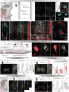
Fig. 2
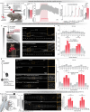
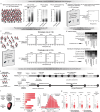
Fig. 2. The neuronal architecture of autonomic dysreflexia.
a, Schematic overview of the experiment. Uniform manifold approximation and projection (UMAP) visualization of 64,739 neuronal nuclei, coloured by neuronal subpopulation identity (left). UMAP visualizations of neuronal subpopulations in the lower thoracic (top) and lumbosacral (bottom) spinal cord (middle). Ranking of neuronal subpopulations most responsive to autonomic dysreflexia with Augur (right). AUC, area under the curve; DE, dorsal excitatory; DI, dorsal inhibitory; MIL, middle inhibitory deep laminate; VEL, ventral excitatory local; VEP, ventral excitatory projecting. b, Schematic overview of the neuronal architecture of autonomic dysreflexia, including the nodes (numbers) that are dissected anatomically and functionally in the subsequent panels. c, Whole-spinal-cord visualization of projections from SCLUMBAR::Vsx2 neurons located in the lumbosacral spinal cord that project to SCTHORACIC::Vsx2 neurons located in the lower thoracic spinal cord. White dashed lines outline the grey matter. D, dorsal; V, ventral. d, CalcaON projections labelled with immunohistochemistry onto SCLUMBAR::Vsx2 neurons in the lumbosacral spinal cord, including insets showing synaptic-like appositions. e, Barplot reporting the severity of autonomic dysreflexia, quantified as the mean change in systolic blood pressure in response to colorectal distension before and after the ablation of CalcaON neurons located in the dorsal root ganglia in CalcaCre::AdvilFlpO::iDTR mice (n = 5; independent samples t-test; t = −6.0; P = 0.0006). f, Severity of autonomic dysreflexia before and after chemogenetic silencing of Vsx2ON neurons located in the lumbosacral spinal cord in Vsx2–Cre mice (n = 5; paired samples t-test; t = −9.47; P = 0.00069). g, Projections from SCLUMBAR::Vsx2 in the lower thoracic spinal cord co-labelled with SCTHORACIC::Vsx2 neurons and their local projections as well as immunohistochemical labelling of ChATON neurons. The insets show synaptic-like appositions from SCLUMBAR::Vsx2 neurons onto SCTHORACIC::Vsx2 neurons, and synaptic-like appositions of projections from SCTHORACIC::Vsx2 neurons to ChATON sympathetic preganglionic neurons located in the intermediolateral column. h, Severity of autonomic dysreflexia before and after chemogenetic silencing of Vsx2ON neurons located in the lower thoracic spinal cord in Vsx2–Cre mice (n = 5; paired samples t-test; t = −9.39; P = 0.00072). i, Severity of autonomic dysreflexia before and after chemogenetic silencing of ChATON neurons located in the lower thoracic spinal cord in ChAT–Cre mice (n = 5; paired samples t-test; t = −8.03; P = 0.00048).
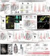
Fig. 3
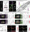
Fig. 3. The neuronal architecture of EES-induced pressor responses.
a, Schematic overview of experiments to trigger pressor responses with EES in mice with SCI. b, Pressor response induced by continuous (40 Hz) EES (black dashed line) in a mouse with SCI. c, UMAP visualization of 21,098 neuronal nuclei, coloured by neuronal subpopulation identity (left). Ranking of neuronal subpopulations most responsive to EES with Augur (right). df, Schematic overview of the successive nodes constituting the neuronal architecture through which EES applied over the lower thoracic spinal cord induces pressor responses. EES-induced pressor responses before and after the ablation of PVON neurons located in the dorsal root ganglia in PVCre::AdvilFlpO::iDTR mice (n = 5; independent samples t-test; t = −5.41; P = 0.0043; d). EES-induced pressor responses before and after chemogenetic silencing of Vsx2ON neurons located in the lower thoracic spinal cord in Vsx2–Cre mice (n = 5; paired samples t-test; t = −4.21; P = 0.014; e). EES-induced pressor responses before and after chemogenetic silencing of ChATON neurons located in the lower thoracic spinal cord in ChAT–Cre mice (n = 5; paired samples t-test; t = −7.07; P = 0.0021; f). g, Photomicrograph of the lower thoracic spinal cord demonstrating vGLUT1 synaptic puncta and synaptic-like appositions from large-diameter afferent neurons onto SCTHORACIC::Vsx2 neurons labelled with in situ hybridization (left) or viral tract tracing (right) in the lower thoracic spinal cord of PVCre::AdvilFlpO::tdTomato mice. Arrowheads mark PVON synaptic-like appositions and L6 projections onto Vsx2ON neurons. White dashed lines outline the grey matter.
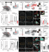
Fig. 4
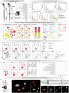
Fig. 4. Competitive neuronal architectures converge on SCTHORACIC::Vsx2 neurons.
a, Schematic overview of autonomic neurorehabilitation and the paradigm to quantify the severity of autonomic dysreflexia. b, Pressor responses (left; individual mice and mean trace) and severity of autonomic dysreflexia (right) in five mice with chronic SCI and five mice that underwent autonomic neurorehabilitation for 4 weeks, starting 1 week after SCI (independent samples t-test; t = −7.45; P = 0.00056). c, Schematic overview illustrating the competitive (overlapping) neuronal architectures of autonomic dysreflexia and EES-induced pressor responses, and their rearrangement after autonomic neurorehabilitation. d, vGLUT1ON synaptic puncta and synaptic-like appositions from SCLUMBAR::Vsx2 neurons onto SCTHORACIC::Vsx2 neurons in mice with SCI and mice with SCI that underwent autonomic neurorehabilitation. The barplots report the mean density of axonal projections from SCLUMBAR::Vsx2 neurons in the thoracic spinal cord in mice with SCI and mice with SCI that underwent autonomic neurorehabilitation (n = 5; independent samples t-test; t = 2.51; P = 0.0369; top). The barplots also report the mean number of vGLUT1ON synaptic puncta apposing SCTHORACIC::Vsx2 neurons (n = 5; independent samples t-test; t = 4.44; P = 0.0055; bottom). e, Schematic overview of experiments in which EES was applied daily over the lumbosacral spinal cord of mice with SCI, and the paradigm to quantify the severity of autonomic dysreflexia. f, As in panel b, but for mice with SCI that were subjected to the daily application of EES over the lumbosacral spinal cord (n = 5; independent samples t-test; t = 5.82; P = 0.00070). Grey shaded region indicates the colorectal distension period.
Fig. 5
Fig. 5. Reduced severity of autonomic dysreflexia in people with chronic SCI following EES targeting the haemodynamic hotspot.
a, Prevalence of autonomic dysreflexia and management efficacy quantified in 1,479 individuals with SCI from the Rick Hansen Spinal Cord Injury Registry,. b, Percentage of individuals with tetraplegia experiencing each symptom of autonomic dysreflexia scored in the ADFSCI across various daily activities (n = 107). c, Implantable system to regulate blood pressure with EES, including a paddle lead with optimized electrode configurations to target the dorsal roots projecting to the haemodynamic hotspot, an implantable pulse generator, communication hub and external smartwatch to operate the various programs of the therapy. d, Post-operative reconstruction of the final position of the electrodes following the implantation of the paddle lead. e, Changes in blood pressure from a representative participant during an orthostatic challenge without EES and with continuous EES applied over the haemodynamic hotspot. The barplots report the average drop in systolic blood pressure during orthostatic challenge (n = 11, paired samples two-tailed t-test; t = 4.7774; P = 0.00101) and the average tilt duration without EES and with EES applied over the haemodynamic hotspot (n = 11, paired samples two-tailed t-test; t = 14.33100; P < 0.001). The Kaplan–Meier plot shows exposure status to time, segregated by the presence or absence of EES. Data are derived from a companion article. f, ADFSCI autonomic dysreflexia score before implantation and after at least 6 months but up to 2 years after implantation of the system and daily use to regulate blood pressure (n = 11, paired samples one-tailed t-test; t = 2.3256, d.f. = 10, P = 0.02118). g, Percentage of individuals (n = 11) experiencing each symptom described in the ADFSCI autonomic dysreflexia section before implantation (before) and at the latest timepoint of ARCIM therapy (after).
Extended Data Fig. 1
Extended Data Fig. 1. Autonomic dysreflexia triggers transcriptional activity in the lumbosacral and thoracic spinal cord.
a, Experimental model of autonomic dysreflexia in mice with SCI and timeline of the experiment and final assessments. Blood pressure responses were monitored beat-by-beat using a blood pressure catheter inserted into the carotid artery. Autonomic dysreflexia was elicited using controlled colorectal distension in mice with upper-thoracic SCI. b, Baseline systolic blood pressure measured at different timepoints after SCI. Raw data and statistics provided in Supplementary Table 1. c, Blood pressure recording before, during and after a controlled colorectal distension. d, Pressor responses (Left; bold line represents mean trace ± sem for each group and individual line traces are from each animal) and severity of autonomic dysreflexia (Right) measured by the change in systolic blood pressure during colorectal distension at different timepoints after SCI (n = 5 per timepoint). Raw data and statistics provided in Supplementary Table 1. e, Overview of experimental protocol to identify the regions of the spinal cord activated during autonomic dysreflexia. Thirty days after receiving SCI, mice underwent repetitive episodes of autonomic dysreflexia over 90 min, consisting of 30 s, and then deflated for 60 s. Tissues were collected one hour after the exposure to autonomic dysreflexia, and then processed to visualize the expression of cFos in neurons. Bottom, CLARITY-optimized light sheet microscopy of the cleared spinal cord enabled visualisation of cFos immunoreactivity over the entire thoracolumbosacral spinal cord, in mice with SCI and mice with SCI that underwent repetitive episodes of autonomic dysreflexia. f, Quantification of cFos immunoreactivity in mice with SCI only and mice with SCI that underwent repetitive episodes of autonomic dysreflexia (mixed effect linear model; t = 6.60; p-value = 0.000213). g, Quantifications of cFos expression over the whole spinal cord were confirmed with immunohistochemistry and labelling for cFos on longitudinal sections of spinal cord, as illustrated in the representative photomicrographs of spinal cord sections from mice with SCI and mice with SCI that underwent repetitive episodes of autonomic dysreflexia (mixed effect linear model; t = 6.11; p-value = 0.000287). h, Schematic overview of experiment to assess blood pressure responses to colorectal distension in mice with SCI after bilateral dorsal rhizotomies at T11-T13. i, Severity of autonomic dysreflexia measured by the change in systolic blood pressure during colorectal distension in injured mice with and without bilateral dorsal rhizotomies at T11-T13 (n = 4; independent samples two-tailed t-test; t = 2.1255; p-value = 0.079). j, Quantifications of cFos expression over the whole spinal cord were confirmed with immunohistochemistry and labelling for cFos on longitudinal sections of spinal cord, as illustrated in the representative photomicrographs of spinal cord sections from mice with SCI and mice with SCI that underwent repetitive episodes of autonomic dysreflexia (mixed effect linear model; t = 14.082; p-value < 0.0001).
Extended Data Fig. 2
Extended Data Fig. 2. The neurons activated by autonomic dysreflexia.
a, Schematic overview of experiments to reveal the phenotype of the neurons that are activated during autonomic dysreflexia. We subjected Vglut2Cre::Ai9(RCL-tdT), VgatCre::Ai9(RCL-tdT) and ChatCre::Ai9(RCL-tdT) to repetitive episodes of autonomic dysreflexia at 30 days post-injury. Longitudinal sections of the spinal cord from T9 to L3 and L4 to S4 were immunohistochemically stained for cFos. Right, Quantification of colocalisation of cFos-labelled neurons and endogenous fluorescence-tagged neurons (Vglut2ON, VgatON and ChatON) was performed with automated spot detection (Imaris, Bitplane v.9.8.2). b, We next performed experiments to determine the role of these neuronal subpopulations in triggering autonomic dysreflexia. To manipulate the activity of Vglut2ON and VgatON neurons, AAV5-hSyn-DIO-hm4D(Gi)-mCherry was infused in either the lower thoracic spinal cord (T11-T13) or lumbosacral spinal cord (L5-S1) of either Vglut2Cre or VgatCre mice prior to performing the SCI to express DREADDs in glutamatergic or gabaergic neurons. Photomicrographs of coronal sections from either the lower thoracic or lumbosacral spinal cord in Vglut2Cre and VgatCre mice reveal the robust expression of mCherry and thus DREADDs in the targeted neurons. c, Chemogenetic inactivation restricted to Vglut2ON neurons located in the lumbosacral (n = 5; paired samples t-test; t = −10.4; p = 0.00048) or lower thoracic spinal cord (n = 5; paired samples t-test; t = −17.5; p = 0.00001) blunted autonomic dysreflexia. In contrast, chemogenetic silencing of VgatON neurons in the lumbosacral (n = 3; paired samples t-test; t = −1.53; p = 0.267) or lower thoracic spinal cord (n = 4; paired samples t-test; t = 0.269; p = 0.805) failed to modulate the severity of autonomic dysreflexia. Pressor responses (Left; bold line represents mean trace ± sem for each group and individual line traces are from each animal) and severity of autonomic dysreflexia (bar graph, Right) measured by the change in systolic blood pressure during colorectal distension. d, We next aimed to expose the projections from Vglut2ON neurons located in the lumbosacral spinal cord to the Vglut2ON neurons located in the lower thoracic spinal cord. For this, we labeled the axons and synapses of Vglut2ON neurons with infusions of AAV-DJ-hSyn-flex-mGFP-2A-synaptophysin-mRuby into the lumbosacral spinal cord of Vglut2Cre. The cell bodies in the lumbosacral spinal cord in both intact and injured mice were quantified (n = 4; independent samples two-tailed t-test; t = 0.473; p-value = 0.654). Photomicrographs of the lower thoracic spinal cord from representative mice demonstrate increases in the density of axons (Left; n = 4; independent samples two-tailed t-test; t = 5.92; p-value = 0.0047) and synaptic puncta (Right; n = 4; independent samples two-tailed t-test; t = 3.47; p-value = 0.027) emanating from Vglut2ON neurons located in the lumbosacral spinal cord after SCI.
Extended Data Fig. 3
Extended Data Fig. 3. Comparative single-nucleus RNA sequencing atlas of perturbation-responsive neuronal subpopulations during autonomic dysreflexia.
a, Scheme illustrating our experimental protocol followed by single-nucleus RNA sequencing. Mice received upper-thoracic SCI. After 30 days, half of the mice underwent repetitive episodes of autonomic dysreflexia during 90 min. The lumbosacral spinal cord and the lower thoracic were dissected from the mice according to standard procedures. b, We obtained high-quality transcriptomes from 64,739 nuclei that were evenly represented across experimental conditions and spatial locations. c, Quality control from 64,739 single-nucleus transcriptomes. Number of unique molecular identifiers (UMIs) per nucleus. Inset text shows the median number of UMIs. d, Number of genes detected per nucleus. Inset text shows the median number of genes detected. e, Proportion of mitochondrial counts per nucleus. Inset text shows the median proportion of mitochondrial counts. f, Number of UMIs quantified per nucleus in each major cell type of the mouse spinal cord. g, Number of genes detected per nucleus in each major cell type of the mouse spinal cord. h, Proportion of mitochondrial counts per nucleus in each major cell type of the mouse spinal cord. i, UMAP visualization of 64,739 nuclei colored by major cell type, segregated by the location of spinal cord tissues (L6, T12) and experimental conditions (SCI only, exposure to repeated episode of autonomic dysreflexia, AD). j, Proportions of nuclei from each major cell type depending on the location of spinal cord tissues and experimental conditions. k, UMAP visualization showing expression of key marker genes for the major cell types of the mouse spinal cord. l, UMAP visualization of 29,144 neuronal nuclei colored by neuronal subpopulations, split by experimental condition. m, UMAP visualization showing expression of key marker genes for the major neuronal subpopulation classifications of the mouse spinal cord. n, UMAP visualization and dendrograms showing cell type prioritizations assigned by Augur across the neuronal taxonomy of the lower thoracic (Top) and lumbosacral (Bottom) spinal cord. Raw AUC values and confidence values are provided in Supplementary Table 2. o, Lollipop plot illustrating the statistical significance of upregulated Gene Ontology (GO) modules associated with circuit reorganization and increased neuronal excitability in Vsx2ON neurons. p, Photomicrographs of the lower thoracic and lumbosacral spinal cord after repetitive episodes of autonomic dysreflexia. Vsx2ON neurons were labelled with immunohistochemistry. Long distance projecting (Zfhx3, lumbosacral spinal cord) and locally-projecting (Nfib, lower thoracic spinal cord) neurons were additionally colocalized with immunohistochemistry labelling of cFos.
Extended Data Fig. 4
Extended Data Fig. 4. The first and second nodes of the neuronal architecture of autonomic dysreflexia.
a, Schematic overview of the neuronal architecture of autonomic dysreflexia. b, Zoom on the first node of the neuronal architecture of autonomic dysreflexia that involves the growth of projections from CalcaON neurons onto Vsx2ON neurons with long-distance projections, named SCLUMBAR::Vsx2 neurons. This growth was assessed on tissues collected 30 days after SCI in wild-type mice. c, Photomicrograph taken at L6 spinal segment from a mouse with an intact spinal cord and a mouse with a chronic SCI in which CalcaON axons were labelled with immunohistochemistry. d, Bar plots reporting the density of CalcaON axonal projections into the intermediate laminae of the spinal cord in uninjured mice and mice with chronic SCI (n = 4; independent samples two-tailed t-test; t = 9.38; p-value = 0.000086). e, Overview of the experimental protocol to test the severity of autonomic dysreflexia after the ablation of CalcaON and PVON neurons. To achieve the ablation of these neurons exclusively in the dorsal root ganglia, we used a Cre- and Flp-dependent strategy in CalcaCre::AvilFlpO::iDTR and PVCre::AvilFlpO::iDTR mice that allowed the expression of diphtheria toxin receptors (DTR) in the CalcaON and PVON neurons located in the dorsal root ganglia, respectively. f, Pressor responses (Left; bold line represents mean trace ± sem for each group and individual line traces are from each mouse) and severity of autonomic dysreflexia (Right) measured by the change in systolic blood pressure during colorectal distension in mice without diphtheria toxin-induced ablation of either CalcaON neurons or PVON neurons, mice with diphtheria toxin-induced ablation of CalcaON neurons and mice with diphtheria toxin-induced ablation of PVON neurons (n = 5; independent samples t-test two-tailed; t = −5.9998; p-value = 0.00064, independent samples two-tailed t-test; t = −9.3261; p-value = 0.00014). g, Zoom on the second node of the neuronal architecture of autonomic dysreflexia that involves SCLUMBAR::Vsx2 neurons projecting to the low thoracic spinal cord. An intersectional viral labelling strategy was used to label the axons of SCLUMBAR::Vsx2 neurons located in the lumbosacral spinal cord and that establish projections in the lower thoracic spinal cord. Vsx2Cre mice received SCI and were injected with Retro-AAV-DIO-FlpO into the lower thoracic spinal cord and AAV8-Con/Fon-EYFP into the lumbosacral spinal cord. h, Photomicrograph of the L6 spinal segment from a Vsx2Cre mouse with an intact spinal cord and a Vsx2Cre mouse with a chronic SCI that received intersectional viral tracing to label SCLUMBAR::Vsx2 neurons. Axons from CalcaON neurons were also labelled with immunohistochemistry. Insets show synaptic-like appositions of axons from CalcaON neurons onto SCLUMBAR::Vsx2 neurons. i, The necessary role of SCLUMBAR::Vsx2 neurons in autonomic dysreflexia was evaluated using Cre-dependent expression of Gi DREADDs in SCLUMBAR::Vsx2 neurons. j, Photomicrograph showing the expression of DREADD (Gi) receptors in SCLUMBAR::Vsx2 neurons. k, Left, changes in systolic blood pressure in response to colorectal distension (shared area). Bold line represents mean trace ± sem for each group and individual line traces are from each mouse) and severity of autonomic dysreflexia. Right, Severity of autonomic dysreflexia in Vsx2Cre mice before and after chemogenetic silencing of Vsx2ON neurons located in the lumbosacral spinal cord (n = 5; paired samples t-test; t = −9.47; p-value = 0.00069). l, The sufficient role of SCLUMBAR::Vsx2 neurons in triggering autonomic dysreflexia was evaluated using optogenetic activation of SCLUMBAR::Vsx2 neurons in Vsx2Cre mice injected with AAV-Syn-flex-ChrimsonR-tdTomato into the lumbosacral spinal cord. 30 days after SCI, blood pressure responses were monitored beat-by-beat using a blood pressure catheter inserted into the carotid artery. Red-shifted light was shined over the lumbosacral spinal cord for 60 s during each trial. m, Photomicrograph showing the expression of ChrimsonR in SCLUMBAR::Vsx2 neurons. n, Left, Changes in systolic blood pressure in response to the photostimulation of SCLUMBAR::Vsx2 neurons in mice with intact spinal cord and with chronic SCI. Bold line represents mean trace ± sem for each group and individual line traces are from each mouse) and blood pressure responses due to optogenetic activation of SCLUMBAR::Vsx2 neurons. Right, Bar plots reporting mean changes in blood pressure in Vsx2Cre mice with intact spinal cord and with SCI during optogenetic activation of SCLUMBAR::Vsx2 neurons (n = 5; independent samples two-tailed t-test; t = 5.14; p-value = 0.00496).
Extended Data Fig. 5
Extended Data Fig. 5. The third and fourth nodes of the neuronal architecture of autonomic dysreflexia.
a, Zoom on the third node of the neuronal architecture of autonomic dysreflexia that involves SCTHORACIC::Vsx2 neurons located in the lower thoracic spinal cord. b, Overview of intersectional viral tracing strategy to label projections from SCLUMBAR::Vsx2 into the lower thoracic spinal cord concomitantly to the labelling of SCLUMBAR::Vsx2 Step 1, AAV5-hSyn-flex-tdTomato was infused into the lower thoracic spinal cord of Vsx2-Cre mice to label SCTHORACIC::Vsx2. Step 2, Retro-AAV-DIO-FlpO was infused into the lower thoracic spinal cord and AAV8-Con/Fon-EYP into the lumbosacral spinal cord to label the projections from SCLUMBAR::Vsx2 neurons located in the lumbosacral spinal cord and that project in the lower thoracic spinal cord. c, Photomicrographs of the lower thoracic spinal cord showing projections from SCLUMBAR::Vsx2 onto SCTHORACIC::Vsx2 neurons in a mouse with an intact spinal cord and a mouse with a chronic SCI. d, Bar plots reporting the mean density of projections from SCLUMBAR::Vsx2 neurons in the grey matter of the lower thoracic spinal cord in mice with an intact spinal cord and with chronic SCI (n = 5; independent samples two-tailed t-test; t = −3.09; p-value = 0.0162). e, Whole spinal cord visualization of projections from SCLUMBAR::Vsx2 neurons located in the lumbosacral spinal cord (red) and visualization of SCTHORACIC::Vsx2 neurons (blue) located in the lower thoracic spinal cord in mice with chronic SCI. f, Schematic overview of the experimental protocol to monosynaptically label SCLUMBAR::Vsx2 after lower thoracic spinal cord infusions targeting the virus to SCTHORACIC::Vsx2 neurons. Step 1, AAV8-hSyn-dlox-TVA950-2A-EGFP was infused into the lower thoracic spinal cord of Vsx2Cre mice to express TVA950 (Avian Tumor Virus A Receptor variant), EGFP (enhanced green fluorescent protein), and oGrev (optimized rabies virus glycoprotein) simultaneously. Step 2, EnvA-ΔG-Rabies-mCherry was infused two weeks after to monosynaptically restrict EnvA pseudotyped and G-protein deleted rabies expressing mCherry and label direct connections from SCLUMBAR::Vsx2 neurons onto SCTHORACIC::Vsx2 neurons. Representative photomicrographs of two neurons infected with the EnvA-ΔG-Rabies-mCherry and positive to Vsx2 immunohistochemistry. g, The necessary role of SCTHORACIC::Vsx2 neurons in autonomic dysreflexia was evaluated using Cre-dependent expression of Gi DREADDs in SCTHORACIC::Vsx2 neurons. h, Photomicrograph showing the expression of Gi DREADD receptors in SCTHORACIC::Vsx2 neurons. i, Left, changes in systolic blood pressure in response to colorectal distension. Bold line represents mean trace ± sem for each group and individual line traces are from each mouse and severity of autonomic dysreflexia. Right, Severity of autonomic dysreflexia in Vsx2Cre mice before and after chemogenetic silencing of Vsx2ON neurons located in the lower thoracic spinal cord (n = 5; paired samples t-test; t = −9.39; p-value = 0.00072). j, The sufficient role of SCTHORACIC::Vsx2 neurons in autonomic dysreflexia was evaluated using optogenetic activation of SCTHORACIC::Vsx2 neurons in Vsx2Cre mice injected with AAV-Syn-flex-ChrimsonR-tdTomato into the lower thoracic spinal cord. 30 days after SCI, blood pressure responses were monitored beat-by-beat using a blood pressure catheter inserted into the carotid artery. Red-shifted light was shined over the lumbosacral spinal cord for 60 s during each trial. k, Photomicrograph showing the expression of ChrimsonR in SCTHORACIC::Vsx2 neurons. l, Left, changes in systolic blood pressure in response to colorectal distension. Bold line represents mean trace ± sem for each group and individual line traces are from each mouse and blood pressure responses due to optogenetic activation of SCTHORACIC::Vsx2 neurons. Right, Blood pressure responses in Vsx2Cre mice with intact spinal cord and with chronic SCI during optogenetic activation of SCTHORACIC::Vsx2 neurons (n = 5; independent samples two-tailed t-test; t = 15.4; p-value = 0.0000148). m, Zoom on the fourth node of the neuronal architecture of autonomic dysreflexia that involves ChatON preganglionic sympathetic neurons. n, Overview of experimental protocol to label projections from SCTHORACIC::Vsx2 neurons located in the lower thoracic spinal cord in Vsx2Cre mice with SCI. Thirty days after SCI and viral tracing, the spinal cord tissues were collected and processed. o, Photomicrograph of the lower thoracic spinal cord from a mouse with an intact spinal cord and a mouse with chronic SCI in which the projections of SCTHORACIC::Vsx2 neurons were labelled concomitantly to the immunohistochemical labelling of ChatON neurons. p, The necessary role of ChatON neurons in autonomic dysreflexia was evaluated using Cre-dependent expression of Gi DREADDs in ChatON neurons. q, Photomicrograph illustrating the expression of Gi DREADD receptors in SCTHORACIC::Vsx2 neurons. r, As in h, for ChatON neurons located in the lower thoracic spinal cord (n = 5; paired samples t-test; t = −8.03; p-value = 0.00048).
Extended Data Fig. 6
Extended Data Fig. 6. Comparative single-nucleus RNA sequencing atlas of perturbation-responsive neuronal subpopulations during epidural electrical stimulation.
a, Scheme illustrating the experimental protocol followed by single-nucleus RNA sequencing. Days after SCI for half of the mice, EES was applied continuously over the lower thoracic spinal cord during 45 min. The lower thoracic spinal cord was harvested from the mice according to standard procedures. b, We obtained high-quality transcriptomes from 21,098 nuclei that were evenly represented across experimental conditions and spatial locations. c, Number of unique molecular identifiers (UMIs) per nucleus. Inset text shows the median number of UMIs. d, Number of genes detected per nucleus. Inset text shows the median number of genes detected. e, Proportion of mitochondrial counts per nucleus. Inset text shows the median proportion of mitochondrial counts. f, Number of UMIs quantified per nucleus in each major cell type of the mouse spinal cord. g, Number of genes detected per nucleus in each major cell type of the mouse spinal cord. h, Proportion of mitochondrial counts per nucleus in each major cell type of the mouse spinal cord. i, UMAP visualization of 21,098 nuclei colored by major cell type, split by experimental condition. j, Proportions of nuclei from each major cell type across all experimental conditions. k, UMAP visualization showing expression of key marker genes for the major cell types of the mouse spinal cord. l, UMAP visualization of 8,471 neuronal nuclei colored by neuronal subpopulations, split by experimental condition. m, UMAP visualization showing expression of key marker genes for the major neuronal subpopulation classifications of the mouse spinal cord. n, UMAP visualization and dendrograms showing cell type prioritizations assigned by Augur across the neuronal taxonomy of the lower thoracic spinal cord. Raw AUC values and confidence values are provided in Supplementary Table 2.
Extended Data Fig. 7
Extended Data Fig. 7. The neuronal architecture activated by EES to induce pressor response.
a, Schematic overview of the neuronal architecture through which EES induces pressor responses. b, Zoom on the first node of the neuronal architecture of EES-induced pressor responses that involves PVON neurons. c, Overview of the experimental protocol to test the involvement of afferent fibers from PVON and CalcaON neurons in EES-induced pressor responses. To achieve the ablation of these neurons exclusively in the dorsal root ganglia, we used a Cre- and Flp-dependent strategy in CalcaCre::AvilFlpO::iDTR and PVCre::AvilFlpO::iDTR mice that allowed the expression of diphtheria toxin receptors (DTR) in these specific neurons. d, EES-induced pressor responses (Left; bold line represents mean trace ± sem for each group and individual line traces are from each mouse) (Right) measured by the change in systolic blood pressure during EES in mice without any ablation, mice with diphtheria toxin-induced ablation of PVON neurons and mice with diphtheria toxin-induced ablation of CalcaON neurons (n = 5; independent samples two-tailed t-test; t = −5.4141; p-value = 0.0043, independent samples two-tailed t-test; t = 6.3166; p-value = 0.0020). e, Overview of the experiment strategy to visualize large-diameter PVON afferent fibers in PVCre::AdvilFlpO::Ai9(RCL-tdT) mice and confirmed that they established vGlut1ON synaptic-appositions onto SCTHORACIC::Vsx2 neurons. Thirty days after SCI, spinal cord tissues were collected and processed. f, Photomicrograph of the lower thoracic spinal cord showing large-diameter afferent neurons (PVCre::AdvilFlpO::tdTomato mice), SCTHORACIC::Vsx2 neurons labelled with in situ hybridization and ChatON neurons labelled with immunohistochemistry. g, Photomicrograph of the SCTHORACIC::Vsx2 neurons labelled with viral tract tracing and vGlut1ON synapses labelled with immunohistochemistry. Quantification of vGlut1ON synaptic-appositions onto SCTHORACIC::Vsx2 neurons and ChatON neurons in PVCre::AdvilFlpO::Ai9(RCL-tdT) mice with an intact spinal cord and with a chronic SCI. h, Schematic overview of the experimental protocol to monosynaptically label PVON neurons in the dorsal root ganglia after lower thoracic spinal cord infusions targeting the virus to SCTHORACIC::Vsx2 neurons. Step 1, AAV8-hSyn-dlox-TVA950-2A-EGFP was infused into the lower thoracic spinal cord of Vsx2Cre mice to express TVA950 (Avian Tumor Virus A Receptor variant), EGFP (enhanced green fluorescent protein), and oGrev (optimized rabies virus glycoprotein) simultaneously. Step 2, EnvA-ΔG-Rabies-mCherry was infused two weeks after to monosynaptically restrict EnvA pseudotyped and G-protein deleted rabies expressing mCherry and label direct connections from PVON neurons onto SCTHORACIC::Vsx2 neurons. Representative photomicrograph showing neurons in the lower thoracic dorsal root ganglion infected with the EnvA-mCherry and labelled with fluorescence in situ hybridization. i, Zoom on the second node of the neuronal architecture of EES-induced pressor responses that involves SCTHORACIC::Vsx2. The necessary role of SCTHORACIC::Vsx2 neurons in EES-induced pressor response was evaluated using Cre-dependent expression of Gi DREADDs in SCTHORACIC::Vsx2 neurons. j, EES-induced pressor responses (Left; bold line represents mean trace ± sem for each group and individual line traces are from each mouse) (Right) measured by the change in systolic blood pressure during EES in the same mice before and after chemogenetic silencing of Vsx2ON neurons located in the lower thoracic spinal cord (n = 5; paired samples t-test; t = −4.21; p-value = 0.014). k, Zoom on the third node of the neuronal architecture of EES-induced pressor responses that involves ChatON preganglionic sympathetic neurons. As in h, for ChatON neurons located in the lower thoracic spinal cord. l, As in j, for ChatON neurons located in the lower thoracic spinal cord (n = 5; paired samples t-test; t = −7.07; p-value = 0.0021). m, Photomicrograph showing the expression of ChrimsonR in Vsx2ON neurons and the tract resulting from the insertion of one electrode shank. n, Schematic overview of experiments to record the activity of SCTHORACIC::Vsx2 during the application of EES and during episodes of autonomic dysreflexia. o, Top, the waveforms display spikes and firing rate evoked by optogenetic stimulation of VsxON neurons by the application of continuous EES over the lower thoracic spinal cord, and by colorectal distention. Heatmap of neuronal clusters activated by EES, activated by EES and colorectal distension, activated by EES and tagged as Vsx2ON neurons by optogenetic stimulation and EES, and activated by EES and colorectal distension and tagged as Vsx2ON neurons activated by optogenetic stimulation.
Extended Data Fig. 8
Extended Data Fig. 8. Autonomic neurorehabilitation reversed autonomic dyresflexia in mice with SCI.
a, Overview of the experimental protocol to deliver autonomic neurorehabilitation in mice with SCI. Step 1. Mice received a complete transection of the spinal cord at the level of the T4 segment. Step 2.Intersectional viral tracing by infusing Retro-AAV-DIO-FlpO into the lower thoracic spinal cord and AAV8-Con/Fon-EYP into the lumbosacral spinal cord to label SCLUMBAR::Vsx2 neurons located in the lumbosacral spinal cord that project onto SCTHORACIC::Vsx2 neurons located in the lower thoracic spinal cord. Step 3. One week after SCI, electrodes were implanted over the T12 spinal segment to deliver EES. Step 4. EES was applied for 30 min everyday for 4 weeks. Step 5. F of autonomic dysreflexia was assessed during terminal experiments conducted in mice with chronic SCI and mice with chronic SCI that underwent autonomic neurorehabilitation. Step 6. Spinal cord tissues were collected and processed. b, Changes in systolic blood pressure (Left; bold line represents mean trace ± sem for each group and individual line traces are from each mouse) and severity of autonomic dysreflexia (Right) measured by the change in systolic blood pressure during colorectal distension in mice with chronic SCI and mice with chronic SCI that underwent autonomic neurorehabilitation (n = 5; independent samples two-tailed t-test; t = −7.45; p-value = 0.00056). c, (Left) Photomicrographs of the lower thoracic spinal cord in mice with chronic SCI and mice with chronic that underwent autonomic neurorehabilitation in which SCLUMBAR::Vsx2 neurons located in the lumbosacral spinal cord were labelled with an intersection virus strategy concomitantly to the labelling of SCTHORACIC::Vsx2 neurons. (Right) Photomicrographs of the lower thoracic spinal cord with intersectional viral labelling combined with immunohistochemical labelling of vGlut1ON synapses in mice with chronic SCI and mice with chronic SCI that underwent autonomic neurorehabilitation. vGlut1ON synaptic puncta and synaptic-like appositions from SCLUMBAR::Vsx2 neurons onto SCTHORACIC::Vsx2 neurons in mice with chronic SCI and mice with chronic SCI that underwent autonomic neurorehabilitation. d, (Left) Bar plots reporting the mean number of vGlut1ON synaptic puncta apposing SCTHORACIC::Vsx2 neurons (n = 5; independent samples two-tailed t-test; t = 4.44; p-value = 0.0055). (Right) and the mean density of axonal projections from SCLUMBAR::Vsx2 neurons in the grey matter of the lower thoracic spinal cord in mice with chronic SCI and mice with chronic SCI that underwent autonomic neurorehabilitation (n = 5; independent samples two-tailed t-test; t = 2.51; p-value = 0.0369). e, As in a, for mice subjected to daily application of EES over the lumbosacral spinal cord. f, As in b, for mice subjected to daily application of EES over the lumbosacral spinal cord (n = 5; independent samples two-tailed t-test; t = 5.82; p-value = 0.00070). g, As in c, for mice subjected to daily application of EES over the lumbosacral spinal cord. h, As in d, for mice subjected to daily application of EES over the lumbosacral spinal cord.
Extended Data Fig. 9
Extended Data Fig. 9. Autonomic neurorehabilitation reversed autonomic dysreflexia in rats with contusion SCI.
a, Overview of the experimental protocol to deliver autonomic neurorehabilitation in rats with SCI. Step 1. Rats received a severe contusion (380 Kdyn) of the spinal cord at the level of T3 segment. Step 2. AAV-DJ-hSyn-flex-mGFP-2A-Synaptophysin-mRuby and an AAV-Cre were co-infused into the L6 segment of the spinal cord to label the projections from neurons located in the lumbosacral spinal cord. Step 3. A wireless telemeter recording system, including a blood pressure cannula inserted into the abdominal aorta and microelectrodes sutured over the sympathetic renal nerve, was implanted chronically to monitor hemodynamics and sympathetic nerve activity, respectively. Step 4. Seven days after SCI, an electronic dura mater (e-dura) designed to target the dorsal roots projecting to the T11, T12, and T13 spinal segments was implanted over the hemodynamic hotspot to regulate blood pressure. Step 5. EES was applied for 30 min everyday during 6 weeks using a proportional-integral (PI) controller that adjusted the amplitude of EES in closed-loop to augment the systolic blood pressure to a target range. Step 6. The severity of autonomic dysreflexia, induced by colorectal distension, was assessed every week for 6 weeks. Step 7. After 6 weeks of autonomic neurorehabilitation, a final assessment was performed to test the severity of autonomic dysreflexia in all groups, which included rats with intact spinal cord, rats with chronic SCI and rats with chronic SCI that underwent autonomic neurorehabilitation. Step 8. Spinal cords were collected and processed. b, Changes in systolic blood pressure in response to colorectal distension (Left; bold line represents mean trace ± sem for each group and individual line traces are from each rat) and bar plots reporting the severity of autonomic dysreflexia (Right) measured by the change in systolic blood pressure during colorectal distension over the course of 6 weeks in rats with intact spinal cord, rats with chronic SCI and rats with chronic SCI that underwent autonomic neurorehabilitation. Raw data and statistics provided in Supplementary Table 1. c, Whole spinal cord visualization of projections from neurons located in the lumbosacral spinal cord. d, Plots reporting density of axonal projections (top) and synaptic punta (bottom) from neurons located in the lumbosacral spinal cord into the grey matter of the lower thoracic spinal cord in rats with intact spinal cord, rats with chronic SCI and rats with chronic SCI that underwent autonomic neurorehabilitation. e, Micrographs of the lower thoracic spinal cord in which the axonal projections and synaptic puncta from neurons located in the lumbosacral spinal cord are labelled for the three groups of rats. f, Bar plots reporting the mean density of axonal projections and synaptic puncta from neurons located in the lumbosacral spinal cord into the grey matter of the lower thoracic spinal cord for the three groups of rats. Raw data and statistics are provided in Supplementary Table 1. g, Micrographs of the lower thoracic spinal cord in which axonal projections and synaptic puncta from neurons located in the lumbosacral spinal cord are labelled concomitantly to vGlut1ON synapses from large-diameter afferents and Vsx2ON neurons. The density of lumbosacral-originating and vGlut1ON synapses onto Vsx2ON neurons is reconstructed for a rat with chronic SCI and a rat with chronic SCI that underwent autonomic neurorehabilitation. h, Bar plots reporting the density of synaptic-like appositions from neurons located in the lumbosacral spinal cord onto Vsx2ON neurons in rats with intact spinal cord, rats with chronic SCI, and rats with chronic SCI that underwent autonomic neurorehabilitation. Raw data and statistics are provided in Supplementary Table 1. i, As in i, for vGlut2ON synaptic puncta onto Vsx2ON neurons. Raw data and statistics are provided in Supplementary Table 1. j, Quantification of vGlut1ON synaptic puncta from large-diameter afferents onto Vsx2ON neurons in rats with chronic SCI and rats with chronic SCI that underwent autonomic neurorehabilitation (n = 5; independent samples t-test; t = 12.71; p-value = 2.78e-06).
Extended Data Fig. 10
Extended Data Fig. 10. Population-level data of self-reported experiences of autonomic dysreflexia symptoms and clinical validation of autonomic neurorehabilitation.
a, The prevalence of autonomic dysreflexia and management efficacy in people with SCI (n = 1479) acquired with the Spinal Cord Injury Community Survey (SCICS). b, Percentage of individuals with SCI experiencing autonomic dysreflexia scored in the ADFSCI (n = 107). c, Percentage of autonomic dysreflexia in individuals with spinal cord injuries split between tetraplegia (top) (n = 52) and paraplegia (bottom) (n = 34) acquired with the SCICS. d, Percentage of individuals experiencing each symptom described in the ADFSCI autonomic dysreflexia section split between all participants (top) and tetraplegic individuals with complete SCI (bottom). e, Timeline of the two clinical trials conducted in Lausanne, Switzerland and in Calgary, Canada. f, Bar plots reporting the average daily usage of the system per participant (left), and the usage of the system throughout the hours of the day (right) for the 7 participants. g, Bar plots reporting the ADFSCI autonomic dysreflexia score for each symptom before implantation and at the latest timepoint of home use of the system to regulate blood pressure (n = 11, paired samples one tailed t-test; p-value = 0.0885, p-value = 0.01833, p-value = 0.08896, p-value = 0.05529, p-value = 0.08963).

References

    1. Phillips, A. A. & Krassioukov, A. V. Contemporary cardiovascular concerns after spinal cord injury: mechanisms, maladaptations, and management. J. Neurotrauma32, 1927–1942 (2015). - PubMed
    1. Squair, J. W., Phillips, A. A., Harmon, M. & Krassioukov, A. V. Emergency management of autonomic dysreflexia with neurologic complications. Can. Med. Assoc. J.188, 1100–1103 (2016). - PMC - PubMed
    1. Wan, D. & Krassioukov, A. V. Life-threatening outcomes associated with autonomic dysreflexia: a clinical review. J. Spinal Cord Med.37, 2–10 (2014). - PMC - PubMed
    1. Cragg, J. J., Noonan, V. K., Krassioukov, A. & Borisoff, J. Cardiovascular disease and spinal cord injury: results from a national population health survey. Neurology81, 723–728 (2013). - PMC - PubMed
    1. Wu, J.-C. et al. Increased risk of stroke after spinal cord injury: a nationwide 4-year follow-up cohort study. Neurology78, 1051–1057 (2012). - PubMed

MeSH terms

LinkOut - more resources